. 2025 Jul 1;15(1):21245.
doi: 10.1038/s41598-025-05307-3.

Topical application of the HSP90 inhibitor 17-AAG reduces skin inflammation and partially restores microbial balance: implications for atopic dermatitis therapy

Affiliations

Topical application of the HSP90 inhibitor 17-AAG reduces skin inflammation and partially restores microbial balance: implications for atopic dermatitis therapy

Krzysztof Sitko et al. Sci Rep. .

Abstract

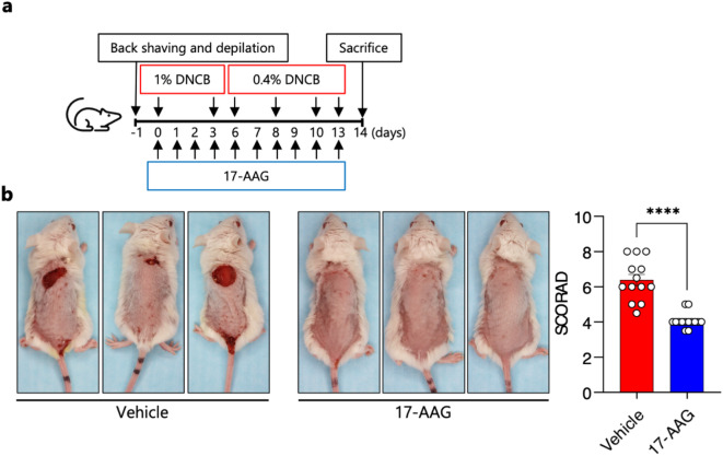
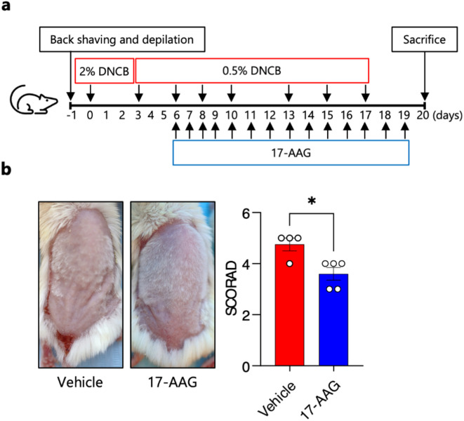
Heat shock proteins belonging to the HSP90 family promote inflammation and are potential therapeutic targets in inflammatory and autoimmune diseases. Here the effects of the HSP90 inhibitor 17-AAG applied topically were evaluated in a DNCB-induced murine model of atopic dermatitis (AD). The use of 17-AAG improved clinical disease activity without causing toxicity in the animals. Topical application of 17-AAG resulted in reduced epidermal hyperplasia, decreased expression of TSLP, IL-5, and IL-6, as well as reduced activation of NF-κB in the skin. In addition, the eosinophil proportion in the blood and eosinophil peroxidase (EPX) activity in the skin were significantly reduced in 17-AAG-treated AD mice. The inhibitory effects of 17-AAG on the production of epidermal alarmins, T-helper cell-associated cytokines, and ROS release were demonstrated in cultures of activated human keratinocytes, CD4+ T lymphocytes, and eosinophils, respectively. Finally, next-generation sequencing metagenomic approaches revealed that topical application of 17-AAG partially restored the normal gut microbiome in AD mice. Moreover, 17-AAG inhibited Staphylococcus aureus biofilm formation in vitro. The findings of this study, combined with the observed increase in HSP90 and EPX activity in the leukocytes of the analyzed cohort of AD patients, support the potential therapeutic use of HSP90 inhibitors in individuals with AD.

Keywords: Staphylococcus aureus; Eosinophiles; Heat shock proteins; Keratinocytes.; Microbiota; Mouse model.


Conflict of interest statement

Declarations. Competing interests: The authors declare no competing interests. Ethical approval: The study was conducted in accordance with the Declaration of Helsinki and was approved by the Independent Bioethics Committee for Scientific Research at the Medical University of Gdańsk, Poland (approval number NKBBN/209/2021). The animal study was reviewed and approved by the local authorities of the Animal Care and Use Committee in Bydgoszcz, Poland (approval number 53/2021).

Figures

Fig. 1
Fig. 1
Topical 17-AAG treatment reduces disease activity in an experimental mouse model of DNCB-induced human-like AD. (a) Schematic representation of experimental design. AD-like skin inflammation was induced in female BALB/c mice by dinitrochlorobenzene (DNCB) application to the shaved and depilated back skin (day − 1) as follows: 1% DNCB on days 0 and 3, followed by 0.4% DNCB on days 6, 8, 10 and 13. 17-AAG (0.5 µM) or vehicle (DMSO: acetone, 1:40 vol/vol) was applied each day with a 2-h interval to DNCB. (b) Representative images of vehicle- and 17-AAG-treated mice at the end of the 14-day treatment period and clinical disease severity of vehicle and 17-AAG-treated mice represented as cumulative SCORAD index. Data are expressed as mean ± SEM (with individual values) of three independent experiments. **** P < 0.0001.
Fig. 2
Fig. 2
Topical Application of 17-AAG Influences the Type 2 Immune Response in Mice with AD. (a) H&E- stained skin sections of naïve, vehicle- and 17-AAG-treated mice. Bars = 50 μm. (b) Relative expression of TSLP, IL-5, and IL-6 in the biopsies of mouse back skin, by qPCR. ELISA test results of (c) IgE level in the serum of mice and (d) NF-κB phosphorylation in the biopsies of mouse back skin. (e) Histological skin infiltration, as well as (f) the number of infiltrating leukocytes (CD45+), including (g) eosinophils (CD45+Siglec-F+) and (h) T helper cells (CD45+CD4+), were assessed in dissociated skin biopsies using flow cytometry. (i) Toluidine blue- stained skin sections of naïve, vehicle- and 17-AAG-treated mice. Bars = 50 μm. (j) Eosinophil proportion (% of WBC) in blood via hematology analyzer. (k) Histamine level in mouse serum, by ELISA. (l) Eosinophil peroxidase (EPX) activity, as measured by OPD assay in the back skin biopsies. Data are expressed as mean ± SEM (with individual values) or mean ± SD (in the case of epidermal thickness) of three independent experiments. * P < 0.05, ** P < 0.01, *** P < 0.001, **** P < 0.0001; ns, no significance; H&E, Hematoxylin and Eosin; OPD, o-phenylenediamine dihydrochloride; OD, optical density; WBC, white blood cells; e, epidermis; d, dermis.
Fig. 3
Fig. 3
17-AAG inhibits the expression of IL-33, the secretion of IL-6, T-helper cell-associated cytokines, and reactive oxygen species in cultures of activated human keratinocytes, CD4 + T lymphocytes, and eosinophils, respectively. TNF-α/IFN-γ- stimulated human keratinocytes (HaCaT) cells were cultured in presence of DMSO 0.1% (Vehicle) or various doses of 17-AAG. (a) Cell proliferation ELISA results as BrdU incorporation (%) after 6 h of incubation. (b) Relative expression of IL-33 and TSLP, by qPCR. (c) IL-6 levels in culture supernatant, by ELISA. (d) Phosphorylated STAT-1, STAT-3, and STAT-6 protein levels, corrected for β-Actin, were analyzed in HaCaT cell lysates following 1-hour or 24-hour activation using Western blot. Representative bands are presented. (e) NF-κB phosphorylation measured by ELISA. (f) FLG relative expression analyzed by qPCR. (g) IL-5, IL-17 A, and IL-22 secretion in the culture supernatant of anti-CD3/CD28 antibody-stimulated (1 µg/mL) CD4+ T cells derived from healthy donors, assessed by ELISA. (h) Luminol-enhanced ROS release in eosinophils derived from healthy donors, activated with fMLP (2 µM) or IL-33 (100 ng/mL). Data are presented as mean ± SD. * P < 0.05, ** P < 0.01, *** P < 0.001, **** P < 0.0001; ns, no significance; BrdU, BromodeoxyUridine; RLU, Relative Light Unit; AUC, Area Under Curve; ROS, Reactive Oxygen Species; OD, optical density.
Fig. 4
Fig. 4
Topical Application of 17-AAG Restores Both Skin and Gut Microbiota in a Mouse Model of AD. (a) Relative abundances of identified phyla and genera in back skin microbiome samples from naïve, vehicle-, and 17-AAG-treated AD mice, with (b) alpha diversity indices (Shannon, Chao1, and Simpson’s) calculated and (c) weighted UniFrac PCoA analysis presented. (d) Effects of 17-AAG on biofilm formation and (e) growth rate of S. aureus ATCC 25,923 and RA532, expressed as min-max range and mean ± SD, respectively. (f) Relative abundances of identified phyla and genera in gut microbiome samples from naïve, vehicle-treated, and 17-AAG-treated AD mice, with (g) alpha diversity indices (Shannon, Chao1, and Simpson’s) calculated and (h) weighted UniFrac PCoA analysis presented. Data expressed as mean (min-max range) or mean ± SD, respectively. Taxa with mean abundances < 1% across all groups are aggregated as “Low abundance.” * P < 0.05, ** P < 0.01, *** P < 0.001, **** P < 0.0001; OD, optical density.
Fig. 5
Fig. 5
Therapeutic treatment with topically applied 17-AAG reduces disease activity in an experimental mouse model of DNCB-induced human-like AD. (a) Schematic representation of the experimental design. AD-like skin inflammation was induced in female BALB/c mice by applying dinitrochlorobenzene (DNCB) to the shaved and depilated back skin (day − 1) as follows: 2% DNCB on day 0, followed by 0.5% DNCB on days 3, 6, 8, 10, 13, 15 and 17. 17-AAG (0.1 µM) or vehicle (DMSO: acetone, 1:40 vol/vol) was applied daily, with a 2-hour interval following DNCB application. (b) Representative images of vehicle- and 17-AAG-treated mice, and clinical disease severity, represented by the cumulative SCORAD index at day 20. Data are expressed as mean ± SEM (with individual values) from one experiment. * P < 0.05.
Fig. 6
Fig. 6
Elevated Activity of HSP90 and EPX in Leukocytes of Individuals with AD. (a) Relative HSP90 homolog expression measured in duplicate from 10 consecutive tape strips sampled from lesional and non-lesional skin of patients with AD. Data are expressed as mean ± SEM (with individual values). (b) HSP90 isoform level relative to total protein content of peripheral blood mononuclear cells. (c) HSP90 isoform level relative to total protein content of polymorphonuclear leukocytes. Both cell populations were derived from patients with AD and age- and gender-matched healthy individuals. Data are expressed as box-and-whisker plots (Tukey). (d) ELISA results showing acetyl-HSP90AA levels in PBMCs from AD patients compared to age- and gender-matched healthy controls. (e) β-Actin-corrected levels of acetyl-HSP90 in activated (TNF-α/IFN-γ, 10 ng/mL) human keratinocyte (HaCaT) cells treated with 0.1% DMSO or non-toxic concentrations of 17-AAG, analyzed by Western blot with representative bands presented. (f) Eosinophil peroxidase (EPX) activity in PMNs from AD patients and age- and gender-matched healthy controls. Data are expressed as box-and-whisker plots (Tukey) or mean ± SD. * P < 0.05, ** P < 0.01. ns, no significance.

Similar articles

References

    1. Backe, S. J., Sager, R. A., Woodford, M. R., Makedon, A. M. & Mollapour, M. Post-translational modifications of Hsp90 and translating the chaperone code. J. Biol. Chem.295, 11099–11117. 10.1074/jbc.REV120.011833 (2020). - PMC - PubMed
    1. Li, Z. N. & Luo, Y. HSP90 inhibitors and cancer: prospects for use in targeted therapies (Review). Oncol. Rep.4910.3892/or.2022.8443 (2023). - PMC - PubMed
    1. Stetz, G., Tse, A. & Verkhivker, G. M. Dissecting Structure-Encoded determinants of allosteric Cross-Talk between Post-Translational modification sites in the Hsp90 chaperones. Sci. Rep.8, 6899. 10.1038/s41598-018-25329-4 (2018). - PMC - PubMed
    1. Schopf, F. H., Biebl, M. M. & Buchner, J. The HSP90 chaperone machinery. Nat. Rev. Mol. Cell. Biol.18, 345–360. 10.1038/nrm.2017.20 (2017). - PubMed
    1. Tukaj, S. & Węgrzyn, G. Anti-Hsp90 therapy in autoimmune and inflammatory diseases: a review of preclinical studies. Cell. Stress Chaperones. 21, 213–218. 10.1007/s12192-016-0670-z (2016). - PMC - PubMed

MeSH terms

LinkOut - more resources