. 2025 Jul 1;15(1):22103.
doi: 10.1038/s41598-025-06485-w.

Impaired intestinal calcium absorption and osteopathy in ICR/Mlac-hydro mice with hypoparathyroidism and severe hydronephrosis

Affiliations

Impaired intestinal calcium absorption and osteopathy in ICR/Mlac-hydro mice with hypoparathyroidism and severe hydronephrosis

Panan Suntornsaratoon et al. Sci Rep. .

Abstract

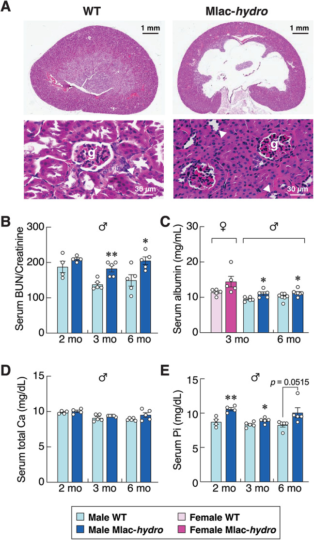
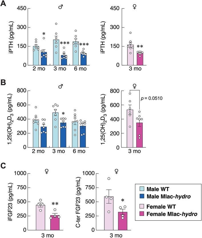
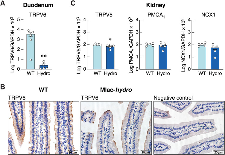
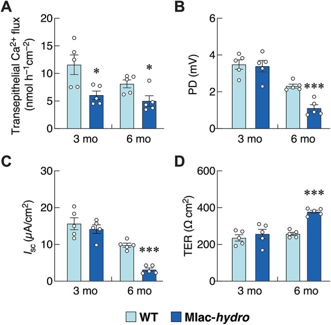
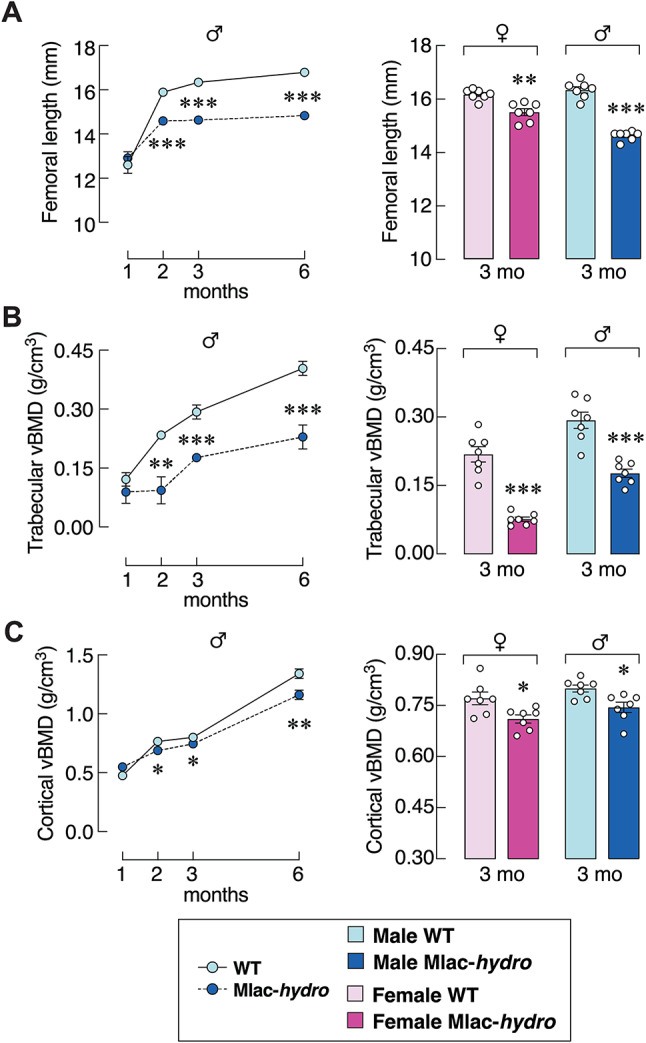
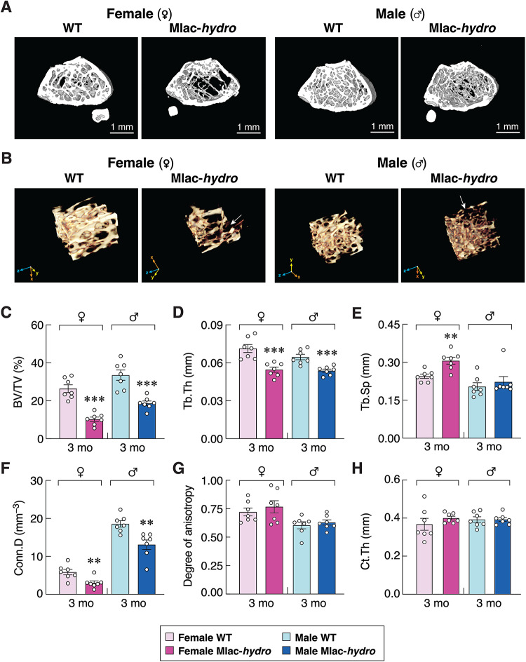
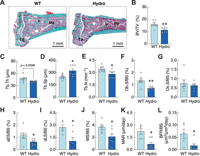
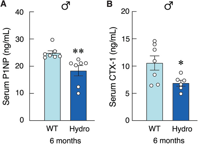
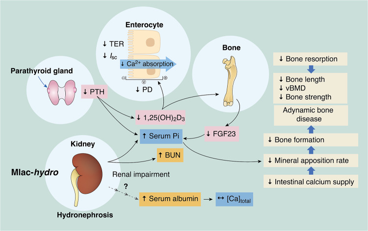
Abnormal fluid accumulation in the renal pelvis and calyces, with enlargement of the pelvicalyceal system, leads to a devastating disease known as hydronephrosis, which subsequently induces progressive renal impairment and mineral imbalance. Since the renal tubular cells play a role in the conversion of 25-hydroxyvitamin D3 to 1,25-dihydroxyvitamin D3 [1,25(OH)2D3], an important calciotropic hormone, we hypothesized that the ICR/Mlac-hydro mice with bilateral non-obstructive hydronephrosis and hypoparathyroidism plausibly manifested derangements of calcium and bone metabolism. The results showed that Mlac-hydro mice had reductions in the levels of intact parathyroid hormone, 1,25(OH)2D3 and fibroblast growth factor-23, along with downregulated TRPV6 expression in the duodenum and ~ 50% reduction in calcium flux as determined by 45Ca radioactive tracer. Aberrant duodenal electrical properties, i.e., decreased potential difference and increased transepithelial resistance, were also observed, indicating reduced intestinal ion permeability. Both male and female Mlac-hydro mice had shorter femoral lengths and lower volumetric bone mineral density than wild-type mice. Ultra-high resolution micro-computed tomography further revealed defects in the trabecular bone microstructure, consistent with several abnormalities of bone histomorphometric parameters, e.g., reductions in osteoblast surface, active erosion surface, mineral apposition rate and bone formation rate. Bone mechanical properties, i.e., maximum load, yield load, and stiffness, were also impaired in both male and female Mlac-hydro mice, as evaluated by the three-point bending test. In conclusion, Mlac-hydro mice with hydronephrosis and hypoparathyroidism exhibited several features of calcium dysregulation and bone defects, e.g., impaired intestinal calcium absorption, poor bone mechanical properties, and low bone turnover, the latter of which suggested an association between adynamic bone disease and hydronephrosis. Our data, therefore, provide relevant information essential for the future development of drugs or treatments for hydronephrotic patients.

Keywords: Bone loss; Calcium absorption; Hydronephrosis; Micro-computed tomography (µCT); Vitamin D.


Conflict of interest statement

Declarations. Competing interests: The authors declare no competing interests.

Figures

Fig. 1
Fig. 1
(A) Representative photomicrographs of cross-sectional renal sections stained with hematoxylin and eosin. Kidneys are collected from 3-month-old male Mlac-hydro mice and wild-type mice (WT). Glomerulus (g) and renal tubules (arrow heads) are identified. Scale bars of low magnification and high magnification images are 1 mm and 30 μm, respectively. Serum levels of (B) blood urea nitrogen (BUN)/creatinine, (C) albumin, (D) total calcium, (E) inorganic phosphate of three age groups, i.e., 2-, 3- and 6-month-old male ICR/Mlac-hydro mice (Mlac-hydro) and wild-type mice (WT). Serum albumin obtained from 3-month-old female mice are shown in panel C. n = 4–7; *P < 0.05, **P < 0.01 vs. age-matched WT control group.
Fig. 2
Fig. 2
Serum levels of (A) intact PTH (iPTH), (B) 1,25(OH)2D3 of three age groups, i.e., 2-, 3- and 6-month-old male ICR/Mlac-hydro mice (Mlac-hydro) and wild-type mice (WT). Serum levels of iPTH and 1,25(OH)2D3 of 3-month-old female mice are also shown in the right panel (n = 7). (C) Serum levels of intact FGF23 (iFGF23) and C-terminal FGF23 (C-ter FGF23) of 3-month-old female Mlac-hydro mice and WT mice (n = 4). *P < 0.05, **P < 0.01, ***P < 0.001 vs. age-matched WT control group.
Fig. 3
Fig. 3
(A) mRNA expression of TRPV6 in duodenal tissue from 3-month-old male ICR/Mlac-hydro mice (Hydro) and wild-type (WT) mice (n = 5). (B) Representative immunohistochemistry photomicrographs of TRPV6 protein in WT and Mlac-hydro mice. Positive red-brown signals of TRPV6 protein are predominantly localized at the apical membrane and within the cytoplasm of villous absorptive epithelial cells. Scale bars, 20 μm. (C) mRNA expression of TRPV5, PMCA1b, and NCX1 in kidney tissue from 3-month-old male Hydro and WT mice (n = 5). *P < 0.05, **P < 0.01 vs. age-matched WT control group. TRPV5, transient receptor potential cation channel subfamily V member 5; TRPV6, transient receptor potential cation channel subfamily V member 6; PMCA1b, plasma membrane Ca2+-ATPase 1b; NCX1, Na+/Ca2+ exchanger 1.
Fig. 4
Fig. 4
Transepithelial calcium flux (A) and epithelial electrical parameters [potential difference (PD; B), short-circuit current (Isc; C), and transepithelial electrical resistance (TER; D)] in the duodenum obtained from male ICR/Mlac-hydro mice (Mlac-hydro) and wild-type mice (WT). n = 5; *P < 0.05, ***P < 0.001 vs. WT control group.
Fig. 5
Fig. 5
(A) Femoral length, (B) trabecular and (C) cortical volumetric bone mineral density (vBMD) in 1-, 2-, 3- and 6-month-old male ICR/Mlac-hydro mice (Mlac-hydro) and wild-type mice (WT) analyzed by ex vivo µCT. Femoral length, trabecular and cortical vBMD of 3-month-old female and male mice are shown on the right panels. n = 7; *P < 0.05, **P < 0.01, ***P < 0.001 vs. age-matched WT control group.
Fig. 6
Fig. 6
(A) Representative µCT images (scale bars, 1 mm) and (B) ultra-high-resolution nanoCT images of the femoral metaphyses. The sites of trabecular separation are indicated by arrows. Microstructural analysis of distal femoral metaphysis in 3-month-old male and female ICR/Mlac-hydro and wild-type mice (WT) as determined by µCT, i.e., (C) trabecular bone volume normalized by tissue volume (BV/TV), (D) trabecular thickness (Tb.Th), (E) trabecular separation (Tb.Sp), (F) connectivity density (Conn.D), (G) degree of anisotropy. (H) Cortical thickness (Ct.Th) of femoral midshaft in 3-month-old male and female Mlac-hydro and WT determined by µCT. n = 7; **P < 0.01, ***P < 0.001 vs. age-matched WT control group.
Fig. 7
Fig. 7
Femoral mechanical properties, i.e., (A) maximum load, (B) yield load, (C) stiffness in 1-, 2-, 3- and 6-month-old male ICR/Mlac-hydro mice (Mlac-hydro) and wild-type mice (WT) analyzed by 3-point bending test. Maximum load, yield load, and stiffness of 3-month-old female and male mice are shown on the right panels. n = 7; *P < 0.05, **P < 0.01, ***P < 0.001 vs. age-matched WT control group.
Fig. 8
Fig. 8
(A) Representative photomicrographs of proximal tibial metaphysis of 6-month-old male ICR/Mlac-hydro mice (Hydro) and wild-type mice (WT) stained with Goldner’s trichrome. Scale bars, 1 mm. Epiphyseal plate (Ep), bone trabeculae (arrows), and bone marrow (Ma) are identified. Mineralized bone matrix, erythrocytes, and cytoplasm are stained green, orange, and red, respectively. (B–H) Microstructural analysis of proximal tibial metaphysis in 6-month-old male Hydro and WT mice determined by static bone histomorphometry. (B) trabecular bone volume normalized by tissue volume (BV/TV), (C) trabecular thickness (Tb.Th), (D) trabecular separation (Tb.Sp), (E) trabecular number (Tb.N), (F) osteoblast surface (Ob.S) normalized by bone surface (BS), (G) osteoclast surface (Oc.S) normalized by BS, (H) active erosion surface (aES) normalized by BS. (I–L) Bone microstructural analysis by dynamic bone histomorphometry. (I) double-labeled surface (dLS) normalized by BS, (J) mineralizing surface (MS) normalized by BS, (K) mineral apposition rate (MAR), (L) bone formation rate (BFR) normalized by BS. n = 7; *P < 0.05, **P < 0.01 vs. WT control group.
Fig. 9
Fig. 9
Serum levels of (A) procollagen type I N-terminal propeptide (PINP)—biomarker for bone formation, and (B) C-terminal telopeptide-1 (CTX-1)—biomarker for bone resorption, from 6-month-old male ICR/Mlac-hydro mice (Hydro) and wild-type mice (WT). n = 7; *P < 0.05, **P < 0.01, vs. WT control group.
Fig. 10
Fig. 10
Graphical illustration demonstrated effects of hydronephrosis on bone and calcium metabolism in ICR/Mlac-hydro mice. Degeneration of renal parenchyma in hydronephrotic mice could impair renal function as manifested by increase BUN and serum phosphate. The unaltered total calcium level in ICR/Mlac-hydro mice might be resulted from elevated serum albumin with unknown mechanism (dash arrow). Hydronephrotic mice manifests sub-normal iPTH, 1,25(OH)2D3 and FGF23 levels, that may lead to impairment of intestinal calcium absorption. Impaired intestinal calcium absorption prevents bone calcium accretion and mineral apposition, resulting in low bone formation and vBMD, deteriorated bone microstructure, defective bone elongation, eventually leading to compromised bone strength.

Similar articles

References

    1. Chang, A. & Laszik, Z. G. in Robbins & Cotran pathologic basis of disease (eds V. Kumar, A.K. Abbas, & J.C. Aster) Ch. 20, 895–952 (Elsevier, 2021).
    1. Pain, V. M. & Strandhoy, J. W. in In Campbell-Walsh Urology. 1227–1273 (eds Assimis, D. G.) (Saunders Elsevier, 2007).
    1. Nakada, T., Miyauchi, T., Sumiya, H. & Shimazaki, J. Nonobstructive urinary tract dilatation in nephrogenic diabetes insipidus. Int. Urol. Nephrol.22, 419–427 (1990). - PubMed
    1. Uribarri, J. & Kaskas, M. Hereditary nephrogenic diabetes insipidus and bilateral nonobstructive hydronephrosis. Nephron65, 346–349 (1993). - PubMed
    1. Fleet, J. C. Vitamin D-mediated regulation of intestinal calcium absorption. Nutrients14, 3351 (2022). - PMC - PubMed

MeSH terms

LinkOut - more resources