. 2025 Jul 1;8(1):194.
doi: 10.1038/s42004-025-01581-4.

A small molecule enhances arrestin-3 binding to the β2-adrenergic receptor

Affiliations

A small molecule enhances arrestin-3 binding to the β2-adrenergic receptor

Han Kurt et al. Commun Chem. .

Abstract

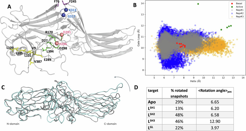
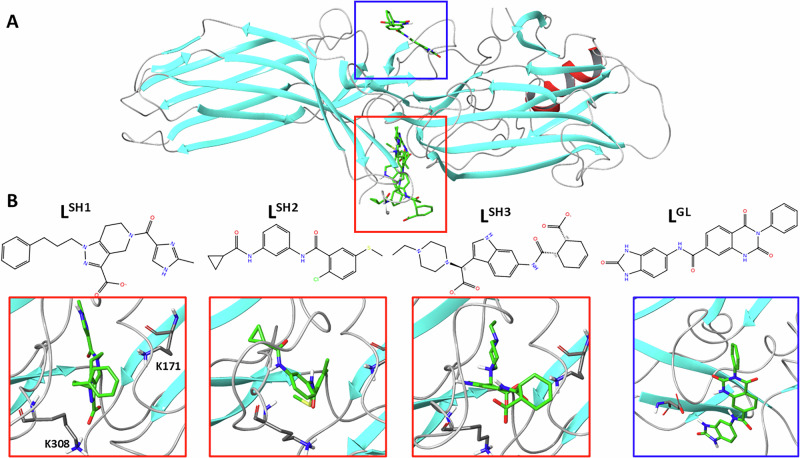
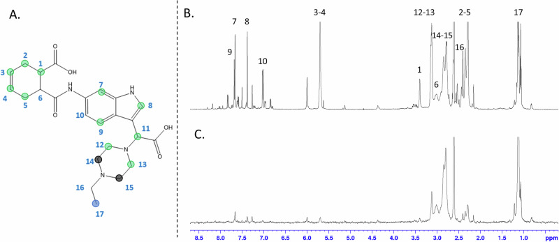
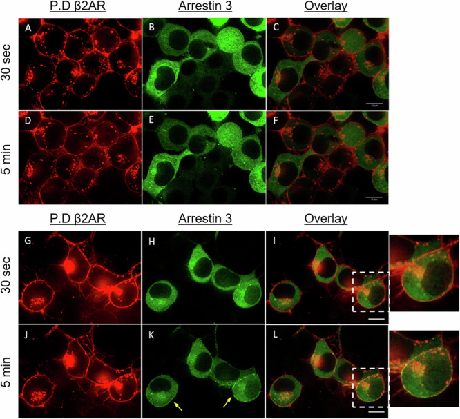
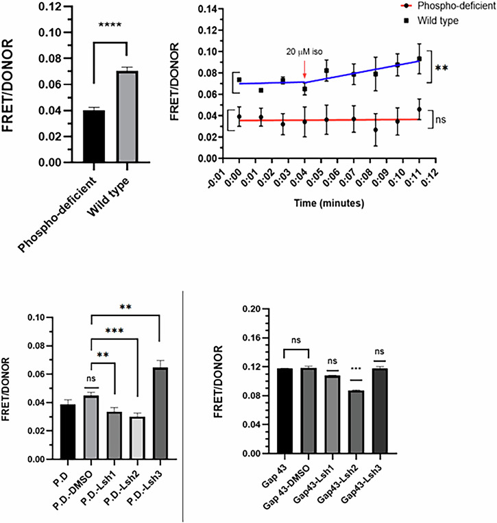
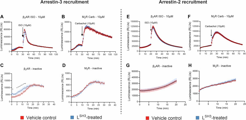
Excessive signaling by various GPCRs underlies a variety of human disorders. Suppression of GPCRs by "enhanced" arrestin mutants was proposed as therapy. We hypothesized that GPCR binding of endogenous arrestins can be increased by small molecules stabilizing pre-activated conformation. Using molecular dynamics, we identified potentially druggable pockets in pre-activated conformation of arrestin-3 and discovered a compound targeting one of these pockets. Saturation-transfer difference NMR data showed that the compound binds at the back loop of arrestin-3. FRET- and NanoBiT-based assays in living cells showed that the compound increased in-cell arrestin-3, but not arrestin-2, binding to basal β2-adrenergic receptor and its phosphorylation-deficient mutant, but not to muscarinic M2 receptor. These experiments demonstrated the feasibility of enhancing the binding of endogenous wild type arrestin-3 to GPCRs in a receptor-specific and arrestin-subtype selective manner.


Conflict of interest statement

Competing interests: The authors declare no competing interests.

Figures

Fig. 1
Fig. 1. Conformational states and N-C rotation angles of arrestin-3 complexes revealed by MD simulations.
A Residue positions K313 and G317 at the termini of the back loop (blue), and D291 and H296 for gate loop (pink) are shown on the 3D structure of arrestin-3 (PDB ID: 3P2D) together with the residues of the polar core (green) and three-element region (yellow) as well as inter-domain aromatic core (purple). B Distribution of the distances used to identify the conformational state of the gate loop and the back loop in arrestin-3 trajectories and those measured in active/pre-activated and inactive structures of arrestins (See Supplementary Table 1 for the complete list of structures). Values for inactive and receptor-bound structures are shown in red and green dots, respectively. Notably, inositol hexakisphosphate-bound arrestin-3 (PDB ID:5TV1) adopts an extremely long distance (See green dot outlier) at the gate loop, due to the perturbation induced by the IP6 molecule near the polar core. C Superposition of traces of two extreme conformations from the principal component analysis of arrestin-3 structures, aligned on the N terminal domain, showing the rotation axis and direction (See Methods). D Table summarizing the occurrence of rotated states in arrestin-3-compound complexes and the average rotation angle.
Fig. 2
Fig. 2. The interfaces targeted by compounds and chemical structures of the compounds.
A All atom structure of basal arrestin-3 highlighting the two interface regions targeted by drug discovery: the back loop characterized by the short helix (red box) and the gate loop (blue box). B Structures of the candidate compounds selected after virtual screening and close-up of complexes: from left to right, LSH-1, LSH-2, LSH-3, LGL.
Fig. 3
Fig. 3. The epitope map achieved by LSH-3.
A Epitope map of LSH-3 with relative STD percentages at initial slope conveyed by color code, acquired at saturation time of 2 s, irradiating at 0 ppm. Dark blue dots indicate the most intense signal (100% relative STD), light blue dots between 20% and 80% and green dots under 20%. B 1H spectrum of LSH-3 C. STD spectrum of LSH-3 in the presence of arrestin-3.
Fig. 4
Fig. 4. (A-F) Spinning disc confocal microscopy images of N2a cells transfected with β2AR-P.D.-mCh., arrestin-3-177-mEGFP and wild-type GRK2.
A Taken from mCh. channel after 30 s Iso stimulation. B Taken from EGFP channel after 30 s Iso stimulation. C Overlay of A and B. D Taken from mCh. channel after 5 min Iso stimulation E Taken from EGFP channel After 5 min Iso stimulation. F Overlay of (D) and (E). (G–L) Spinning disc confocal microscopy images of N2a cells transfected with β2AR- mCh., arrestin-3-177 mEGFP, and wild-type GRK2. Spinning disc confocal microscopy images of N2a cells transfected with β2AR- mCh., arrestin-3-177 mEGFP, and GRK2. G Taken from mCh. channel after 30 s Iso stimulation. H Taken from EGFP channel after 30 s Iso stimulation. I Overlay of (G) and (H). J Taken from mCh. channel after 5 min Iso stimulation K Taken from EGFP channel After 5 min Iso stimulation. Yellow arrows show arrestin-3 colocalizations with β2AR. L Overlay of J and K. Magnification 63X, scale bars are 10 µm.
Fig. 5
Fig. 5. FRET levels in N2a cells measured under different stimulation conditions.
Upper left panel: Basal FRET levels in N2a cells expressing phosphorylation-deficient β2AR-mCherry, arrestin-3-177-mEGFP, GRK2, or β2AR-mCherry, arrestin-3-177-mEGFP, GRK2. Upper right panel: Calculated FRET levels in N2a cell before and after 20 µM Iso stimulation. Means ± s.e.m. are shown. **p < 0005, ****p < 0.0001 statistically significant differences. ns: no significant differences. Lower left panel: LSH-3 chemical (1H-Indole-3-acetic acid, 6-[[(6-carboxy-3-cyclohexen-1-yl) carbonyl] amino] - α- (4-ethyl- 1- piperazinyl) increases FRET between b2AR phosphorylation-deficient–mCh., and arrestin-3-177-mEGFP. Each chemical was tested in two separate experiments n = 6. Means ± s.e.m. are shown. **p < 0005, ***p < 0001, statistically significant differences. ns: no significant differences. Lower right panel: Chemical interaction with membrane targeting sequence fused mCh. and mEGFP transfected N2a cells. Means ± s.e.m. are shown. ***p < 0.001, statistically significant differences, ns: no significant differences. Chemical-treated samples were compared with Gap 43 vehicle (1% DMSO).
Fig. 6
Fig. 6. Arrestin-3 recruitment to inactive β2-adrenergic receptor (β2AR).
HEK293 arrestin2/3 KO cells were co-transfected with plasmids encoding arrestin-3 (A–D) or arrestin-2 (E–H), both with N-terminal SmBiT and β2AR (A, C, E, and G) or M2R (B, D, F, and H) with C-terminal LgBiT. The cells were treated with 100 mM LSH3 or DMSO (vehicle control) for 30 min prior to the addition of luciferase substrate (time point 0). Agonist (10 mM isoproterenol (ISO) for β2AR or 10 mM carbachol for M2R) was added at 30 min. A Arrestin-3 recruitment to β2AR. B Arrestin-3 recruitment to M2R. C Arrestin-3 recruitment to inactive β2AR (no agonist). D Arrestin-3 recruitment to inactive M2R. E Arrestin-2 recruitment to β2AR. F Arrestin-2 recruitment to M2R. G Arrestin-2 recruitment to inactive β2AR. H Arrestin-2 recruitment to inactive M2R. Data are presented as means ± SEM (N = 6). Statistical significance was determined by two-way repeat measures ANOVA followed by Fisher’s LSD post hoc test and indicated, as follows: *, p < 0.05.

Update of

Similar articles

References

    1. Hauser, A. S., Attwood, M. M., Rask-Andersen, M., Schiöth, H. B. & Gloriam, D. E. Trends in GPCR drug discovery: new agents, targets and indications. Nat. Rev. Drug Discov.16, 829–842 (2017). - PMC - PubMed
    1. Weis, W. I. & Kobilka, B. K. The molecular basis of G protein-coupled receptor activation. Annu. Rev. Biochem.87, 897–919 (2018). - PMC - PubMed
    1. Gimenez, L. E. et al. Role of receptor-attached phosphates in binding of visual and non-visual arrestins to G Protein-coupled receptors. J. Biol. Chem.287, 9028–9040 (2012). - PMC - PubMed
    1. Lohse, M. J., Benovic, J. L., Codina, J., Caron, M. G. & Lefkowitz, R. J. β-arrestin: a protein that regulates β-adrenergic receptor function. Science248, 1547–1550 (1990). - PubMed
    1. Moore, C. A. C., Milano, S. K. & Benovic, J. L. Regulation of receptor trafficking by GRKs and arrestins. Annu. Rev. Physiol.69, 451–482 (2007). - PubMed

LinkOut - more resources