

Using Phantomless QCT for evaluating BMD evolution in maintenance hemodialysis patients
- PMID: 40593118
- PMCID: PMC12216142
- DOI: 10.1038/s41598-025-07025-2
Using Phantomless QCT for evaluating BMD evolution in maintenance hemodialysis patients
Abstract
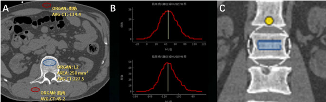
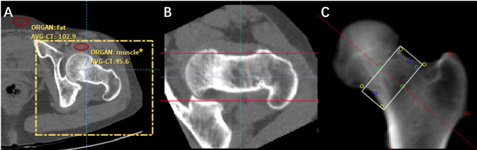
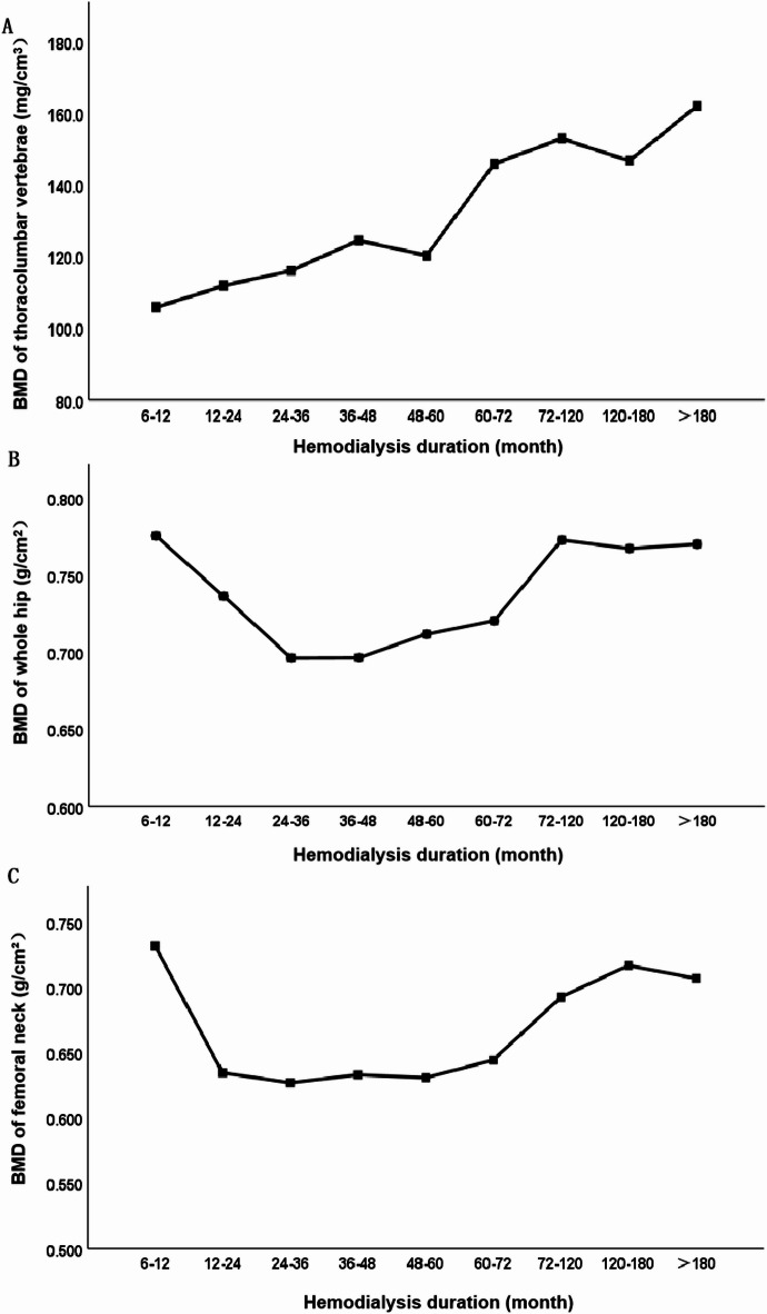
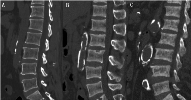
This study was aimed to investigate the evolution of bone mineral density (BMD) in patients with maintenance hemodialysis (MHD) by using phantom-less quantitative computed tomography (PL-QCT). We collected patients with MHD in Suzhou Hospital of Nanjing Medical University from September 2020 to December 2023 as the prospective observation group. BMD of thoracolumbar vertebra, total hip and femoral neck were measured by PL-QCT. Patients with MHD were divided into 9 groups according to hemodialysis duration. Chest CT scans of patients in prospective observation group were collected during the first three months of MHD and 1 year, 2 years, 3 years after dialysis between January 2017 and December 2023 as the retrospective observation group, and BMD of the twelfth thoracic vertebra was measured. According to the BMD changes among the prospective observation group and the retrospective observation group, the evolution of thoracolumbar vertebral BMD, whole hip BMD and femoral neck BMD were comprehensively analyzed. BMD of thoracolumbar vertebra gradually decreased within 36 months in patients with MHD. Thoracolumbar vertebral BMD tended to increase when hemodialysis duration was more than 36-48 months, and thoracolumbar vertebral BMD increased significantly with hemodialysis duration when hemodialysis duration was more than 60 months, and significantly exceeded the BMD before MHD. BMD of total hip and femoral neck gradually decreased within 36 months in patients with MHD. BMD of total hip and femoral neck increased with hemodialysis duration when hemodialysis duration was more than 72 months, but was almost the same as that of the first year of MHD. In the follow-up evaluation of BMD in MHD patients, it is recommended to use QCT to measure BMD in thoracolumbar vertebrae or hip the first 3 years of MHD, and use QCT to measure BMD in thoracolumbar vertebrae to evaluate changes over 5 years of MHD.
Keywords: Bone mineral density; Evolution; Hemodialysis duration; Maintenance hemodialysis; Phantom-less quantitative computed tomography.
© 2025. The Author(s).
Conflict of interest statement
Declarations. Competing interests: The authors declare no competing interests.
Figures
Similar articles
-
Relationship of lumbar vertebral volumetric bone mineral density with trunk muscle density assessed by quantitative computed tomography in maintenance hemodialysis patients.Ren Fail. 2025 Dec;47(1):2534844. doi: 10.1080/0886022X.2025.2534844. Epub 2025 Jul 23. Ren Fail. 2025. PMID: 40697034 Free PMC article.
-
Does Augmenting Irradiated Autografts With Free Vascularized Fibula Graft in Patients With Bone Loss From a Malignant Tumor Achieve Union, Function, and Complication Rate Comparably to Patients Without Bone Loss and Augmentation When Reconstructing Intercalary Resections in the Lower Extremity?Clin Orthop Relat Res. 2025 Jun 26. doi: 10.1097/CORR.0000000000003599. Online ahead of print. Clin Orthop Relat Res. 2025. PMID: 40569278
-
Two-year follow-up study (PRIMROSE 3) to assess bone mineral density in subjects with uterine fibroids completing the PRIMROSE 1 and PRIMROSE 2 linzagolix trials.Hum Reprod Open. 2025 May 13;2025(3):hoaf025. doi: 10.1093/hropen/hoaf025. eCollection 2025. Hum Reprod Open. 2025. PMID: 40575397 Free PMC article.
-
Calcium and vitamin D for increasing bone mineral density in premenopausal women.Cochrane Database Syst Rev. 2023 Jan 27;1(1):CD012664. doi: 10.1002/14651858.CD012664.pub2. Cochrane Database Syst Rev. 2023. PMID: 36705288 Free PMC article.
-
Intravenous magnesium sulphate and sotalol for prevention of atrial fibrillation after coronary artery bypass surgery: a systematic review and economic evaluation.Health Technol Assess. 2008 Jun;12(28):iii-iv, ix-95. doi: 10.3310/hta12280. Health Technol Assess. 2008. PMID: 18547499
References
-
- Isakova, T. et al. KDOQI US commentary on the 2017 KDIGO clinical practice guideline update for the diagnosis, evaluation, prevention, and treatment of chronic kidney Disease-Mineral and bone disorder (CKD-MBD). Am. J. Kidney Dis.70, 737–751. 10.1053/j.ajkd.2017.07.019 (2017). - PubMed
MeSH terms
Grants and funding
LinkOut - more resources
Full Text Sources
Medical
