. 2025 Jul 1;15(1):20678.
doi: 10.1038/s41598-025-07121-3.

TRBP modulates RLR signaling by inhibiting PKR-mediated antiviral stress granule formation

Affiliations

TRBP modulates RLR signaling by inhibiting PKR-mediated antiviral stress granule formation

Koji Onomoto et al. Sci Rep. .

Abstract

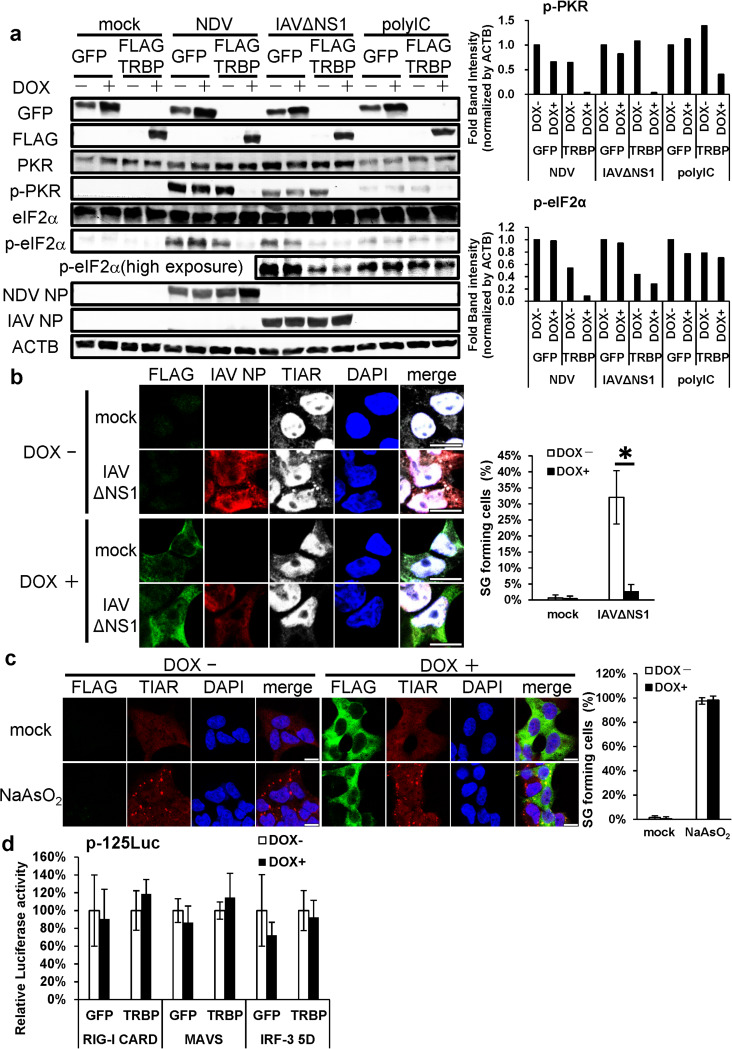
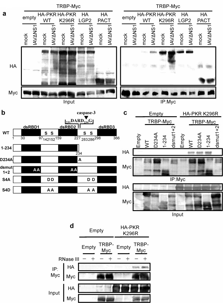
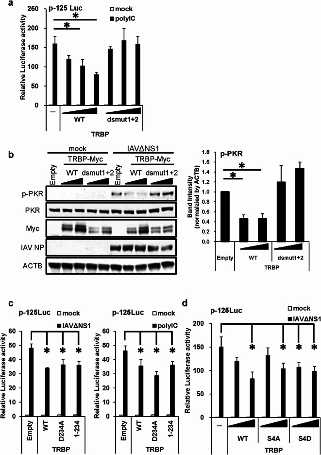
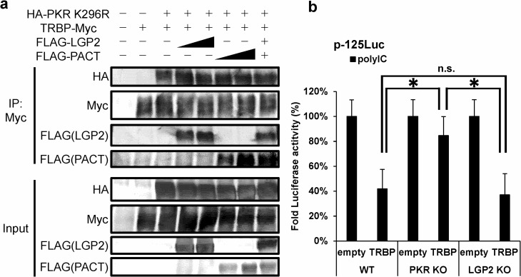
Stress granules (SGs) are dense aggregates of RNA and proteins that form in response to various cellular stresses. Virus-induced SGs, known as antiviral SGs (avSGs), play a crucial role in regulating retinoic acid-inducible gene I-like receptors (RLRs)-mediated antiviral innate immunity. However, the regulation of avSG formation remains not fully understood. In this study, we demonstrate that TAR-RNA binding protein (TRBP), an RNA silencing regulator, negatively regulates type I interferon (IFN) expression by inhibiting avSG formation in response to RNA virus infection. Overexpression of TRBP inhibits both IFN-β promoter activity and avSG formation following viral infection or the viral RNA mimic, polyinosinic-polycytidylic acid transfection. TRBP knockout cells exhibit enhanced phosphorylation and activation of IFN regulatory factor-3 (IRF-3) and increased IFN-β mRNA expression compared to wild-type cells. Additionally, depletion of G3BP1 and G3BP2, which are essential for SG formation, abolishes the inhibitory effect of TRBP on IRF-3 phosphorylation. Mechanistically, TRBP physically interacts with double-stranded RNA (dsRNA)-dependent protein kinase R (PKR), a key kinase involved in avSG formation, via its dsRNA-binding domains, and inhibits PKR activation. In summary, our findings reveal a novel function for TRBP as a negative regulator of RLR-mediated signaling through PKR-dependent inhibition of avSG formation.

Keywords: Antiviral innate immunity; IFN; RLR; Stress granule; TRBP.


Conflict of interest statement

Declarations. Competing interests: The authors declare no competing interests.

Figures

Fig. 1
Fig. 1
TRBP negatively regulates RLR-mediated IFN signal. (a–c) L929 cells were transfected with the IFN reporter gene (p-125Luc) and internal control (pRL-tk) vectors, together with the expression vector for Empty or TRBP. After 48 h transfection, the cells were mock-treated (mock) or stimulated by IAVΔNS1 infection (a), polyIC transfection (b), and SenV infection (c) for 12 h and subjected to a dual-luciferase assay. (d) HeLa WT and TRBP KO cells (#1, #2, #3) were mock-treated (mock) or transfected with polyIC for 9 h, and IFN-β mRNA expression levels were determined by qRT-PCR. (e) HeLa WT, TRBP KO (#2, #3), and FLAG-TRBP- expressing TRBP KO cells were infected with IAVΔNS1. After 18 h incubation, cell extracts were prepared and subjected to SDS-PAGE, and immunoblotted using antibodies against phospho-IRF-3 (Ser386), IRF-3, TRBP, FLAG-tag, IAV NP, and β-actin (ACTB). The intensities of the p-IRF-3 bands were normalized against ACTB, measured using ImageJ software. The data represent the mean results of three independent experiments. (f) Flp-In 293 CBP-SBP-GFP (GFP) or FLAG-TRBP (TRBP) cells were treated with doxycycline (Dox) 0 or 1.0 µg/mL for 16 h. The cells were infected with IAVΔNS1 for 9 h, and IFN-β mRNA level was determined by qRT-PCR. (g) Flp-In 293 CBP-SBP-GFP (GFP) (left) or FLAG-TRBP (FLAG) (right) cells were treated with DOX for 16 h and then the cells were mock-transfected or transfected with short or long polyIC for 9 h. IFN-β mRNA expression levels were determined by qRT-PCR. At least two independent experiments represent similar results, and the bars indicate one representative; each bar shows the means and ± the standard deviation (S.D.) of the technical triplicate. P-values were determined by Student’s t-test (*P < 0.05).
Fig. 2
Fig. 2
TRBP inhibits PKR activation and avSG formation. (a) Flp-In 293 CBP-SBP-GFP (GFP) or FLAG-TRBP cells were treated with doxycycline (Dox) 0 or 1.0 µg/mL for 14 h. The cells were mock-treated (mock), infected with NDV or IAVΔNS1, or transfected with polyIC. After 12 h incubation, cell extracts were prepared and subjected to SDS-PAGE, and immunoblotted using antibodies against GFP, FLAG-tag, PKR, phospho-PKR (Thr 446), eIF2α, phospho-eIF2α (Ser 51), NDV NP, IAV NP, and ACTB. The intensity of the p-PKR and p-eIF2α bands was normalized against ACTB, measured using ImageJ software. (b) Flp-In 293 FLAG-TRBP cells were treated with DOX 0 or 1.0 µg/mL for 24 h, and infected with IAVΔNS1 for 12 h. The cells were stained with anti-FLAG (Green = TRBP), anti-IAV NP (Red), anti-TIAR (Gray) antibodies, and DAPI (Blue). The white scale bar corresponds to 10 μm. The percentages of SG-containing cells were calculated in more than 10 randomly chosen fields for each slide (n > 660 cells). (c) Flp-In 293 FLAG-TRBP cells were treated with DOX 0 or 1.0 µg/mL for 24 h, and treated with NaAsO2 (0.5 mM) for 30 min and stained with anti-FLAG (Green = TRBP), anti-TIAR (Red) antibodies and DAPI (Blue). The white scale bar corresponds to 10 μm. The percentages of SG-containing cells were calculated in more than six randomly chosen fields for each slide (n > 280 cells). (d) Flp-In 293 CBP-SBP-GFP (GFP) or FLAG-TRBP (TRBP) cells were transiently transfected with p-125Luc and pRL-tk reporter vectors, together with the RIG-I CARD, MAVS, and IRF-3-5D expressing vectors, respectively. After 24 h transfection, the cells were treated with DOX 0 (-) or 1.0 (+) µg/mL for 24 h and subjected to a dual-luciferase assay. Luciferase activity is normalized against each DOX (-) as 100%. At least two independent experiments represent similar results, and the bars indicate the one representative; each bar shows the means and ± the standard deviation (S.D.) of the technical triplicate. P-values were determined by Student’s t-test (*P < 0.05).
Fig. 3
Fig. 3
avSG formation is indispensable for the negative regulation of IFN signal via TRBP. (a) HEK293T WT and G3BP1/2 dKO cells were mock-treated, infected with IAVΔNS1 for 9 h, or treated with NaAsO2 (0.5 mM) for 30 min. The cells were stained with anti-Caprin1 (Green), anti-TIAR (Red) antibodies, and DAPI (Blue). A merged image at the original magnification is shown in the rightmost panel. The magnified images correspond to the boxed region in each panel. The white scale bar corresponds to 40 μm. (b) HEK293T WT and G3BP1/2 dKO cells were transfected with empty vector (-) or Myc-tagged TRBP (+) expression vector. The cells were mock-treated or infected with IAVΔNS1. After 12 h, the cell extracts were subjected to SDS-PAGE, and immunoblotted using antibodies against phospho-PKR (Thr 446), PKR, phospho-IRF-3 (Ser386), IRF-3, G3BP1, G3BP2, Myc-tag, IAV NP, RIG-I, MDA5 and ACTB. The intensities of the p-PKR and p-IRF-3 bands were normalized against ACTB, measured using ImageJ software. The data represents the mean results and ± the standard deviation (S.D.) of five independent experiments. P-values were determined by Student’s t-test (*P < 0.05).
Fig. 4
Fig. 4
TRBP interacts with PKR without dsRNA through dsRBD. (a) HEK293T cells were transfected with Myc-tagged TRBP (TRBP-Myc) and empty vector, HA-tagged PKR WT, PKR K296R, LGP2, or PACT. After 48 h incubation, the cells were infected with IAVΔNS1 for 12 h and cell extracts were subjected to immunoprecipitation (IP) with anti-Myc-antibody. Input and IP samples were subjected to SDS-PAGE and immunoblotted using antibodies against Myc-tag and HA-tag. (b) Schematic diagram of TRBP domain structure and the mutants. The lysine (K) at positions 80 and 81 in double-stranded RNA-binding domain (dsRBD) 1 and 210 and 211 in dsRBD2 were substituted with alanine (A) in the TRBP-dsRBDmt1 + 2 mutant (dsmut1 + 2). The serine (S) at positions 142, 152, 283, and 286 were substituted with alanine (A) or aspartic acid (D) in the TRBP S4A and S4D mutants, respectively. The arrowheads indicate the caspase-3 cleavage site. (c) HEK293T cells were transfected with empty vector, Myc-tagged TRBP WT, D234A, 1-234, or dsmut1 + 2 together with empty vector or HA-tagged PKR K296R. After 48 h incubation, cell extracts were subjected to IP with anti-Myc-antibody. Input and IP samples were subjected to SDS-PAGE and immunoblotted using antibodies against Myc-tag and HA-tag. (d) HEK293T cells were transfected with empty vector, Myc-tagged TRBP WT, and HA-tagged PKR K296R. After 48 h incubation, cell extracts were subjected to IP with anti-Myc-antibody. RNase III was added to the lysis buffer to remove dsRNA. The input and IP samples were subjected to SDS-PAGE and immunoblotted using antibodies against Myc-tag and HA-tag.
Fig. 5
Fig. 5
dsRBDs of TRBP are essential to regulate avSG-mediated IFN signal. (a) L929 cells were transfected with p-125Luc and pRL-tk reporter vectors, together with the empty (-), TRBP WT, or TRBP dsmut1 + 2 (0.25, 0.625, or 1.25 µg) expression vectors. The cells were mock-treated or infected with polyIC for 12 h and subjected to a dual-luciferase assay. Data represent relative firefly luciferase activity, normalized to Renilla luciferase activity. (b) HEK293T cells were transfected with Myc-tagged TRBP (TRBP-Myc) WT or dsmut 1 + 2. The cells were mock-treated or infected with IAVΔNS1 for 18 h. Cell extracts were prepared and subjected to SDS-PAGE, and immunoblotted using antibodies against phospho-PKR (Thr 446), PKR, IAV NP, Myc-tag, and ACTB. The intensities of the p-PKR bands were normalized against ACTB, measured using ImageJ software. The data represent the means of three independent experiments. (c) L929 cells were transfected with p-125Luc and pRL-tk reporter vectors, together with the empty, TRBP WT, D234A, or 1-234 expression vectors. The cells were mock-treated or infected with IAVΔNS1 (left) or transfected with polyIC (right) for 12 h and subjected to a dual-luciferase assay. Data represent relative firefly luciferase activity, normalized to Renilla luciferase activity. (d) L929 cells were transfected with p-125Luc and pRL-tk reporter vector, together with the empty, TRBP WT, S4A, or S4D (0.25 or 1.25 µg) expression vectors. The cells were mock-treated or infected with IAVΔNS1 for 12 h and subjected to a dual-luciferase assay. Data represent relative firefly luciferase activity, normalized to Renilla luciferase activity. At least two independent experiments represent similar results, and the bars indicate one representative; each bar shows the means and ± the standard deviation (S.D.) of the technical triplicate. P-values were determined by Student’s t-test (*P < 0.05).
Fig. 6
Fig. 6
LGP2 and PACT are not involved in the interaction between TRBP and PKR. (a) HEK293T cells were transfected with empty vector, Myc-tagged TRBP WT, or HA-tagged PKR K296R together with empty vector, FLAG-LGP2, FLAG-PACT, or both. After 48 h incubation, cell extracts were subjected to IP with anti-Myc-antibody. Input and IP samples were subjected to SDS-PAGE and immunoblotted using antibodies against HA-tag, Myc-tag, and FLAG-tag. (b) WT, PKR KO, or LGP2 KO MEF cells were transfected with p-125Luc and pRL-tk reporter vectors, together with the empty or TRBP WT expression vector. The cells were transfected with polyIC for 12 h and subjected to a dual-luciferase assay. Data represent luciferase activity normalized against MEF transfected with empty vector followed by polyIC transfection as 100%. At least two independent experiments represent similar results, and the bars indicate one representative; each bar shows the means and ± the standard deviation (S.D.) of the technical triplicate. P-values were determined by Student’s t-test (*P < 0.05).

Similar articles

References

    1. Yoneyama, M. et al. The RNA helicase RIG-I has an essential function in double-stranded RNA-induced innate antiviral responses. Nat. Immunol.5, 730–737. 10.1038/ni1087 (2004). - PubMed
    1. Yoneyama, M. et al. Shared and unique functions of the DExD/H-box helicases RIG-I, MDA5, and LGP2 in antiviral innate immunity. J. Immunol.175, 2851–2858. 10.4049/jimmunol.175.5.2851 (2005). - PubMed
    1. Onomoto, K., Onoguchi, K. & Yoneyama, M. Regulation of RIG-I-like receptor-mediated signaling: interaction between host and viral factors. Cell. Mol. Immunol.18, 539–555. 10.1038/s41423-020-00602-7 (2021). - PMC - PubMed
    1. Takahasi, K. et al. Nonself RNA-sensing mechanism of RIG-I helicase and activation of antiviral immune responses. Mol. Cell.29, 428–440. 10.1016/j.molcel.2007.11.028 (2008). - PubMed
    1. Takahasi, K. et al. Solution structures of cytosolic RNA sensor MDA5 and LGP2 C-terminal domains: identification of the RNA recognition loop in RIG-I-like receptors. J. Biol. Chem.284, 17465–17474. 10.1074/jbc.M109.007179 (2009). - PMC - PubMed

MeSH terms

LinkOut - more resources