. 2025 Jul 1;15(1):21129.
doi: 10.1038/s41598-025-08329-z.

CXCL10-dependent epithelial-vascular cross-talk for endothelial activation following SARS-CoV-2 infection

Affiliations

CXCL10-dependent epithelial-vascular cross-talk for endothelial activation following SARS-CoV-2 infection

Chaillot Laura et al. Sci Rep. .

Abstract

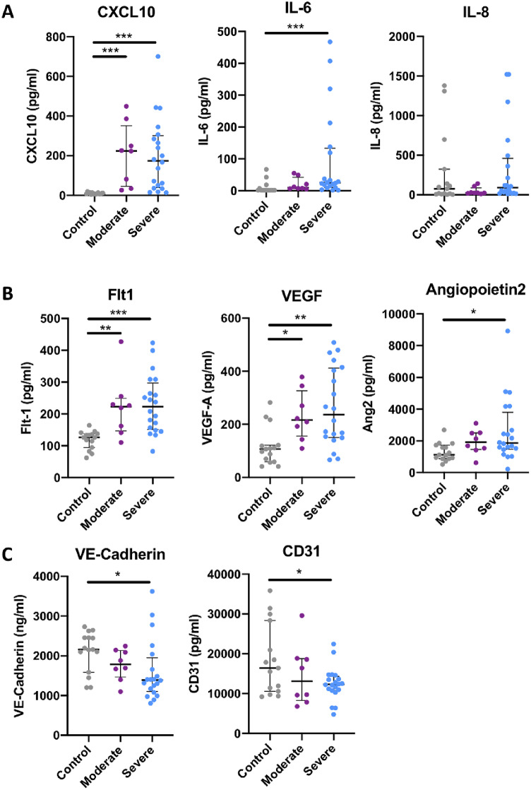
The blood vessel network is heavily impacted by SARS-CoV-2 infection. How SARS-CoV-2 contributes to vascular inflammation and whether epithelio-endothelial cross-talk is involved remain unclear. We investigated in detail the interaction between SARS-CoV-2 and the vasculature using 2D and 3D vesseloid in vitro models. We first assessed whether SARS-CoV-2 is able to directly infect endothelial cells. In the absence of ACE2 in endothelial cells, no productive infection was detected. Low uptake of viral particles by ACE2-overexpressing endothelial cells was observed without efficient viral production. Thus, the indirect effect of SARS-CoV-2 infection may involve epithelio-endothelial cell cross-talk. After infection of the epithelial cells, a significant inflammatory response was detected in the endothelial cells. CXCL10 was the most highly expressed proinflammatory cytokine involved in this intercellular communication, and its function was subsequently explored. Finally, the clinical relevance of our findings was confirmed in two patient cohorts.

Keywords: CXCL10; Chemokines; Endothelium; SARS-CoV-2.


Conflict of interest statement

Declarations. Competing interests: The authors declare no competing interests.

Figures

Fig. 1
Fig. 1
Absence of direct effects of SARS-CoV-2 on the vascular endothelium. (A) Western blot analysis of the ACE2 protein in HUVECs, HUAECs, HCAECs, Calu-3 cells and ACE2 overexpressing HUVECs cells. Tubulin was used as a control. (B) RT‒qPCR quantification of N gene RNA in media after vesseloid or Calu-3 cell SARS-CoV-2 infection. Analysis was performed at 3- or 4-days post infection (MOI = 0.1). The values were normalized to a range of RNA extracted from the virus we use to infect, for which we know the TCID50 and PFU. The results are presented as the mean ± SEM (one-way ANOVA *P < 0.05). (C) Relative N gene RNA expression in endothelial cells from vesseloids or Calu-3 cells at 3 days post infection with SARS-CoV-2 (MOI = 0.1). (D) Immunofluorescence analysis of cleaved-Caspase-3 (c-Caspase-3) in vesseloids 3 days post infection (MOI = 0.1). The fluorescence intensity per nucleus was quantified. The results are presented as the means ± SEMs.
Fig. 2
Fig. 2
The inflammatory response of ECs is mediated by epithelial-derived CXCL10. (A) Calu-3 cells were infected with SARS-CoV-2 (MOI = 1) for 4 h and plated at the bottom of the transwell chamber before they were cocultured with vesseloids placed in the upper chamber for 3 days. (B) Relative VCAM-1 mRNA expression in vesseloids infected with SARS-CoV-2 alone or after coculture with Calu-3 cells infected with SARS-CoV-2. Analyze was performed 3 days post infection (MOI = 0.1). The values were normalized according to tubulin mRNA expression. The results are presented as the means ± SEMs (unpaired t test; *P < 0.05). (C) Heatmap of pixel density quantification from the cytokine array assay for selected chemokines and cytokines from vesseloids infected alone or after coculture with Calu-3 cells. SARS-CoV-2 was added to the culture supernatant, which was incubated for 4 h and analyzed 3 days post infection (MOI = 1). (D) Relative CXCL10 mRNA expression in SARS-CoV-2-infected vesseloids alone or after coculture with infected Calu-3 cells or infected Calu-3 cells alone. The data were analyzed 3 days post infection by SARS-CoV-2 (MOI = 0.1). The results are presented as the means ± SEMs (unpaired t test *P < 0.05). (E) CXCL10 concentration in the supernatant of infected vesseloids alone or after coculture with infected Calu-3 cells or infected Calu-3 cells alone. Analyze of the cells was performed 3 days post infection (MOI = 0.1). The results are presented as the means ± SEMs (one-way ANOVA, *P < 0.05).
Fig. 3
Fig. 3
Epithelial-derived CXCL10 opens inter-endothelial junctions. (A) HUVECs were seeded in the upper well of a transwell chamber and grown to confluency. Cells were treated with 1 µg/ml CXCL10 or 1 µg/ml VEGF-A for 1 h after 25 µg/ml FITC-dextran was added to the upper well. Every 20 min, after the addition of dextran, the fluorescence in the lower compartments was measured. The results are presented as the means ± SEMs (one-way ANOVA, *P < 0.05). (B) For quantification of VE-cadherin junctions, HUVECs were seeded, grown to confluency and treated for 4 h with 1 µg/ml CXCL10 or 1 µg/ml VEGF-A and VE-cadherin, and actin was stained. The results are presented as the mean ± SEM (one-way ANOVA, **P < 0.01). (C) VE-cadherin immunostaining after stimulation for 4 h with 1 µg/ml CXCL10 or 1 µg/ml VEGF-A. Green indicates VE-cadherin, red indicates actin, and blue indicates DAPI.
Fig. 4
Fig. 4
Epithelial derived CXCL10 activates endothelial cells and allows myeloid cells trans-endothelial migration through a CXCR3 / Src kinase dependent signaling. (A) Calu-3 cells were seeded at the bottom of the transwell chamber for 48 h before they were cocultured with confluent HUVEC endothelial cells placed in the upper chamber, together with a THP1 monocytes suspension for 24 h. (B) Pictures of activated THP-1 cells which have transmigrate through HUVEC confluent monolayer. The number of transmigrating cells was then counted. (C) Quantification of (B). The results are presented as the means ± SDs (one-way ANOVA; *P < 0.05, ***P < 0.001). (D) Western blot analysis of p-Src, Src, p-ERK and the ERK protein. Vinculin was used as a loading control. (E) Quantification of the data in (D). Bar graphs are presented as the means ± SDs. (F) THP-1 cells were placed in the upper chamber, and migration was stimulated by CXCL10 in the lower chamber. The number of transmigrating cells was then counted after DAPI staining. Treatment with 1 µg/ml CXCL10 alone (blue) or in combination with 6 nM SCH546738 or 1 nM PP2 compared to the control. (G) ELISA dosage of CXCL10 in the supernatant of HUVECs, control scrambled Calu-3 cells, Calu-3 cells transfected with CXCL10 expressing lentivirus alone or in coculture with HUVECs. The results are presented as the means ± SDs (one-way ANOVA; *P < 0.05, **P < 0.01, and ***P < 0.001).
Fig. 5
Fig. 5
Clinically relevant serum samples and bioinformatics data from SARS-CoV-2-infected patients. (A) CXCL10, IL6 and IL8 protein levels in the plasma of SARS-CoV-2-infected patients (severe or moderate) and SARS-CoV-2-noninfected patients (Bordeaux, COLCOV collection). Moderate = acute respiratory syndrome coronavirus 2 without intensive care unit transfer; severe = severe acute respiratory syndrome coronavirus 2 requiring intensive care unit transfer. The results are presented as the mean ± SEM (one-way ANOVA, ***P < 0.001). (B) Representation of Flt1, VEGF and Ang2 levels in the plasma of SARS-CoV-2-infected patients (severe or moderate) and SARS-CoV-2-noninfected patients. (C) Representation of VE-Cadherin and CD31 levels in the plasma of SARS-CoV-2-infected patients (severe or moderate) and SARS-CoV-2-noninfected patients.

References

    1. Lai, C. C., Shih, T. P., Ko, W. C., Tang, H. J. & Hsueh, P. R. Severe acute respiratory syndrome coronavirus 2 (SARSCoV-2) and coronavirus disease-2019 (COVID-19): the epidemic and the challenges. Int. J. Antimicrob. Agents. 55 (3), 105924 (2020). - PMC - PubMed
    1. Copin, M. C. et al. Time to consider histologic pattern of lung injury to treat critically ill patients with COVID-19 infection. Intensive Care Med.46 (6), 1124–1126. 10.1007/s00134-020-06057-8 (2020). Epub 2020 Apr 23. PMID: 32328726; PMCID: PMC7178098. - PMC - PubMed
    1. Su, H. et al. Renal histopathological analysis of 26 postmortem findings of patients with COVID-19 in China. Kidney Int.98 (1), 219–227 (2020). Epub 2020 Apr 9. PMID: 32327202; PMCID: PMC7194105. - PMC - PubMed
    1. Cipolloni, L. et al. Preliminary post-mortem COVID-19 evidence of endothelial injury and factor VIII hyperexpression. Diagnostics (Basel). 10 (8), 575. 10.3390/diagnostics10080575 (2020). PMID: 32784826; PMCID: PMC7460315. - PMC - PubMed
    1. Birnhuber, A. et al. Between inflammation and thrombosis: endothelial cells in COVID-19. Eur. Respir J.58 (3), 2100377 (2021). PMID: 33958433; PMCID: PMC8112008. - PMC - PubMed

MeSH terms