

Relationship between zygomatic process morphology and maxillary sinus pneumatization
- PMID: 40593371
- PMCID: PMC12214160
- DOI: 10.1007/s44445-025-00031-0
Relationship between zygomatic process morphology and maxillary sinus pneumatization
Abstract
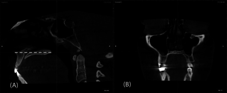
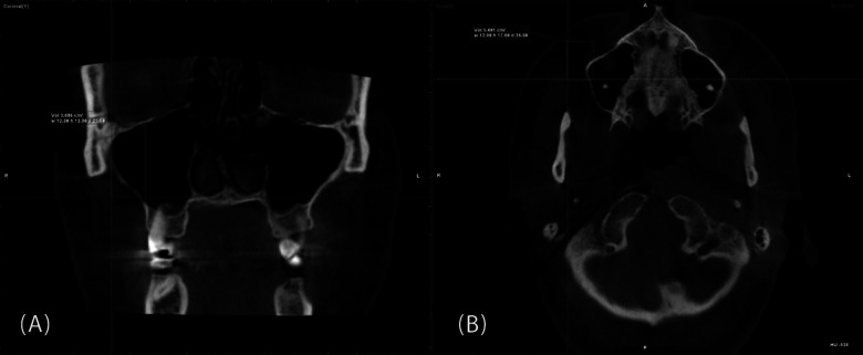
Recognizing the characteristics of the zygomatic process of the maxilla (ZPM) has several applications in implant dentistry and orthodontics. The present study evaluated the relationship between ZPM morphology, as obtained from panoramic images, and sinus pneumatization within the ZPM, according to cone-beam computed tomography (CBCT). CBCT and panoramic images of 300 patients were obtained, and the patients were divided into three age groups: Group I (15-34 years), Group II (35-55 years), and Group III (≥ 55 years). ZPM morphology was classified into J-shaped and non-J-shaped types based on panoramic images. The ZPM volume, maxillary sinus volume, and extent of sinus pneumatization within the ZPM were measured using CBCT scans, and their relationship to ZPM morphology classification was evaluated. Statistical analyses were performed using the Shapiro-Wilk test, one-way ANOVA, independent sample t-test, Mann-Whitney U test, Kruskal-Wallis test, and chi-square test (α = 0.05). Participants ranged in age from 17 to 82 years, and 67% were female. Most ZPMs were J-shaped (75%). J-shaped ZPMs had significantly greater sinus volume (1.83 ± 0.81 cm3), sinus pneumatization (53.56 ± 12.60%), and ZPM volume (3.50 ± 0.98 cm3) compared to non-J-shaped ZPMs (P < 0.05). Male participants had significantly larger ZPM volumes than females (P = 0.039). Sinus pneumatization and maxillary sinus volume were comparable across age groups and between gender. The presence of a non-J-shaped ZPM might be associated with less sinus pneumatization than J-shaped ZPMs, regardless of the patient's age or gender. Therefore, non-J-shaped ZPMs might offer more bone availability for various applications in dentistry.
Keywords: Cone-beam computed tomography; Maxillary sinus pneumatization; Panoramic; Radiography; Zygomatic process of the maxilla.
© 2025. The Author(s).
Conflict of interest statement
Declarations. Ethics approval and consent to participate: All methods were carried out in accordance with relevant guidelines and regulations. The protocol of this study was approved by the Ethics Committee of Mashhad University of Medical Sciences (code: IR.MUMS.DENTISTRY.REC.1403.105). Consent for publication: Not applicable. Competing interests: The authors declare that they have no competing interests.
Figures


References
-
- Abdinian M, Faghihian R, Safi M (2017) Comparison of linear and angular measurement accuracy between cone beam computed tomography images and panoramic radiography. SRM J Res Dent Sci 8:14–20. 10.4103/srmjrds.srmjrds_26_16 - DOI
-
- Anbiaee N, Bagherpour A, AhmadianYazdi A, Khodabakhsh R (2018) Relationship of Maxillary Sinus Volume and Pneumatization with Age and Gender. J Mashhad Dent School 42:200–194. 10.22038/jmds.2018.11455 - DOI
Grants and funding
LinkOut - more resources
Full Text Sources