. 2025 Jul 1;16(1):466.
doi: 10.1038/s41419-025-07789-5.

Inhibition of Wnt/β-catenin increases anti-tumor activity by synergizing with sorafenib in hepatocellular carcinoma

Affiliations

Inhibition of Wnt/β-catenin increases anti-tumor activity by synergizing with sorafenib in hepatocellular carcinoma

Yuchao He et al. Cell Death Dis. .

Abstract

Hepatocellular carcinoma (HCC) poses a major global health challenge owing to limited treatment efficacy and drug resistance to therapies such as the tyrosine kinase inhibitor (TKI) sorafenib. We utilized a microfluidic three-dimensional (3D) drug testing system to assess drug responses in 37 fresh clinical samples and performed immunohistochemical analysis of 41 tumor tissue samples that received sorafenib therapy. Results revealed that Wnt/β-catenin activation was associated with sorafenib resistance, with higher nuclear β-catenin levels predicting poor response. Targeting Wnt/β-catenin via genetic intervention enhanced TKI sensitivity by promoting apoptosis and reducing clonogenicity. Through a large scale of drug and inhibitor library screening, we identified PRI-724, a potent CREB-binding protein (CBP)/β-catenin transcription antagonist, which synergistically induces apoptosis with sorafenib in vitro and in vivo by inhibiting β-catenin/CBP/c-myc, β-catenin nuclear localization and ERK/AKT signaling. The microfluidic 3D drug testing system confirmed the synergistic anti-tumor effects of this combination, underscoring its clinical application potential. Conclusively, our study provides a new combination therapy with sorafenib and PRI-724 to overcome TKI resistance and improve clinical outcomes in patients with HCC. Schematic representation of the speculative molecular mechanism model. Our study revealed that β-catenin activation drives sorafenib resistance in HCC, and disrupting β-catenin enhances sorafenib efficacy by promoting apoptosis and inhibiting proliferation. The combination of sorafenib and PRI-724, a Wnt/β-catenin inhibitor, showed synergistic anti-tumor effects in vitro across various HCC cell lines, in vivo using xenograft models, ex vivo utilizing MDT chip system to explore clinical applications, offering a novel therapeutic strategy for HCC patients.


Conflict of interest statement

Competing interests: The authors declare no competing interests. Ethics approval and consent to participate: Informed consent was obtained from all participants involved in this study. Approval for this clinical research was granted by the Medical Ethics Committee of Tianjin Medical University Cancer Institute and Hospital (Approval number: bc2021260). And the study-related animal experiments were approved by the Experimental Animal Ethics Committee (Approval number: PMIF-2021077) of Tianjin Medical University Cancer Institute and Hospital, and the study was conducted in compliance with the Declaration of Helsinki.

Figures

None
Schematic representation of the speculative molecular mechanism model. Our study revealed that β-catenin activation drives sorafenib resistance in HCC, and disrupting β-catenin enhances sorafenib efficacy by promoting apoptosis and inhibiting proliferation. The combination of sorafenib and PRI-724, a Wnt/β-catenin inhibitor, showed synergistic anti-tumor effects in vitro across various HCC cell lines, in vivo using xenograft models, ex vivo utilizing MDT chip system to explore clinical applications, offering a novel therapeutic strategy for HCC patients.
Fig. 1
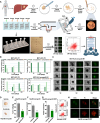
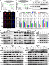
Fig. 1. 3D drug testing work system: micro-dissected tumor tissues (MDT) on a chip.
A The workflow of the microfluidic device for 3D MDT culture and drug testing. B Annexin V-PE/7-AAD staining by flow cytometry was performed to detect the mean apoptosis rates of MDTs from patients (P1, P2, and P3) and HLE xenograft tumor tissues (M1). Early apoptosis (Annexin V-PE+/7-AAD-), late apoptosis (Annexin V-PE+/7-AAD+), and necrosis (Annexin V-PE-/7-AAD+). C The viability of MDTs on chip from HLE xenograft tumor tissues by CTG staining. D The tumor images, E tumor weight, and F tumor volume of Hep3B subcutaneous xenografts with or without treatment with 30 mg/kg sorafenib (n = 4/group). G The apoptosis analysis of Hep3B xenograft MDTs treated with or without 5 μM sorafenib. H The viability analysis on chip detected by CTG and PI staining. The error bars indicate the means ± SEM; ** p < 0.01, t test (EG).
Fig. 2
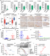
Fig. 2. Wnt/β-catenin activation is associated with sorafenib resistance in HCC.
A Statistical analysis of sorafenib’s suppression effect on MDTs from 37 HCC patients divided into responder and non-responder groups. Five out of 37 MDTs responded to sorafenib based on the viable fraction after 5 μM sorafenib treatment for 7 days. B The ssGSEA score of Wnt pathway activity including liver-related Wnt target genes signal and classical Wnt target genes signal, in the tissues of the 37 MDTs from liver cancer patients. C The representative IHC images of β-catenin in sorafenib responder and non-responder HCC patients. Scale bars, 200 µm for 200×; 100 µm for 400×. D The comparison of the IHC score of nuclear β-catenin between sorafenib responder and non-responder HCC patients. E The comparison of survival time between 41 HCC patients with negative and positive nuclear β-catenin expression. F Identification of independent risk factors for OS in the cohort of 41 HCC cases according to multivariate analysis. G Kaplan–Meier plots comparing 5-year OS (p < 0.001) rates between patients with negative vs. positive nuclear β-catenin expression. H The β-catenin and active β-catenin levels and IC50 of sorafenib in various HCC cell lines detected by western blotting assay and CCK-8 assay, respectively. I The association analysis between active β-catenin level and IC50 of sorafenib in various HCC cell lines. The error bars indicate the means ± SEM; ns: p > 0.05, * p < 0.05, ** p < 0.01, *** p < 0.001, **** p < 0.0001, t test (A, B, and DF).
Fig. 3
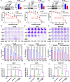
Fig. 3. β-catenin regulates resistance to sorafenib in HCC.
Western blot and the quantified intensities of bands showing β-catenin expression in β-catenin-knockdown Hep3B cells (A), HepG2 cells (B), and MHCC-97H cells (C). β-catenin-knockdown cells and SCR control cells were treated with the indicated concentrations of sorafenib for 48 h and subjected to a cell viability assay. β-catenin-knockdown cells and SCR control cells were treated with or without the indicated concentrations of sorafenib for 8 days in Hep3B cells (D), 14 days in HepG2 (E), and MHCC-97H cells (F), and then subjected to a clonogenic cell survival assay. Quantitation of clonogenic cells from three independent experiments is shown for Hep3B (G), HepG2 (H), and MHCC-97H (I). After treatment with or without sorafenib for 48 h, β-catenin-knockdown cells and SCR control cells were double-stained with Annexin V-PE/7-AAD apoptosis kit and analyzed by flow cytometry: 1.25 μM sorafenib for Hep3B cells (J), 2.5 μM for HepG2 cells (K), and 5 μM for MHCC-97H cells (L). The error bars indicate the means ± SEM; ns: p > 0.05, *p < 0.05, **p < 0.01, ***p < 0.001, ****p < 0.0001, t test (AC) two-way ANOVA (GI), one-way ANOVA (JL).
Fig. 4
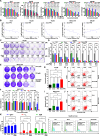
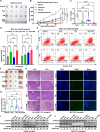
Fig. 4. Synergistic effect of combination treatment of sorafenib and β-catenin inhibitors, PRI-724.
A The cell viability assay was performed for Huh7 cells, Hep3B cells, HepG2 cells, MHCC-97H cells, and SNU387 cells after treatment with the indicated concentrations of sorafenib, PRI-724, and the combination for 48 h (sample size, n = 6). B The synergistic effect of sorafenib and PRI-724 was shown by combination index based on the above cell viability assay results in various HCC cells. CF The synergistic effect of β-catenin inhibitor PRI-724 and sorafenib was measured by a clonogenic cell survival assay following an 8-day treatment in Hep3B cells (C&D) and a 14-day treatment in MHCC-97H cells (E&F). Representative images and quantitative clonogenic cells of three independent experiments are shown. Hep3B cells were treated with 1.25 μM sorafenib, 0.95 μM PRI-724, or their combination (G, H); MHCC-97H cells were treated with 5 μM sorafenib, 5 μM PRI-724, or their combination (I, J). The apoptosis rate and representative images by flow cytometric analysis are shown. K The cell cycle distribution of Hep3B cells treated with 1.25 μM sorafenib, 0.95 μM PRI-724, or their combination. n = 3 for BK. The error bars indicate the means ± SEM; ns: p > 0.05, *p < 0.05, **p < 0.01, ***p < 0.001, ****p < 0.0001, two-way ANOVA (F–J, Q, and S), one-way ANOVA (T and V).
Fig. 5
Fig. 5. Combinations of PRI-724 and sorafenib inhibit both β-catenin/CBP and ERK/AKT signaling and decrease β-catenin nuclear localization in HCC cells.
A GSEA of catenin import into nucleus signal in Hep3B cells with combination treatment with 2.5 μM sorafenib and 1.9 μM PRI-724 compared to the control group. B GSEA of formation of the β-catenin: TCF transactivating complex signal in Hep3B cells with combination treatment with 2.5 μM sorafenib and 1.9 μM PRI-724 compared to the 2.5 μM sorafenib group. C Western blot for β-catenin in the cytoplasmic and nuclear fractions according to the indicated groups in MHCC-97H cells. Sorafenib, 5 μM for 24 h; PRI-724, 5 μM for 24 h. Histone H3 was used as the nuclear marker. D Active β-catenin protein expression patterns and localization were assessed by IF staining in MHCC-97H cells treated with or without 5 μM sorafenib, 5 μM PRI-724, and their combination for 24 h. Scale bars, 10 µm. E Quantitative RT-PCR of the Wnt/β-catenin downregulation target genes mRNA after 24 h treatment of sorafenib (5 μM), PRI-724 (5 μM), and their combination. F Expression of Wnt/β-catenin/CBP, ERK/p-ERK, and AKT/p-AKT signaling and downstream target c-myc were analyzed by western blotting in Hep3B, HepG2, and MHCC-97H cells treated with PRI-724 and sorafenib for 24 h. PRI-724 and sorafenib were 1.9 μM and 2.5 μM for Hep3B and HepG2, and 5 μM and 5 μM for MHCC-97H cells, respectively. G The level of active β-catenin, β-catenin, and AKT/p-AKT with or without the AKT inhibitor LY294002 or the ERK inhibitor PD98059 under the combination treatment condition in Hep3B cells. H Hep3B cells were treated with PRI-724 (1.9 μM) and sorafenib (2.5 μM), and (I) MHCC-97H cells were treated with PRI-724 (5 μM) and sorafenib (5 μM) for 4, 48, and 72 h. The protein expression was assessed by western blotting analysis, including β-catenin, ERK/p-ERK, AKT/p-AKT, and P38/p-P38 signaling. The error bars indicate the means ± SEM; ns: p > 0.05, *p < 0.05, **p < 0.01, ***p < 0.001, ****p < 0.0001, two-way ANOVA (E).
Fig. 6
Fig. 6. Inhibition of β-catenin activity enhances the antitumor activity of sorafenib against HCC xenografts.
A Image of the xenograft tumors formed by Hep3B cells treated with 30 mg/kg sorafenib and 50 mg/kg PRI-724 or their combination (n = 5). “×” indicates that the mouse tumor disappeared by the end of the drug treatment. Additionally, in the combination group, one mouse died due to the infusion technique on the second day of administration. B The growth curve and (C) tumor weight of the Hep3B xenograft tumors in the indicated 4 groups. D, E Annexin V assays followed by flow cytometry were used to evaluate the apoptosis rate of MDTs from Hep3B xenograft tissues treated with PRI-724 (3.8 μM) and sorafenib (5 μM) for 4 and 7 days. F Image of the xenograft tumors formed by MHCC-97H cells treated with 30 mg/kg sorafenib and 50 mg/kg PRI-724 or their combination (n = 3). G Tumor weight of MHCC-97H xenograft tumors at endpoint in the indicated 4 groups. H H&E and TUNEL staining image in MHCC-97H xenograft tumor samples after treatment with sorafenib, PRI-724, or the combination. Scale bars, 400 µm for 100×; 200 µm for 200×. The total and active β-catenin levels were assessed by western blotting assay in Hep3B xenograft tumor tissues (I) and MHCC-97H tumor tissues (J) after the indicated treatment (sample numbers = 12, n = 3/group). The error bars indicate the means ± SEM; ns: p > 0.05, *p < 0.05, **p < 0.01, ***p < 0.001, ****p < 0.0001, two-way ANOVA (BD, G).
Fig. 7
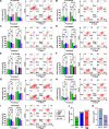
Fig. 7. Exploration in clinical application of combination of sorafenib and PRI-724.
AI The MDT samples from 37 HCC patients were subjected to 3D culture and drug sensitivity testing using the MDT chip system treated with PRI-724 (5 μM) and sorafenib (5 μM) for 4 and 7 days. The results for the 9 cases that responded to single or combination drug treatments are shown by the apoptosis rates analysis. J The response rate of sorafenib, PRI-724, and combination therapy in 37 MDTs from HCC patients. K Statistical analysis of response rates shows that combination therapy is superior to single-agent treatments with sorafenib or PRI-724. The error bars indicate the means ± SEM; ns: p > 0.05, *p < 0.05, **p < 0.01, ***p < 0.001, ****p < 0.0001, two-way ANOVA (AI).

Similar articles

References

    1. Rumgay H, Arnold M, Ferlay J, Lesi O, Cabasag CJ, Vignat J, et al. Global burden of primary liver cancer in 2020 and predictions to 2040. J Hepatol. 2022;77:1598–606. - PMC - PubMed
    1. Llovet JM, Kelley RK, Villanueva A, Singal AG, Pikarsky E, Roayaie S, et al. Hepatocellular carcinoma. Nat Rev Dis Prim. 2021;7:6. - PubMed
    1. Llovet JM, Ricci S, Mazzaferro V, Hilgard P, Gane E, Blanc JF, et al. Sorafenib in advanced hepatocellular carcinoma. N Engl J Med. 2008;359:378–90. - PubMed
    1. Bruix J, da Fonseca LG, Reig M. Insights into the success and failure of systemic therapy for hepatocellular carcinoma. Nat Rev Gastroenterol Hepatol. 2019;16:617–30. - PubMed
    1. Tang W, Chen Z, Zhang W, Cheng Y, Zhang B, Wu F, et al. The mechanisms of sorafenib resistance in hepatocellular carcinoma: theoretical basis and therapeutic aspects. Signal Transduct Target Ther. 2020;5:87. - PMC - PubMed

MeSH terms