. 2025 Jul 1;16(1):5630.
doi: 10.1038/s41467-025-60565-z.

Echs1-mediated histone crotonylation facilitates zygotic genome activation and expression of repetitive elements in early mammalian embryos

Affiliations

Echs1-mediated histone crotonylation facilitates zygotic genome activation and expression of repetitive elements in early mammalian embryos

Yong-Feng Wang et al. Nat Commun. .

Abstract

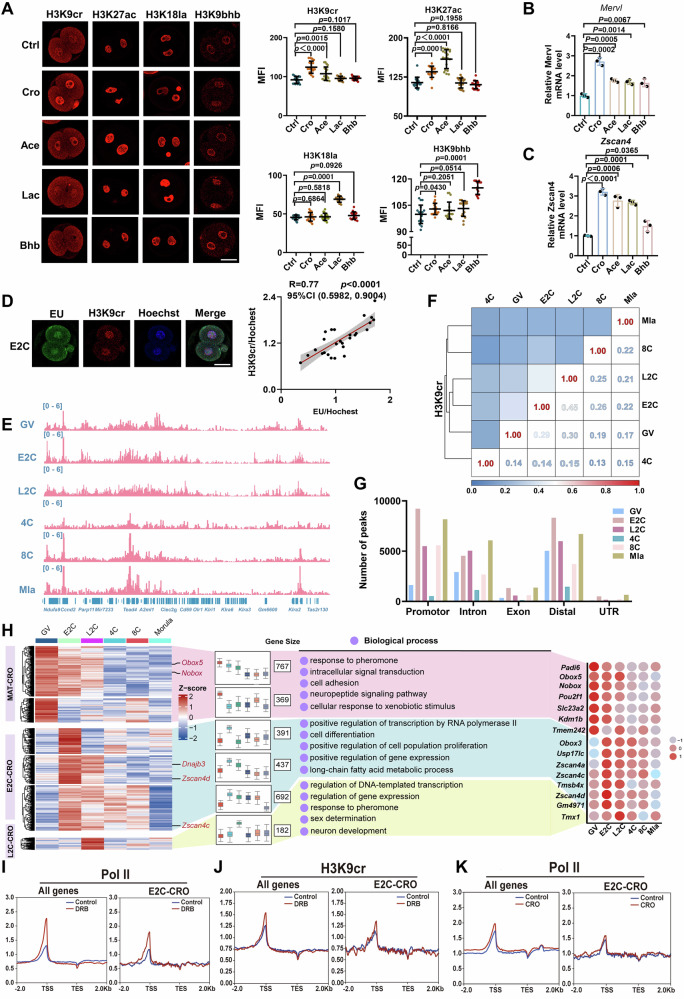
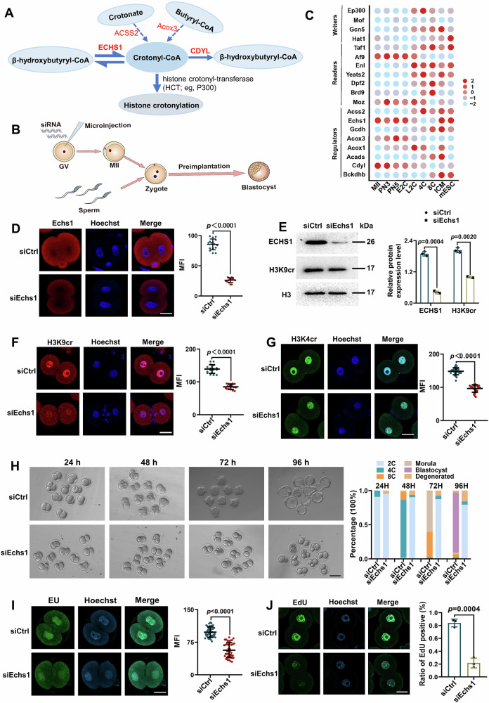
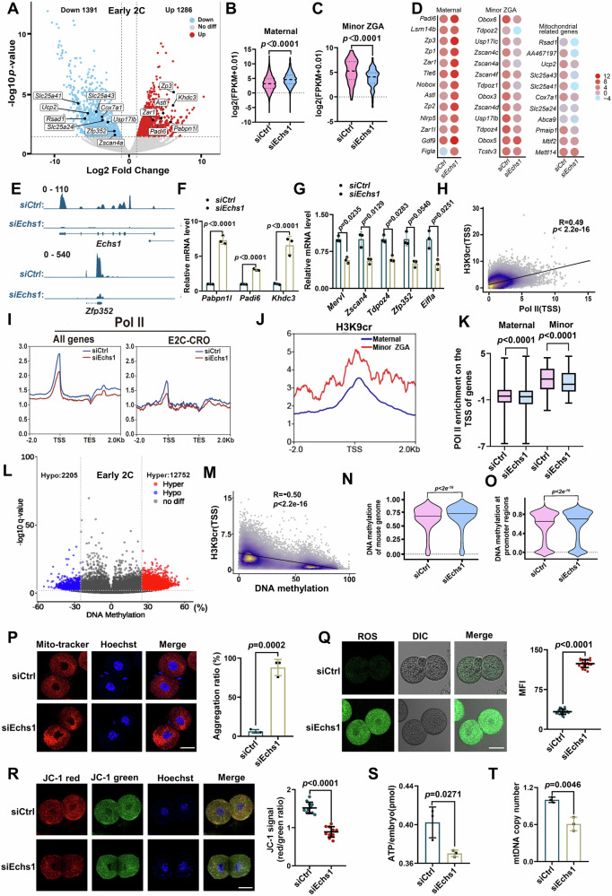
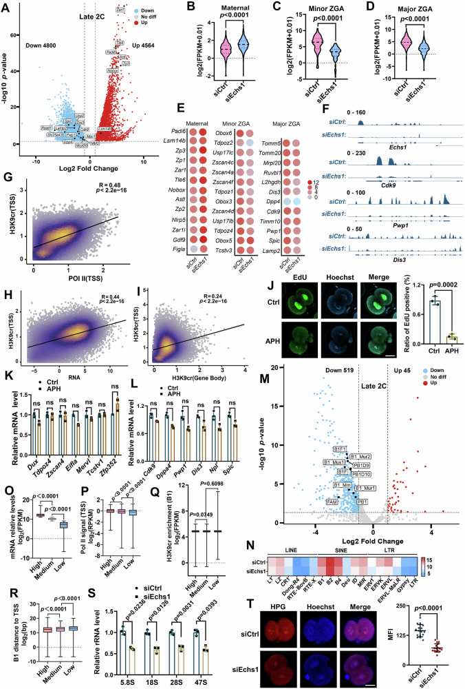
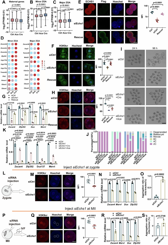
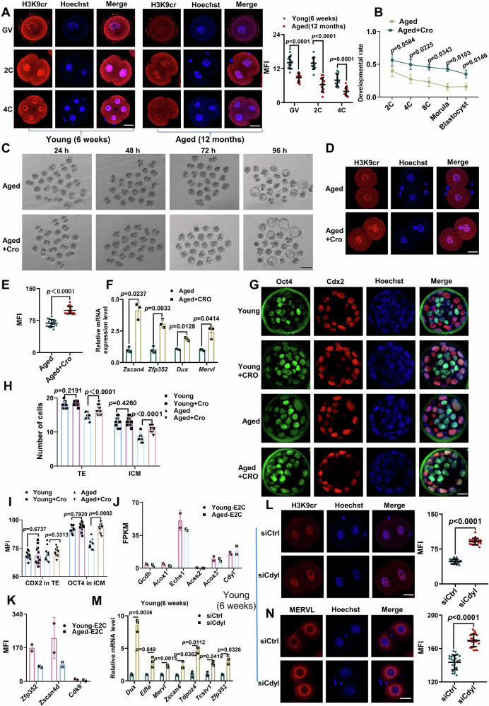
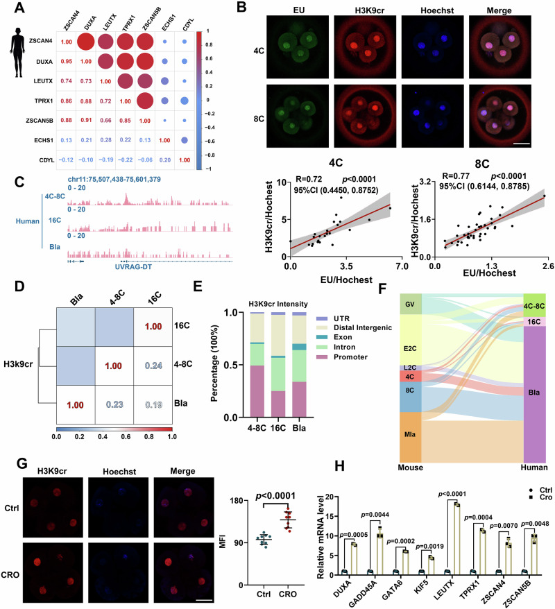
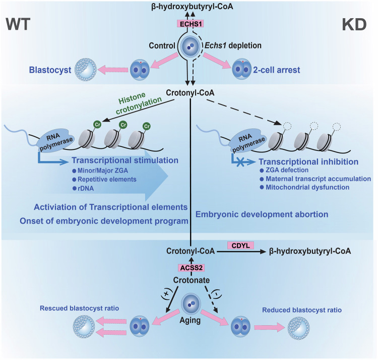
Histone crotonylation, a conserved post-translational histone modification, plays a crucial role in transcriptional regulation. However, its function in early embryonic development remains largely unexplored. Here, we perform genome-wide mapping of histone crotonylation in mouse and human early embryos. Our analysis reveals that histone crotonylation is highly enriched at promoter regions and exhibits distinct dynamic patterns throughout embryogenesis. Notably, strong histone crotonylation signals are observed at the mouse 2-cell and human 4-to-8-cell stages, coinciding with zygotic genome activation. In mice, Echs1 knockdown in oocytes, which suppresses histone crotonylation, results in developmental arrest at the 2-cell stage. Further investigation demonstrates that reduced histone crotonylation impairs transcriptional activity at zygotic genome activation genes, retrotransposon elements, and ribosomal DNA loci. Moreover, early embryos from aged female mice exhibit significantly diminished histone crotonylation, while supplementation with exogenous sodium crotonate enhances blastocyst formation. Collectively, our findings establish histone crotonylation as a key regulatory mechanism in early mammalian embryogenesis by facilitating transcriptional activation of zygotic genome activation genes and repetitive elements.


Conflict of interest statement

Competing interests: The authors declare no competing interests.

Figures

Fig. 1
Fig. 1. Histone Kcr is critical for early embryonic development in mice.
A Immunofluorescence staining and mean fluorescence intensities (MFI) of H3K9cr, H3K27ac, H3K18la and H3K9bhb in early 2-cell embryos treated by sodium crotonate (Cro), sodium acetate (Ace), sodium lactate (Lac), or β-hydroxybutyric acid (Bhb) from early zygote stage (n = 18). Scale bars: 20 μm. B, C qRT-PCR showed the expression of Mervl (B) and Zscan4 (C) in early 2-cell embryos treated with epigenetic metabolites (n = 30). D Immunofluorescence co-staining and Pearson correlation analysis of EU and H3K9cr signals in early 2-cell embryos, the experiment was conducted with three independent biological replicates, (n = 28). CI: confidence interval. Scale bars: 20 μm. E Genomic view of H3K9cr occupancy in GV oocyte and preimplantation embryo by IGV browser. F Spearman correlation analysis of genome-wide H3K9cr read counts at various developmental stages. G The number of H3K9cr peaks at different genomic regions including promoter, intron, exon, UTR and distal regions across various developmental stages. H Heatmap shows hierarchical clustering of H3K9cr intensity at promoter regions across developmental stages, with row-normalized values color-coded. Representative genes in each cluster were marked. Three gene clusters with similar H3K9cr change patterns were named ‘MAT-CRO’, ‘E2C-CRO’, and ‘L2C-CRO’. Boxplots display H3K9cr intensity trends for each cluster during embryonic development, the boxplot composition (centre line: median; box limits: quartile 1 and quartile 3, and whiskers showing the maximum and minimum values). On the right are GO analysis results for the three gene classes and a dot plot of H3K9cr intensity normalized count changes for typical maternal and ZGA genes. I–K Metaplots display the average Pol II (I) or H3K9cr (J) enrichment signals at TSS and TES of all genes or ‘E2C-CRO’ group genes in early 2-cell embryos treated with DRB. In early 2-cell embryos treated with sodium crotonate (K), metaplots showed higher enrichment signals of Pol II at TSS regions of all genes or ‘E2C-CRO’ group genes. In (A), (B) and (C), data were presented as Mean ± SD of three independent replicates, and statistical evaluation was performed by two-tailed unpaired Student’s t-test. Source data are provided as a Source Data file.
Fig. 2
Fig. 2. Echs1 is the key regulator of histone Kcr in early mouse embryos.
A Model diagram of key factors controlling histone crotonylation modification. Crotonyl-CoA and β-hydroxybutyryl-CoA can be interconverted by Echs1. In addition, crotonyl-CoA can be generated from crotonic acid catalyzed by Acss2 or from Butyryl-CoA catalyzed by Acox3, and ultimately converted unidirectionally to β-hydroxybutyryl-CoA catalyzed by Cdyl. B Experimental design schematic for Echs1 knockdown in oocytes, IVM, IVF, and preimplantation development of mouse embryos. Generally, siRNA was injected into GV oocytes for IVM, then MII oocytes were obtained for IVF, the zygotic and preimplantation embryos were obtained through culturing in KSOM medium. C Dotplot indicated expression changes of known histone Kcr writers, readers, and regulators in mouse oocytes and preimplantation embryos (log2FPKM). The transcriptome data was derived from GSE165782. D Immunofluorescence staining and MFI of ECHS1 protein in early 2-cell embryos of siCtrl (control siRNA was injected) and siEchs1 (siRNA against Echs1 was injected) groups (n = 16). Scale bar: 20 μm. E Examination and quantification analysis of ECHS1, H3K9cr, and total histone H3 protein levels in siCtrl and siEchs1 early 2-cell embryos by immunoblotting. The blots were derived from the same experiment and processed in parallel. Total histone H3 level was set as an internal reference (n = 300). F, G Immunofluorescence staining and MFI of H3K9cr (F) (n = 16) and H3K4cr (G) (n = 27) in siCtrl and siEchs1 embryos. Scale bar: 20 μm. H Representative developmental progression images of early embryos. Different stage embryo percentages were shown to average on the right (n  =  3 independent replicates). Scale bar: 100 mm. I, J Confocal images showed newly synthesized DNA by EU staining (I) (n = 40) and newly synthesized RNA by EdU staining (J) in siCtrl and siEchs1 embryos (n  =  3 independent replicates), scale bar: 20 μm, MFI and the percentages of positive EdU embryos was shown on the right of each image. In (D), (E), (F), (G), (I) and (J), data were presented as Mean ± SD of three independent replicates and statistical evaluation was performed by two-tailed unpaired Student’s t-test. Source data are provided as a Source Data file.
Fig. 3
Fig. 3. Echs1-mediated histone Kcr is critical for minor ZGA in early 2-cell embryos in mice.
A Volcano of transcript changes in siEchs1 versus siCtrl early 2-cell embryos (p-value were calculated using DESeq2). Representative mitochondria-related genes, ‘maternal RNA’ and ‘minor ZGA’ genes were indicated. B, C Violin plots demonstrated transcriptional differences (Mean ± SD) of ‘maternal RNA’ (B) and ‘minor ZGA’ (C) genes (GSE169632) between early 2-cell embryos. D Dotplots indicated different expressed genes of early 2-cell embryos. E Genome browser view of RNA- seq results of early 2-cell embryos by IGV. F, G qRT-PCR results of ‘maternal RNA’ (F) and ‘minor ZGA’ genes (G) in early 2-cell embryos (n = 30). H Pearson analysis of correlation between H3K9cr and Pol II occupancy at promoter regions in early 2-cell embryos. I Metaplots showed average Pol II enrichment signals at TSS and TES for all genes and ‘E2C-CRO’ genes. J Metaplot of H3K9cr enrichment at TSSs of ‘maternal RNA’ and ‘minor ZGA’ genes in early 2-cell embryos. K Boxplots showed Pol II enrichment at TSSs of ‘maternal RNA’ and ‘minor ZGA’ genes in early 2-cell embryos, the boxplot composition (center line: median; box limits: quartile 1 and quartile 3, and whiskers showing the maximum and minimum values). L Volcano plot of show differential methylation between siCtrl and siEchs1 early 2-cell embryos (calculated using methylKit). M Pearson analysis of correlation between H3K9cr density and DNA methylation level at gene promoter regions in early 2-cell embryos. N, O Different methylation levels in early 2-cell embryos at the global genome (N) and the promoter regions (O), the median is represented by a horizontal line in the violin plot. P Quantification of mislocalized mitochondria in early 2-cell embryos (n = 12). Scale bar: 20 μm. Q, R Examination of ROS (n = 19) and JC-1 (n = 11) levels in early 2-cell embryos. S, T Relative ATP levels (S) and mtDNA copy numbers (T) in early 2-cell embryos (n = 20). In (F), (G), (H), (P), (Q), (R), (S) and (T), data were presented as Mean ± SD of three independent replicates and examined by two-tailed unpaired Student’s t-test. In (B), (C), (K), (N), and (O), data were examined by the Mann-Whitney U test. Source data are provided as a Source Data file.
Fig. 4
Fig. 4. Histone Kcr is essential for major ZGA and repetitive elements in late 2-cell stage in mice.
A Volcano of transcript changes in siEchs1 versus siCtrl late 2-cell embryos (p-value were calculated using DESeq2). B–D Violin plots of ‘maternal RNA’ (B), ‘minor ZGA’ (C) and ‘major ZGA’ (D) genes (GSE169632) expression in late 2-cell embryos. E Dotplots indicated representative genes expression in late 2-cell embryos. F Genomic browser view of RNA-seq results by IGV in late 2-cell embryos. G Pearson analysis between H3K9cr and Pol II density (GSE215813) at promoter in late 2-cell embryos. H, I Pearson analysis H3K9cr density between promoter and corresponding gene expression (H) or gene body (I) in late 2-cell embryos. J Images of EdU staining in 2-cell embryos treated with (APH)/without APH (Ctrl) (n = 12), scale bar: 20 μm. K, L qRT-PCR of ‘minor ZGA’ (K) and ‘major ZGA’ (L) genes expression in late 2-cell embryos (n = 30). Two-way ANOVA for analysis (Mean ± SD). M Volcano showed retrotransposon expression changes between siEchs1 and siCtrl late 2-cell embryos (p-value were calculated using DESeq2), SINE/B1 family with changed expression were indicated. N Heatmap illustrated expression of retrotransposon subfamilies at late 2-cell embryo stage. O SINE/B1 adjacent genes were categorized into ‘high’, ‘medium’, and ‘low’ groups by transcription level in late 2-cell embryos. P Boxplot showing Pol II intensity at promoter in ‘high’, ‘medium’, and ‘low’ groups. Q Boxplot showing enrichment of H3K9cr at adjacent SINE/B1 elements of ‘high’, ‘medium’, and ‘low’ group genes. R Boxplot showing the distances between adjacent SINE/B1 elements and TSS of ‘high’, ‘medium’, and ‘low’ group genes. S qRT-PCR analysis the rRNA level in siCtrl and siEchs1 late 2-cell embryos (n = 30). T HPG fluorescent staining in late 2-cell embryos (n = 16). Scale bar: 20 μm. In (J), (S) and (T), data were presented as Mean ± SD of three independent replicates and examined by two-tailed unpaired Student’s t-test, ns indicates P > 0.05. In (B), (C) and (D), data were presented as Mean ± SD and examined by Mann-Whitney U test. In (O), (P), (Q) and (R), the boxplot exception (centre line: median; box limits: quartile 1 and quartile 3, and whiskers showing the maximum and minimum values). Source data are provided as a Source Data file.
Fig. 5
Fig. 5. Echs1-mediated histone Kcr specifically regulates mouse early development beyond 2-cell stage.
A–C Boxplots demonstrated transcript differences of ‘maternal RNA’ (A), ‘minor ZGA’ (B) and ‘major ZGA’ (C) genes (GSE169632) of early 2-cell embryos cultured without (Ctrl) or with sodium crotonate (Cro) /sodium acetate (Ace). The data (A–C) were examined by Mann–Whitney U test, the boxplot composition (centre line: median; box limits: quartile 1 and quartile 3, and whiskers showing the maximum and minimum values). D Dotplots indicated expression of representative genes in early 2-cell embryos. E, F Immunofluorescence staining and MFI of ECHS1 protein (E) (n = 12) and H3K9cr (F) (n = 22) in siCtrl, siEchs1 and exogenous Echs1 co-expressed (rescued) embryos. Scale bars: 20 μm. G qRT-PCR result of representative ‘minor ZGA’ genes in siCtrl, siEchs1 and rescued early 2-cell embryos (n = 30). H Immunofluorescence staining of H3K9ac in siCtrl, siEchs1, and rescued early 2-cell embryos (n = 10). Scale bars: 20 μm. I Representative developmental progression images of siCtrl, siEchs1, sodium crotonate rescued (siEchs1+Cro), and sodium acetate rescued (siEchs1+Ace) embryos. Scale bar: 100 mm. (J) Percentages of embryos at different stages (n  =  3 independent replicates). K qRT-PCR result of representative ‘minor ZGA’ genes in siCtrl, siEchs1, siEchs1+Cro, and siEchs1+Ace early 2-cell embryos (n = 30). L–O Schematic (L) for Echs1 knockdown in zygotes and experimental results (M–O). M Immunofluorescence staining and MFI of H3K9cr in early 2-cell embryos (n = 12). Scale bar: 20 mm. N qRT-PCR result of representative ‘minor ZGA’ genes in early 2-cell embryos (n = 30). O Aggregation ratio of mislocalized mitochondria in early 2-cell embryos (n = 12). P–S Schematic (P) for Echs1 knockdown in MII oocytes and experimental results (Q–S). Q Immunofluorescence staining of H3K9cr of pronuclei observed in zygotes. Scale bar: 20 mm. R qRT-PCR result of representative ‘minor ZGA’ genes in siCtrl and siEchs1 zygotes (n = 30). S Aggregation ratio of mislocalized mitochondria in zygotes (n = 12). In (E), (F), (G), (H), (J), (K), (M), (N), (O), (Q), (R) and (S), data were presented as Mean ± SD of three independent replicates and statistical evaluation was performed by two-tailed unpaired Student’s t-test. Source data are provided as a Source Data file.
Fig. 6
Fig. 6. Exogenous crotonate improves the blastocyst formation rate in embryos derived from aged female mice.
A Immunofluorescence staining and MFI of H3K9cr in GV oocytes, 2-cell embryos and 4-cell embryos of maternally young/aged mice (n = 12). Scale bars: 20 μm. B Percentages of embryos at different stages were presented as Mean ± SD (n =  3 independent replicates), data statistical evaluation was performed by two-way ANOVA. C Representative developmental progression images of maternally aged embryos which were cultured in KSOM medium with (Aged+Cro) or without sodium crotonate (Aged) (n  =  3 independent replicates). Scale bars: 100 μm. D, E Immunofluorescence staining (D) and MFI (E) of H3K9cr in ‘Aged’ and ‘Aged+Cro’ early 2-cell embryos (n = 19). Scale bars: 20 μm. F qRT-PCR results of representative ‘minor ZGA’ genes in ‘Aged’ and ‘Aged+Cro’ early 2-cell embryos (n = 30). G Representative Oct4 and Cdx2 immunofluorescence images and quantification of maternally young/aged embryos which cultured with or without sodium crotonate. Different lineage cells were defined by fluorescence signals: inner cell mass (ICM, Oct4+/Cdx2-) and trophectoderm (TE, Cdx2+). Scale bars: 20 μm. H Graphs showed the statistical number of TE and ICM cells respectively in maternally young/aged blastocysts cultured with or without sodium crotonate (n = 12). I Graphs showed the MFI of TE and ICM respectively in maternally young/aged blastocysts which cultured with or without sodium crotonate (n = 12). J–K Bar charts compared expression differences (GSE241388) of histone crotonylation regulators (J) and ZGA genes (K) between maternally young/aged mouse early 2-cell embryos, expression of either Cdyl or Echs1 was not significantly changed (P > 0.05 by DESeq2). L Immunofluorescence staining and MFI of H3K9cr in maternally young early 2-cell embryos (n = 19) with (siCdyl) or without (siCtrl) Cdyl knockdown. Scale bars: 20 μm. M qRT-PCR analysis showed expression changes of representative ‘minor ZGA’ genes in maternally young siCdyl early 2-cell embryos (n = 30). N Immunofluorescence staining and MFI of MERVL protein in maternally young siCdyl and siCtrl early 2-cell embryos (n = 24). Scale bars: 20 μm. In (A), (E), (F), (H), (I), (L), (M) and (N), data were presented as Mean ± SD of three independent replicates and statistical evaluation was performed by two-tailed unpaired Student’s t-test. Source data are provided as a Source Data file.
Fig. 7
Fig. 7. Enrichment of histone Kcr during early human embryonic development.
A Pearson correlation analysis on expression of human ZGA genes, ECHS1 and CDYL, using reported single-cell RNA-seq data of human 8-cell stage embryos (GSE36552, GEO repository, NCBI). B Immunofluorescence co-staining of EU and H3K9cr in human 4-cell and 8-cell embryos, and Pearson correlation analysis demonstrated positive correlation between EU and H3K9cr fluorescence signals in both 4-cell and 8-cell embryos, with shaded areas representing the 95% confidence intervals (n = 24). Scale bars: 20 μm. C Genome browser view of representative H3K9cr signals in cleavage embryos and blastocyst by IGV. D Spearman correlation analysis of genome-wide H3K9cr occupancy in various developmental stages of human embryos. E The percentages of H3K9cr peaks assigned to the different genomic regions including promoter, intron, exon, UTR, and intergenic regions across various developmental stages. F Sankey diagram comparing genes with H3K9cr peaks at promoter regions in different developmental stages between human and mouse, with the width of the connecting line being proportional to the strength of the correlation. G Immunofluorescence staining and mean fluorescence intensity of H3K9cr in human 3PN embryos which cultured in G1 medium with (CRO) or without (Ctrl) sodium crotonate for 32 h from 1-cell stage (n = 9). Scale bars: 20 μm. H qRT-PCR result of representative ‘minor ZGA’ genes in human 8-cell embryos which cultured in G1 medium with (CRO) or without (Ctrl) sodium crotonate (n = 20). The data of qRT-PCR were presented as Mean ± SD of three independent replicates. The data statistical evaluation of qRT-PCR and fluorescence intensity quantification was performed by two-tailed unpaired Student’s t-test. Source data are provided as a Source Data file.
Fig. 8
Fig. 8. Working model illustration of Echs1-mediated histone crotonylation regulating early embryo development.
Echs1 supports early embryo development by regulating crotonyl-CoA synthesis to generate histone crotonylation. Under physiological condition, Echs1 sustains normal level of crotonyl-CoA in early embryos, ensuring sufficient histone crotonylation for transcription activation at ZGA genes, retrotransposon elements, and rDNA loci, thereby ensuring normal early embryo development. When Echs1 is deficient, histone crotonylation level decreases, ZGA event fails, maternal mRNA degradation is impaired, mitochondrial dysfunction occurs, and embryonic development is arrested. Additionally, the addition of exogenous crotonate to the culture medium of embryos from aged female mice improves blastocyst formation. In summary, Echs1-mediated histone crotonylation is crucial for transcription activation of ZGA genes and repetitive elements to support early embryo development.

Similar articles

References

    1. Wei, J. et al. Temporospatial hierarchy and allele-specific expression of zygotic genome activation revealed by distant interspecific urochordate hybrids. Nat. Commun.15, 2395 (2024). - PMC - PubMed
    1. Schulz, K. N. & Harrison, M. M. Mechanisms regulating zygotic genome activation. Nat. Rev. Genet.20, 221–234 (2019). - PMC - PubMed
    1. Wang, C. et al. Dynamic nucleosome organization after fertilization reveals regulatory factors for mouse zygotic genome activation. Cell Res.32, 801–813 (2022). - PMC - PubMed
    1. Zhang, B. et al. Widespread enhancer dememorization and promoter priming during parental-to-zygotic transition. Mol. cell72, 673–686.e676 (2018). - PubMed
    1. Li, J. et al. Metabolic control of histone acetylation for precise and timely regulation of minor ZGA in early mammalian embryos. Cell Discov.8, 96 (2022). - PMC - PubMed

LinkOut - more resources