. 2025 Jul 1;16(1):5769.
doi: 10.1038/s41467-025-60924-w.

NONO, SFPQ, and PSPC1 promote telomerase recruitment to the telomere

Affiliations

NONO, SFPQ, and PSPC1 promote telomerase recruitment to the telomere

Alexander P Sobinoff et al. Nat Commun. .

Abstract

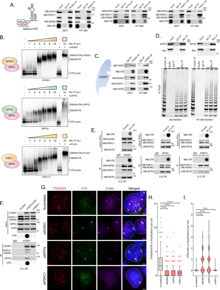
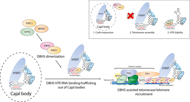
Telomerase is a ribonucleoprotein enzyme that maintains telomeric repeats on chromosome ends in continuously dividing cells. Telomere maintenance via telomerase is dependent on the correct assembly of the enzyme complex, complex stabilization by associated cofactors, and effective recruitment to the telomere. Here, we show that telomerase is regulated in each of these processes by the Drosophila behaviour/human splicing (DBHS) family of RNA/DNA binding proteins (NONO, SFPQ and PSPC1). The DBHS proteins associate with catalytically active telomerase through the hTR RNA template component. Cells lacking the DBHS proteins display telomerase retention in nuclear Cajal bodies and impaired telomerase recruitment to the telomere, with NONO and PSPC1 depletion culminating in progressive telomere shortening in several cell lines, with the exception of long-term NONO depletion in 293 and 293T. Our results reveal the DBHS protein family as components of the telomerase trafficking machinery integral to telomere maintenance.


Conflict of interest statement

Competing interests: The authors declare no competing interests.

Figures

Fig. 1
Fig. 1. DBHS proteins interact with telomerase RNP via hTR and promote telomerase recruitment to telomeres.
A Immunoprecipitation (IP) of DBHS proteins with hTR in 293T and HT1080 cells (n = 3). Indicated proteins assayed by Western blot; hTR assayed by northern dot blot. B EMSA showing DBHS proteins binding to radiolabelled hTR. Overexpressed DBHS proteins were immunopurified using an anti-Myc antibody (n = 2). Bound complex is indicated by gel shift. Super-shift complex comprised of hTR, DBHS proteins and α-DBHS or α-FLAG antibody is indicated by a further shift. C Interaction of DBHS proteins with immunopurified hTERT in 293T and HT1080 cells (n = 3). Indicated proteins assayed by Western blot; hTR assayed by northern dot blot. D TRAP on immunopurified overexpressed NONO (anti-Myc antibody; n = 3). Indicated proteins assayed by Western blot; immunopurified hTERT used as a positive control. Buffer only (buffer) and IgG IP were used as negative controls. E DBHS protein interaction with overexpressed hTR in hTERT negative U-2 OS cells transfected with plasmids expressing wild-type (Wt) hTR and DKC1 (n = 3). Input, indicated proteins assayed by Western blot; hTR assayed by northern dot blot; IP, DBHS proteins and hTR immunopurified using NONO, SFPQ, and PSPC1 antibodies assayed by Western or northern dot blot. IgG IP, non-specific IgG used for IP. F DBHS protein interaction with overexpressed hTERT in hTR negative U-2 OS cells stably transfected with plasmids expressing hTERT and/or Wt hTR and DKC1 (n = 3). Input, indicated proteins assayed by Western blot. IP, DBHS proteins and hTERT immunopurified using hTERT antibodies assayed by Western blot. IgG IP, non-specific IgG used for IP. G Representative images of telomere (red), hTR (green), and coilin (purple) co-localizations in DBHS depleted 293T cells. TTAGGG/hTR co-localizations are indicated by yellow arrows; hTR/coilin co-localizations are indicated by white asterisks. Scale bars are 5 μm. H Tukey boxplots (median, interquartile range, and Tukey whiskers) of TTAGGG/hTR co-localizations in DBHS depleted 293T cells. Out of three experiments, n  =  204 cells scored per treatment, ****p  <  0.0001, Kruskal-Wallis test. I Violin plot of hTR/coilin co-localizations in DBHS depleted 293T cells. Out of three experiments, n  =  150 cells scored per treatment, n.s. = non-significant, ****p  <  0.0001, Kruskal-Wallis test. Source data are provided as a Source data file.
Fig. 2
Fig. 2. NONO/PSPC1 overexpression partially rescues siSFPQ induced hTR sequestration in Cajal bodies.
A Representative images of telomere (red), hTR (green), and coilin (purple) co-localizations in DBHS depleted 293T cells overexpressing NONO (NONO+), SFPQ (SFPQ+) or PSPC1 (PSPC1+). TTAGGG/hTR co-localizations are indicated by yellow arrows; hTR/coilin co-localizations are indicated by white asterisks. Scale bars are 5 μm. BD Tukey boxplots (median, interquartile range, and Tukey whiskers) of TTAGGG/hTR co-localizations in NONO+, SFPQ+, or PSPC1 + DBHS protein depleted 293T cells (left panels). Out of three experiments, n  =  150 cells scored per treatment, ****p  <  0.0001, Kruskal-Wallis test. Violin plots of hTR/coilin co-localizations in NONO+, SFPQ+, or PSPC1 + DBHS protein depleted 293T cells (right panels). Out of three experiments, n  =  150 cells scored per treatment, n.s. = non-significant, ****p  <  0.0001, Kruskal-Wallis test. E Telomere-ChIP against DBHS proteins in 293T cells after DBHS protein knockdown. Values are mean ± SEM from n = 3 experiments, n.s. = non-significant, **p  =  0.0019 between NONO Scrambled and siPSPC1, p  =  0.0023 between SFPQ Scrambled and siNONO, p  =  0.0045 between PSPC1 Scrambled and siSFPQ, ***p  = 0.0008, ****p  <  0.0001, # = no data, Welch’s two-tailed t-test. F Interaction of DBHS proteins with hTERT assayed by IP of hTERT in 293T cells after DBHS protein knockdown (left panel). Indicated proteins assayed by Western blot. Quantitation of DBHS proteins in hTERT IP samples (right panel). Values are mean ± SEM from n = 3 experiments, n.s. = non-significant, *p  =  0.0103 between NONO Scrambled and siNONO, p = 0.0344 between SFPQ Scrambled and siSFPQ, **p =  0.0059, ***p =  0.0003, Welch’s two-tailed t-test. Source data are provided as a Source data file.
Fig. 3
Fig. 3. NONO, SFPQ, and PSPC1 mediated telomerase recruitment is attenuated by mutation of the DBHS region.
A Schematic of DBHS domain functions, mutations, or deletions. B Representative images of telomere (red), hTR (green), and coilin (purple) co-localizations in 293T 3′UTR NONO functional mutant rescue experiments (left panel). TTAGGG/hTR co-localizations are indicated by yellow arrows; hTR/coilin co-localizations are indicated by white asterisks. Scale bars are 5 μm. Tukey boxplots (median, interquartile range, and Tukey whiskers) of TTAGGG/hTR co-localizations (middle panel). Out of three experiments, n  =  198 cells scored per treatment, n.s. = non-significant, *p  =  0.0239, ****p  <  0.0001, Kruskal-Wallis test. Violin plots of hTR/coilin co-localizations (right panel). Out of three experiments, n  =  198 cells scored per treatment, n.s. = non-significant, ****p  <  0.0001, Kruskal-Wallis test. C Representative images of telomere (red), hTR (green), and coilin (purple) co-localizations in 293T siSFPQ functional mutant rescue experiments (left panel). TTAGGG/hTR co-localizations are indicated by yellow arrows; hTR/coilin co-localizations are indicated by white asterisks. Scale bars are 5 μm. Tukey boxplots (median, interquartile range, and Tukey whiskers) of TTAGGG/hTR co-localizations (middle panel). Out of three experiments, n  =  150 cells scored per treatment, n.s. = non-significant, ****p  <  0.0001, Kruskal-Wallis test. Violin plots of hTR/coilin co-localizations (right panel). Out of three experiments, n  =  150 cells scored per treatment, n.s. = non-significant, ****p  <  0.0001, Kruskal-Wallis test. D Tukey boxplots (median, interquartile range, and Tukey whiskers) of TTAGGG/hTR co-localizations in 293T cells overexpressing PSPC1 functional mutants (left panel). Out of three experiments, n  =  150 cells scored per treatment, n.s. = non-significant, *p = 0.0124, ****p  <  0.0001, Kruskal-Wallis test. Violin plots of hTR/coilin co-localizations in 293T cells overexpressing PSPC1 functional mutants (right panel). Out of three experiments, n  =  150 cells scored per treatment, n.s. = non-significant, ***p  =  0.0004, ****p  <  0.0001, Kruskal-Wallis test. E Representative images of telomere (red), hTR (green), and coilin (purple) co-localizations in 293T gPSPC1 cells overexpressing PSPC1 functional mutants (left panel). TTAGGG/hTR co-localizations are indicated by yellow arrows; hTR/coilin co-localizations are indicated by white asterisks. Scale bars are 5 μm. Tukey boxplots (median, interquartile range, and Tukey whiskers) of TTAGGG/hTR co-localizations (middle panel). Out of three experiments, n  =  150 cells scored per treatment, n.s. = non-significant, ****p  <  0.0001, Kruskal-Wallis test. Violin plots of hTR/coilin co-localizations (right panel). Out of three experiments, n  =  150 cells scored per treatment, n.s. = non-significant, **p  =  0.0033, ***p  =  0.0005, ****p  <  0.0001, Kruskal-Wallis test. Source data are provided as a Source data file.
Fig. 4
Fig. 4. Genetic disruption of NONO and PSPC1 causes telomere shortening and reduced telomerase recruitment in HT1080 cells.
A Western blot of DBHS and telomerase biology associated proteins in parental HT1080 and gNONO CRISPR edited clones (left panel). Telomere lengths measured by TRF Southern blot with cell passage (right panel). Pd, population doublings. Peak intensity of telomere length is indicated by red dot. B Western blot of DBHS and telomerase biology associated proteins in parental HT1080 and gPSPC1 CRISPR edited clones (left panel). Telomere lengths measured by TRF Southern blot with cell passage (right panel). Pd, population doublings. Peak intensity of telomere length is indicated by red dot. C Representative images of telomere (red), hTR (green), and coilin (purple) co-localizations in parental HT1080, gNONO, and gPSPC1 CRISPR edited clones (left panel). TTAGGG/hTR co-localizations are indicated by yellow arrows; hTR/coilin co-localizations are indicated by white asterisks. Scale bars are 5 μm. Tukey boxplots (median, interquartile range, and Tukey whiskers) of TTAGGG/hTR co-localizations (middle panel). Out of three experiments, n  =  150 cells scored per treatment, ****p  <  0.0001, Kruskal-Wallis test. Violin plots of hTR/coilin co-localizations (right panel). Out of three experiments, n  =  150 cells scored per treatment, n.s. = non-significant, ****p  <  0.0001, Kruskal-Wallis test. Source data are provided as a Source data file.
Fig. 5
Fig. 5. Model for DBHS mediated telomerase recruitment to telomeres.
The DBHS proteins (NONO, SFPQ, PSPC1) dimerize with each other to facilitate telomerase trafficking out of Cajal bodies by interacting with the hTR telomerase subunit. The DBHS proteins then facilitate telomerase recruitment to the telomere, potentially stabilizing the interaction through polymerization. In addition to recruitment, PSPC1 is also involved in Cajal body formation through coilin expression (1), while NONO and SFPQ play cell line specific roles in reducing telomerase assembly with hTR (2) and hTR stability (3).

Similar articles

References

    1. Denchi, E. L. & de Lange, T. Protection of telomeres through independent control of ATM and ATR by TRF2 and POT1. Nature448, 1068–1071 (2007). - PubMed
    1. Harley, C. B., Futcher, A. B. & Greider, C. W. Telomeres shorten during ageing of human fibroblasts. Nature345, 458–460 (1990). - PubMed
    1. Barbaro, P. M., Ziegler, D. S. & Reddel, R. R. The wide-ranging clinical implications of the short telomere syndromes. Intern. Med. J.46, 393–403 (2016). - PubMed
    1. Vinagre, J. et al. Frequency of TERT promoter mutations in human cancers. Nat. Commun.4, 2185 (2013). - PubMed
    1. Schmidt, J. C. & Cech, T. R. Human telomerase: biogenesis, trafficking, recruitment, and activation. Genes Dev.29, 1095–1105 (2015). - PMC - PubMed

MeSH terms

LinkOut - more resources