. 2025 Jul 1;16(1):5956.
doi: 10.1038/s41467-025-60673-w.

Light-induced structural adaptation of the bundle-shaped phycobilisome from thylakoid-lacking cyanobacterium Gloeobacter violaceus

Affiliations

Light-induced structural adaptation of the bundle-shaped phycobilisome from thylakoid-lacking cyanobacterium Gloeobacter violaceus

Jianfei Ma et al. Nat Commun. .

Abstract

Gloeobacter diverged from other lineages early in cyanobacterial evolution, preferentially growing under low light intensity conditions. Among cyanobacteria, G. violaceus exhibits unique features, including lack of a thylakoid membrane and bundle-shaped antenna phycobilisomes (PBSs), densely packed and well-organized on the plasma membrane. However, without high-resolution structures, it has remained unclear how G. violaceus PBSs assemble into a bundle-shaped configuration. Here we solve the cryo-EM structures of PBSs from G. violaceus cells cultured under low (Sr-PBS) or moderate (Lr-PBS) light intensity. These structures reveal two unique linker proteins, LRC91kDa and LRC81kDa, that play a key role in the PBS architecture. Analysis of the bilin arrangement indicates that the bundle-shaped structure allows efficient energy transfer among rods. Moreover, comparison between Lr-PBS and Sr-PBS uncovers a distinct mode of adaption to increased light intensity wherein the ApcA2-ApcB3-ApcD layer can be blocked from binding to the core by altering structural elements exclusively found in the G. violaceus LCM. This study illustrates previously unrecognized mechanisms of assembly and adaptation to varying light intensity in the bundle-shaped PBS of G. violaceus.


Conflict of interest statement

Competing interests: The authors declare no competing interests.

Figures
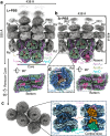
Fig. 1
Fig. 1. Overall structures of PBS from G. violaceus 7421.
a, b Cryo-EM maps of two types of assemblies of PBS (Lr-PBS and Sr-PBS) from face and bottom views. The rods are colored gray. PBS core is presented in different colors. The solid and dashed black circles are represented whether ApcA2-ApcB3-ApcD layer is bond with the core or not in Lr-PBS and Sr-PBS. c Top view of rods. Enlarged view of the boxed area on the right side of (c) shows the cryo-EM map of Rod b after local refinement. The LRCpcC1, LRCpcC2, and LRC91kDa are colored as pink, orange, and lime green. d Enlarged view of the boxed area of (b) shows the cryo-EM map of Rod d after local refinement. The LRT and LRC81kDa are colored as wheat and warm pink.
Fig. 2
Fig. 2. Rod-core linker proteins, LRC91kDa and LRC81kDa.
a Top, diagram of the structural elements of LRC91kDa. Bottom, the structure of LRC91kDa is represented as a cartoon. Three Pfam00427 domains are presented in different colors. Four helices are highlighted in sky blue. b Interactions between LRC91kDa and core. The grooves on the α-subunits that contact the helices are shown in red. c Sequence alignment of the helices of LRC91kDa with LRC from Synechocystis sp. PCC 6803, Synechococcus sp. strain PCC 7002, Nostocaceae PCC 7120, G.pacifica and P.purpureum. The conserved Phenylalanine or Tryptophan is marked as a red triangle. d Top, diagram of the structural elements of LRC81kDa. Bottom, the structure of LRC91kDa is represented as a cartoon. The Pfam00427-1 domain we solved is presented in megenta. The Pfam00427-2 and -3 are presented in a dashed ellipse. The helix is highlighted in sky blue. e Interactions between LRC81kDa and core. The groove on the α-subunit that contacts the helix is shown in red.
Fig. 3
Fig. 3. Phylogenetic relationship of LRC and classifications of Pfam00427.
a The maximum likelihood phylogenetic trees constructed using the full-length protein sequences of LRC from thylakoid-lacking cyanobacterium G. violaceus 7421, G. kilaueensis JS1, A. panamensis, and crown cyanobacteria Nostocaceae PCC 7120, Synechocystis sp. PCC 6803, Synechococcus sp. strain PCC 7002, A. platensis, and red algae G.pacifica and P.purpureum. Bootstrap values are presented on the tree nodes, and the values higher than 0.6 are shown. b Left, phylogenetic trees constructed using the separated Pfam00427 domains, Pfam00427 domains of LR and LRC are labeled in firebrick and black, respectively. The Pfam00427 domains are classified into two types: Type I and II 00427 domains are shown on the purple and light orange background, respectively. The Pfam00427 domains belong to thylakoid-free cyanobacteria are marked with a red star. Right, the structures of type I and II 00427 domains are shown as a cartoon. c Structural alignment of the type I and II 00427 domains. Region of structural change is shown with 70% transparency. Enlarged view of the boxed area of c shows the altered structures including key aromatic residues, representative helix of type II 00427 domain, as well as the shift of bilins.
Fig. 4
Fig. 4. Interactions of the linker proteins LRCpcC2 and LRC91kDa with chromophores in the rod Rb.
a Overall structure of the rod Rb with the hexamers shown in surface representation and the linker proteins shown in cartoon representation. b Structure of the layer Rb2I. Proteins and bilins are shown in cartoon and sticks representations, respectively. Three β subunits are colored differently, and the β82 PCBs are boxed and analyzed in (ce). c No interactions between the LRCpcC2 and the bilin Rb2Iβ182. d The interactions between F224, F235 and F239 and the bilin Rb2Iβ282. e The interactions between Y118 and Rb2Iβ382. f Structure of the layer Rb1I. Proteins and bilins are shown in cartoon and sticks representations, respectively. Three β subunits are colored differently, and the β82 PCBs are boxed and analyzed in (gi). g The interactions between F510, F512, F521 and F528 and the bilin Rb1Iβ182. h The interactions between Y404 and the bilin Rb1Iβ282. i The interactions between F474 and the bilin Rb1Iβ382.
Fig. 5
Fig. 5. Structural differences between LCM/Lr-PBS and LCM/Sr-PBS.
a Structural alignment of LCM/Lr-PBS and LCM/Sr-PBS. LCM/Lr-PBS and LCM/Sr-PBS are presented in hot pink and green colors. ApcA2-ApcB3-ApcD layer attached with LCM/Lr-PBS is shown as a surface view. The altered structures (loop1, loop2, and bulge) are highlighted in dashed rectangles. b Enlarged view of the boxed area of (a) shows that the structural change of loop1 reduces the interactions between loop1 and ApcA. c Electrostatic interaction between loop1/Lr-PBS and ApcA. d Enlarged view of the structural change of loop2 shows the absence of the interactions between loop2/Sr-PBS and LC. loop2/Sr-PBS could form a steric hindrance to prevent LC binding with the core. e Three successive residues (R238-S239-D240) of αLCM could form a “bulge” that would occupy the position of the ApcD.
Fig. 6
Fig. 6. Possible excitation energy transfer (EET) pathways.
a Estimated maps of the energy transfer pathways among bilins based on orientation factor (κ2) and distances (d). The colors correspond to higher (red), middle (blue), and lower (gray) κ2/d6 values, which are used to represent the EET rates. The key bilins of rods are shown as spheres in orange color. Bilins that are more than 44 Å are omitted. The gray and black arrows are represented as the rod-core and inner-core bottleneck, respectively. The energy transfer from top B/B’ to bottom A/A’ is high, represented as red lightning. The absent bilins and pathways in Sr-PBS are shown as yellow spheres and dashes, respectively. The terminal emitters A3αLCM196 (A3αLCM’196) and A4αApcD81 (A4αApcD’81) are shown in sphere representation. b Top view of the energy transfer pathways. The rods are represented as circles in different colors. EET exchange among the rods is represented by black dashed arrows. c The absence of ApcA2-ApcB3-ApcD layer and EET pathways in Sr-PBS is shown as a surface representation. The corresponding bilins are shown as sticks.

Similar articles

References

    1. Nelissen, B., Van De Peer, Y., Wilmotte, A. & De Wachter, R. An early origin of plastids within the cyanobacterial divergence is suggested by evolutionary trees based on complete 16S rRNA sequences. Mol. Biol. Evol.12, 1166–1173 (1995). - PubMed
    1. Blank, C. E. & Sanchez-Baracaldo, P. Timing of morphological and ecological innovations in the cyanobacteria—a key to understanding the rise in atmospheric oxygen. Geobiology8, 1–23 (2010). - PubMed
    1. Sanchez-Baracaldo, P., Hayes, P. K. & Blank, C. E. Morphological and habitat evolution in the cyanobacteria using a compartmentalization approach. Geobiology3, 145–165 (2005).
    1. Schirrmeister, B. E., Gugger, M. & Donoghue, P. C. J. Cyanobacteria and the great oxidation event: evidence from genes and fossils. Palaeontology58, 769–785 (2015). - PMC - PubMed
    1. Rexroth, S. et al. The Plasma Membrane of the cyanobacterium Gloeobacter violaceus contains segregated bioenergetic domains. Plant Cell23, 2379–2390 (2011). - PMC - PubMed

MeSH terms

Supplementary concepts

LinkOut - more resources