. 2025 Jul 1;16(1):5891.
doi: 10.1038/s41467-025-61139-9.

Mosquito immune cells enhance dengue and Zika virus infection in Aedes aegypti

Affiliations

Mosquito immune cells enhance dengue and Zika virus infection in Aedes aegypti

David R Hall et al. Nat Commun. .

Abstract

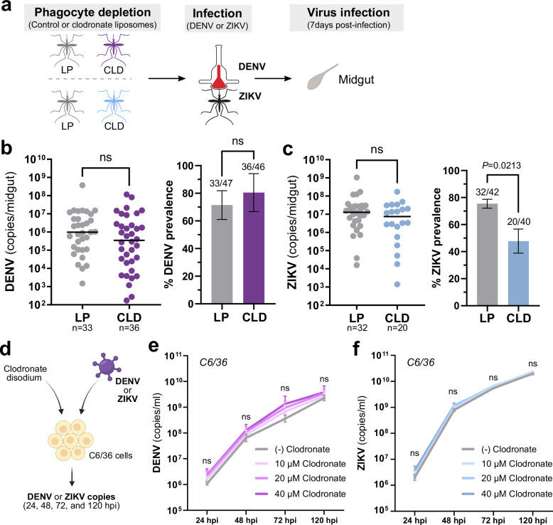
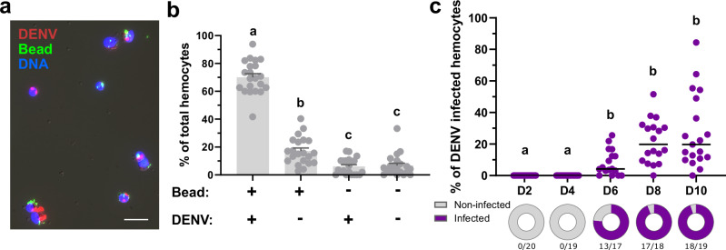
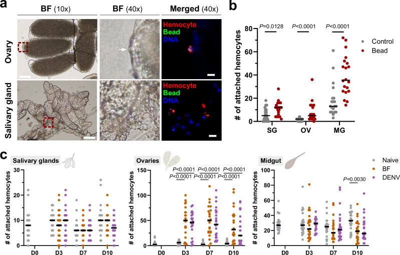
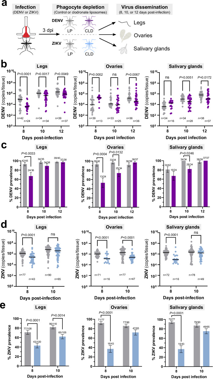
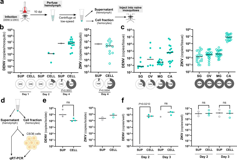
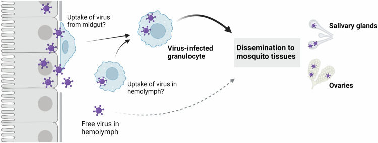
Mosquito-borne arboviruses cause more than 400 million annual infections, yet despite their public health importance, the mechanisms by which arboviruses infect and disseminate in the mosquito host are not well understood. Here, we provide evidence that dengue virus and Zika virus actively infect Aedes aegypti hemocytes and demonstrate, through phagocyte depletion, that hemocytes facilitate virus infection to peripheral tissues including the ovaries and salivary glands. Adoptive transfer experiments further reveal that virus-infected hemocytes efficiently confer virus infection to naïve recipient mosquitoes. Together, these data support a model of arbovirus dissemination where infected hemocytes enhance virus infection of mosquito tissues required for transmission, which parallels vertebrate systems where immune cell populations promote virus dissemination. This study significantly advances our understanding of virus infection dynamics in the mosquito host and highlights potential conserved roles of immune cells in arbovirus infection across vertebrate and invertebrate systems.


Conflict of interest statement

Competing interests: The authors declare no competing interests.

Figures

Fig. 1
Fig. 1. Effects of phagocyte depletion on DENV and ZIKV midgut infection.
Overview of experiments performed in Ae. aegypti where adult female mosquitoes were injected with either control (LP) or clodronate liposomes (CLD) to examine the effects of phagocytic granulocyte depletion on midgut virus infection (a). Approximately 24 h post-injection, mosquitoes were orally infected with DENV or ZIKV, then midguts were dissected at 7 days post-infection to determine infection outcomes. Viral midgut copy numbers and infection prevalence were determined for DENV (b) or ZIKV (c) by qRT-PCR, with each dot representing the viral titer of each individual midgut, with the median marked by the black line. The infection prevalence (number of infected mosquitoes of the total analyzed) is displayed in bar graphs as the mean ± SEM. All infection data were pooled from three or more independent experiments. Viral copy numbers were analyzed using a two-tailed Mann–Whitney test, while prevalence data were examined using a two-sided Fisher’s exact test. Exact P values are displayed in the figure where applicable; ns, not significant. Overview of in vitro experiments performed with C6/36 cells to examine DENV or ZIKV infection in the presence of different concentrations of clodronate disodium salt (d), the active component used in clodronate liposomes. DENV (e) and ZIKV (f) copy numbers were examined at 24, 48, 72, 120 h post-infection (hpi). Virus growth curves were examined using a two-way ANOVA with a Dunnett’s multiple comparison to determine significance. Data in (e) and (f) display the mean ± SEM of a total of six replicates obtained from two independent biological experiments. Statistical analysis was performed using a two-way ANOVA with a Dunnett’s multiple comparisons test. ns not significant. Illustrations in (a) were created by David Hall using Inkscape, while the image in (d) was created in BioRender. Smith, R. (2025) https://biorender.com/miahijk. Source data are provided as a Source Data file.
Fig. 2
Fig. 2. Immunolocalization of DENV in phagocytic granulocytes.
Hemocytes were perfused from DENV-infected mosquitoes 10 days post-infecton after injection with fluorescent beads (green). Following fixation, virus localization was examined by immunofluorescence on fixed hemocytes using an anti-DENV monoclonal antibody (clone 3H5-1) followed by an Alexa Fluor 568 goat anti-mouse IgG secondary antibody (red; scale bar: 10 μm) and mounted in ProLong®Diamond Antifade mountant with DAPI (blue) (a). To examine the abundance of virus in phagocytic granulocyte populations, the presence/absence of DENV was quantified in the context of the presence/absence of beads used to identify phagocytic granulocyte populations. b For each experimental outcome, the percentage of the total number of hemocytes for each phenotype are displayed per individual mosquito (dots, n = 21) and displayed as the mean ± SEM. Data were pooled from two independent biological experiments. c Additional experiments examine the percentage of virus-infected hemocytes following an oral infection with DENV. Hemocytes were perfused at 2, 4, 6, 8, and 10 days (D) post-infection, fixed, then examined by immunofluorescence as outlined above. Circles under each timepoint display the prevalence of infection and the number of individual mosquitoes (n) examined from two independent biological experiments. For both (b) and (c), statistical analysis was performed using Kruskal–Wallis with a Dunn’s multiple comparison test. Letters denote statistically significant differences between sample treatments. Source data and more detailed statistical comparisons, including P values associated with each comparison, are provided as a Source Data file.
Fig. 3
Fig. 3. Phagocytic granulocytes associate with the salivary glands and ovaries of DENV-infected mosquitoes.
To examine hemocyte attachment to mosquito tissues, DENV-infected mosquitoes were injected with CM-DiI (red) and fluorescent beads (green) at 7 days post-infection to identify phagocytic granulocyte populations (a). Following staining in vivo, ovary and salivary gland tissues were dissected to examine granulocyte attachment to each respective tissue and mounted using ProLong®Diamond Antifade mountant with DAPI (blue). Red dashed line boxes denote the field of view at 40× magnification, with white arrows used to indicate attached phagocytic hemocytes in the bright field (BF) image where applicable. Scale bars denote 100 μm for 10× images and 10 μm for 40× images. b The number of hemocytes attached to salivary gland (SG), ovary (OV), and midgut (MG) tissues was quantified in the presence or absence of beads in individual mosquitoes (n = 20 for all tissues, two independent replicates). Statistical analysis were performed using Multiple Mann–Whitney tests with a two-stage step-up (Benjamini, Krieger, and Yekutieli) to correct for multiple comparisons. Exact P values are displayed for each comparison. Since bead injection artificially increased hemocyte attachment (b), similar experiments relying on only CM-DiI staining were used to quantify hemocyte attachment to the salivary glands, ovaries, and midgut under naïve, blood-fed (BF), and DENV infection (c). Attachment was evaluated at 0, 3, 7, and 10 days (D) post blood-feeding, infection, or in age-matched naive controls in individual mosquitoes (n = 20 for all tissues, two independent replicates). Statistical analysis were performed using Multiple Mann–Whitney tests with a two-stage step-up (Benjamini, Krieger, and Yekutieli) to correct for multiple comparisons. Exact P values are displayed for any significant comparisons. For both (b) and (c), dots represent data collected from individual mosquitoes. Tissue images in (c) were created by David Hall using Inkscape. Source data are provided as a Source Data file.
Fig. 4
Fig. 4. Phagocytic granulocyte depletion impairs virus infection of the mosquito legs, ovaries, and salivary glands.
a Overview of virus dissemination experiments. After oral challenge with DENV or ZIKV, mosquitoes were injected with control (LP) or clodronate liposomes (CLD) at 3 days post-infection. Virus dissemination was examined in legs, ovaries, and salivary glands at 8, 10, and 12 days post-infection for DENV (b, c), or 8- and 10-days post-infection for ZIKV (d, e). Virus copy numbers from infected samples are shown for the legs, ovaries, and salivary glands for each time point for DENV (b) or ZIKV (d), with each dot representing the viral titer of each individual mosquito sample (n) and the median marked by the black line. The infection prevalence (number of infected tissues of the total analyzed) is displayed for the legs, ovaries, and salivary glands in bar graphs as the mean ± SEM for DENV (c) or ZIKV (e). Infection data were pooled from four independent experiments for DENV (b) and two independent experiments for ZIKV (d). Viral copy numbers were analyzed using a Multiple Mann–Whitney test with a two-stage step-up (Benjamini, Krieger, and Yekutieli) to correct for multiple comparisons. Exact P values are displayed in the figure where applicable; ns, not significant. Infection prevalence data were analyzed using a two-sided Fisher’s exact test. Exact P values are displayed in the figure for any significant comparisons. Illustrations in (a) were created by David Hall using Inkscape. Source data are provided as a Source Data file.
Fig. 5
Fig. 5. Cellular fractions of the hemolymph are more efficient in transferring a virus infection to naïve mosquitoes.
To assess the respective infectivity of the cellular and acellular fractions of mosquito hemolymph, Ae. aegypti were first infected with DENV or ZIKV, then hemolymph was perfused from mosquitoes at 10 days post-infection (a). Hemolymph was separated into hemocyte-containing cell (CELL) and acellular hemolymph supernatant (SUP) fractions using low-speed centrifugation and then injected into naïve mosquitoes (a). Whole mosquitoes were evaluated for viral titer and infection prevalence by qRT-PCR at 1, 2, and 4 days post-injection for DENV and 4 days post-injection for ZIKV (b). Additional analysis of salivary gland (SG), ovary (OV), midgut (MG), and carcass (CA) tissues at 4 days post-transfer of the CELL fraction confirm DENV and ZIKV infection of each respective tissue (c). For both (b) and (c), dots represent the viral titer of each individual mosquito/tissue, with the black line denoting the median. Due to the low numbers of virus positive samples in certain control and experimental conditions, viral titers were not directly compared. Infection prevalence (number of infected mosquitoes of the total examined, n) is displayed as shaded areas of circles under each experimental condition. Prevalence data were analyzed using a two-sided Fisher’s exact test, with exact P values are displayed in the figure where applicable. Data were pooled from two independent experiments. d Overview of experiments to examine the SUP and CELL fractions, where the DENV and ZIKV viral copy numbers were compared in SUP and CELL fractions (e) or that examine the ability of the SUP and CELL fractions to infect C6/36 cells at Days 2 and 3 post-infection (f). ZIKV viral copy numbers in e represent two independent experiments. All other data in (e) and (f) represents three independent biological experiments, and were analyzed using a two-tailed Student’s t test. Exact P values are displayed in the figure where applicable; ns, not significant. Illustrations in a were created by David Hall using Inkscape. The image in (d) was created in part using BioRender. Smith, R. (2025) https://BioRender.com/9cvyzdr and illustrations created by David Hall using Inkscape. Source data are provided as a Source Data file.
Fig. 6
Fig. 6. Phagocytic granulocyte depletion impairs the transfer of a virus infection to naive mosquitoes.
Mosquitoes were treated with clodronate liposomes (CLD) to deplete phagocytic granulocytes prior to DENV or ZIKV infection to confirm that granulocytes are required for the transfer of virus infection in the cellular fractions of the mosquito hemolymph (a). Empty liposomes (LP) serve as a control. Whole mosquitoes were assessed for viral titer and infection prevalence at 4 days post-injection for both DENV and ZIKV (b). Due to the low numbers of virus positive samples in certain control and experimental conditions, viral titers were not directly compared. Dots represent the viral copy number of individual mosquito samples, with the median displayed by the black line. Shaded areas of circles under each experimental condition correspond to infection prevalence, with the number infected of the total number of mosquitoes examined (n) displayed for each treatment. Prevalence data were analyzed using a two-sided Fisher’s exact test, with exact P values displayed in the figure where applicable. Data were pooled from two independent experiments. The image in a was created in part using BioRender. Smith, R. (2025) https://BioRender.com/4p4w103 and illustrations created by David Hall using Inkscape. Source data are provided as a Source Data file.
Fig. 7
Fig. 7. “Trojan horse” model of virus infection in the mosquito host.
Following virus infection of the mosquito midgut, a subset of phagocytic granulocytes likely acquire virus either through attachment to the virus-infected midgut, the uptake of free virus in the mosquito hemolymph, or the attachment to other infected mosquito tissues such as the trachea (not shown). Through the movement of these immune cells in the hemolymph and their ability to adhere to mosquito tissues, virus-infected granulocytes display increased efficiency to promote virus infection of the salivary glands and ovaries when compared to free virus present in the hemolymph. This suggests that these virus-infected granulocytes act as a “trojan-horse” to potentially enable further virus replication and enhance virus infection. Created in BioRender. Smith, R. (2025) https://BioRender.com/kj6zzk3.

Update of

Similar articles

Cited by

References

    1. Lwande, O. W. et al. Globe-trotting Aedes aegypti and Aedes albopictus: risk factors for arbovirus pandemics. Vector Borne Zoonotic Dis.20, 71–81 (2020). - PMC - PubMed
    1. Kraemer, M. U. G. et al. The global distribution of the arbovirus vectors Aedes aegypti and Ae. albopictus. Elife4, e08347 (2015). - PMC - PubMed
    1. Kraemer, M. U. G. et al. Past and future spread of the arbovirus vectors Aedes aegypti and Aedes albopictus. Nat. Microbiol.4, 854–863 (2019). - PMC - PubMed
    1. Bhatt, S. et al. The global distribution and burden of dengue. Nature496, 504–507 (2013). - PMC - PubMed
    1. Fauci, A. S. & Morens, D. M. Zika virus in the Americas — yet another arbovirus threat. N. Engl. J. Med.374, 601–604 (2016). - PubMed

MeSH terms

LinkOut - more resources