. 2025 Jul 1;16(1):5879.
doi: 10.1038/s41467-025-60984-y.

HIV-1 vif mediates ubiquitination of the proximal protomer in the APOBEC3H dimer to induce degradation

Affiliations

HIV-1 vif mediates ubiquitination of the proximal protomer in the APOBEC3H dimer to induce degradation

Katarzyna A Skorupka et al. Nat Commun. .

Abstract

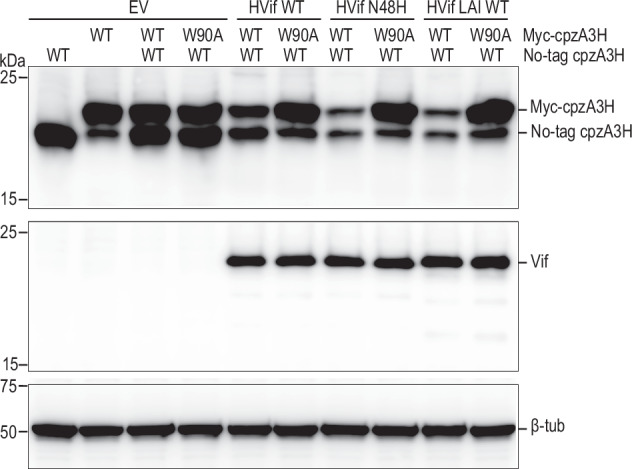
The APOBEC3 family of cytidine deaminases restricts retroviruses like HIV-1 by mutating viral DNA. HIV-1 evades this restriction by producing Vif, which recruits the Cullin-5 (CUL5) E3 ubiquitin ligase complex to promote APOBEC3 degradation. Here we resolve key aspects of this counter-defense mechanism by determining a 3.6 Å cryo-EM structure of chimpanzee APOBEC3H (cpzA3H) in complex with HIV-1 Vif and three components of the CUL5 E3 ligase-CBFβ, EloB, and EloC (VCBC). The structure captures cpzA3H as an RNA-mediated dimer within the cpzA3H-VCBC complex, allowing us to examine the role of dimerization. We find that ubiquitination occurs specifically at two lysine residues on the Vif-proximal protomer, while the distal protomer remains unmodified. The structural model of the active cpzA3H-Vif-CUL5 E3 ligase holoenzyme reveals spatial preferences for ubiquitin transfer to the targeted lysine residues. These findings enhance our understanding of A3H degradation and suggest new antiviral strategies targeting this host-virus interface.


Conflict of interest statement

Competing interests: The authors declare no competing interests.

Figures

Fig. 1
Fig. 1. Cryo-EM structure of the cpzA3H-VCBC complex.
Top panel: 4 Å cryo-EM reconstruction (EM map), the atomic model, and arrangement of protomers (pink and black) of the cpzA3H-VCBC complex without symmetry applied (C1). Two molecules of cpzA3H (dark green and green) complexed with dsRNA (orange), Vif (dark blue), and CBFβ (dark grey) form the black protomer. The pink protomer consists of one molecule of cpzA3H (light green) bound to dsRNA (yellow), Vif (light blue), CBFβ (light grey), and EloBC (white). Representative cryo-EM densities of distal A3H, RNA, and the Zn-coordinating region of proximal A3H are shown in semi-transparent densities superposed with corresponding atomic models. Bottom panel: 3.6 Å cryo-EM reconstruction, the atomic model, and arrangement of protomers of the cpzA3H-VCBC complex with C2 symmetry applied. Representative cryo-EM densities of RNA and the Zn-coordinating region of proximal A3H are shown in semi-transparent densities superposed with corresponding atomic models. Both black and pink protomers contain identical component molecules: one molecule of cpzA3H (dark green in the black protomer, light green in the pink protomer), dsRNA (orange and yellow, respectively), Vif (dark blue and light blue, respectively), and CBFβ (dark grey and light grey, respectively).
Fig. 2
Fig. 2. Molecular details of the cpzA3H-Vif interface.
a, b Atomic model of cpzA3H (green), dsRNA (yellow), Vif (cyan), with close-up views of the interface between cpzA3H and Vif (middle panels). Potential hydrogen bonds are indicated by dashed lines. Part of the atomic model was hidden to show the interacting amino acids clearly. c Schematic of the amino acid residues involved in this cpzA3H-Vif interactions. d Effect of amino acid substitution at the cpzA3H-Vif interface. The individual Vif amino acid residues highlighted in (c) were substituted with alanines, and the degradation of FLAG-tagged cpzA3H (top) was assessed. The intracellular levels of cpzA3H and Vif were probed 40 h post-transfection by Western blotting using anti-FLAG and anti-Vif mAbs. Blotting for ß-tubulin with polyclonal Abs (ß-Tub) was used as a loading control. The cpzA3H stability in the absence of Vif (no, lane 1) and presence of wild-type Vif (WT, lane 2) were used as controls. The results are representative from three independent experiments.
Fig. 3
Fig. 3. Across protomers RNA-Vif and cpzA3H-Vif interaction.
a Atomic model of the cpzA3H-bound dsRNA from protomer 1 (green and yellow, respectively) and Vif from protomer 2 (pink). b Close-up view of the across-protomers interface between cpzA3H and Vif. The amino acids involved in these interfaces are represented in stick model format. c Close-up view of the interface between RNA and Vif amino acid residues across the protomers. d Schematic of the Vif amino acid residues involved in the RNA interactions across protomers. e Effect of amino acid substitutions at these RNA-Vif and cpzA3H-Vif interfaces on cpzA3H degradation. Individual Vif amino acid residues highlighted in (b) and (d) were substituted with alanines, and the degradation of FLAG-tagged cpzA3H was assessed (top). The intracellular levels of cpzA3H and Vif were probed 40 h post-transfection by Western blotting using anti-FLAG and anti-Vif mAbs. β-Tub was used as a loading control. The cpzA3H stability in the absence of Vif (“no”, lane 1) and presence of wild-type Vif (“WT”, lane 2) were used as controls. The results are representative from three independent experiments.
Fig. 4
Fig. 4. Interaction between RNA and Vif.
a Superimposed structures of the cpzA3H-VCBC complex (with cpzA3H in green, RNA in yellow and Vif across protomers in pink) and the Vif (blue) and RNA (brown) molecules from the A3G-VCBC complex (PDB ID: 8H0I). The RMSD between the two Vif molecules is 0.62 Å. In the cpzA3H-VCBC structure, nucleotides 16–19 from 8H0I’s RNA strand were connected to chain G of cpzA3H-bound dsRNA (this study). b The panel expands on the Vif 161-PPLPS-165 region, showing an adenine molecule (from A3G-VCBC structure) with hydrogen bonds to P164, S165, and K168 (cyan dotted lines). c Electrostatic potential of cpzA3H, cpzA3H-bound dsRNA and Vif, including extended RNA strand from (PDB ID: 8H0I) as shown in panel (a). RNA-interacting residues of cpzA3H and Vif form a continuous positively charged surface accommodating the negatively charged RNA. The surface was colored in ChimeraX according to the electrostatic potential, ranging from -10.0 kT/e (red) to +10.0 kT/e (blue).
Fig. 5
Fig. 5. Across protomers Vif-Vif interface.
a Atomic model of the Vif-Vif interface formed between the protomers of the cpzA3H-VCBC complex. b Close-up view of the interface between Vif 1 (cyan) and Vif 2 (pink). The amino acids involved in this interface are represented in stick model format. c Schematic of the amino acid residues involved in Vif-Vif interactions. d Effect of amino acid substitutions at the Vif-Vif interface on cpzA3H degradation. The individual Vif amino acid residues highlighted in (c) were substituted with alanines, and the degradation of FLAG-tagged cpzA3H was assessed (top). The intracellular levels of cpzA3H and Vif were probed 40 h post-transfection by Western blotting using anti-FLAG and anti-Vif mAbs. An anti-ß tubulin polyclonal Abs (β-Tub) was used as a loading control. The cpzA3H stability in the absence of Vif (“no”, lane 1) and presence of wild-type Vif (“WT”, lane 2) were used as controls. The results are representative from three independent experiments.
Fig. 6
Fig. 6. K50 and K51 are critical ubiquitination sites of cpzA3H.
a The schematic overview of Vif-induced ubiquitination of A3H. ARIH2 is responsible for transferring the first Ub molecule onto A3H. The UbcH3 then elongates the Ub chain. b In vitro ubiquitination of cpzA3H by NL4-3 Vif WT (lanes 2–5) and the R41A variant (lanes 6–9). Ubiquitinated cpzA3H proteins were detected by Western blotting using an anti-A3H rabbit polyclonal Abs. c Ubiquitinated lysine (red) of cpzA3H, detected by mass spectrometry, are highlighted in the amino acid sequence of cpzA3H. d Comparison of K50, K51, and K52 ubiquitination. The frequency (%) with which peptides containing ubiquitinated K50, K51, K52, and their combinations are detected in the trypsin-digested K-ɛ-GG-enriched sample is shown in the pie chart. e cpzA3H-7KR contains K27R, K50R, K51R, K52R, K153R, K161R, and K168R mutations and protects cpzA3H from both NL4-3 Vif (left panel) and LAI Vif (right panel) induced degradation (lane 2). Each mutation was reversed within cpzA3H-7KR to generate cpzA3H-7KR-R27K, -R50K, -R51K, -R52K, -R153K, -R161K and -R168K (lanes 3 to 9). The 7KR + R50K (lane 4) and 7KR + R51K (lane 5) variants showed protein degradation efficiency equivalent to wild-type cpzA3H (lane 1). All proteins were expressed without any tag and detected by anti-A3H rabbit polyclonal Abs.
Fig. 7
Fig. 7. Vif induces degradation of proximal/Vif-Bound cpzA3H molecule.
HEK293T cells were co-transfected with either an empty vector (no Vif) or HIV-1 Vif expression plasmids, alongside the untagged cpzA3H (WT) and myc-cpzA3H (WT or W90A) expression plasmids. Independent vectors were used for each cpzA3H construct. At 40 h post-transfection, the intracellular levels of cpzA3H were analyzed by western blotting using an anti-A3H rabbit polyclonal antibody. Vif expression levels were detected with an anti-Vif mAbs. The ß-Tub was used as a loading control. We tested NL4-3 WT Vif (lanes 5 and 6), the NL4-3 N48H Vif variant (lanes 7 and 8), and LAI Vif (lanes 9 and 10) for their ability to degrade cpzA3H. Both myc-tagged and untagged WT cpzA3H were similarly degraded by all three Vif proteins (lanes 5, 7, and 9). In contrast, degradation occurred only for WT cpzA3H, which binds to Vif, when WT and W90A cpzA3H were co-expressed (lanes 6, 8, and 10).
Fig. 8
Fig. 8. Model of cpzA3H-Vif-CUL5 E3 ligase complex.
Model based on the cryo-EM structure from this study and components from PDB IDs 7B5L, 7B5M, 7ONI, 8FVJ,,. a cpzA3H-VCBC dimer structure (this study) is modeled with the CUL5 E3 ligase machinery, with proteins color-coded as follows: cpzA3H in green, Vif in cyan, CBFβ in dark gray, EloBC in light gray, members of CUL5 E3 ligase complex in gray, ARIH in beige, and ubiquitin in dark red. Gly76 of ubiquitin and Lys50 and Lys51 of cpzA3H are shown as spheres. This model depicts transition state 2 that is about to transfer ubiquitin to the target protein. b Distances between the cpzA3H lysine side chains and Gly76 of the E3-bound ubiquitin. Ubiquitination of these lysines, mediated by the Vif-CUL5 E3 ligase machinery, was detected by mass spectrometry.

Similar articles

References

    1. Sheehy, A. M., Gaddis, N. C., Choi, J. D. & Malim, M. H. Isolation of a human gene that inhibits HIV-1 infection and is suppressed by the viral Vif protein. Nature418, 646–650 (2002). - PubMed
    1. Harris, R. S. et al. DNA deamination mediates innate immunity to retroviral infection. Cell113, 803–809 (2003). - PubMed
    1. Kao, S. et al. The human immunodeficiency virus type 1 Vif protein reduces intracellular expression and inhibits packaging of APOBEC3G (CEM15), a cellular inhibitor of virus infectivity. J. Virol.77, 11398–11407 (2003). - PMC - PubMed
    1. Mangeat, B. et al. Broad antiretroviral defence by human APOBEC3G through lethal editing of nascent reverse transcripts. Nature424, 99–103 (2003). - PubMed
    1. Mariani, R. et al. Species-specific exclusion of APOBEC3G from HIV-1 virions by Vif. Cell114, 21–31 (2003). - PubMed

MeSH terms

LinkOut - more resources