. 2025 Jul 1;11(1):123.
doi: 10.1038/s41522-025-00724-8.

Microanatomy related biocidal activity at cellular resolution and bone reconstruction potential of PEG EGaIn nanocapsules

Affiliations

Microanatomy related biocidal activity at cellular resolution and bone reconstruction potential of PEG EGaIn nanocapsules

Kevin H Mwangi et al. NPJ Biofilms Microbiomes. .

Abstract

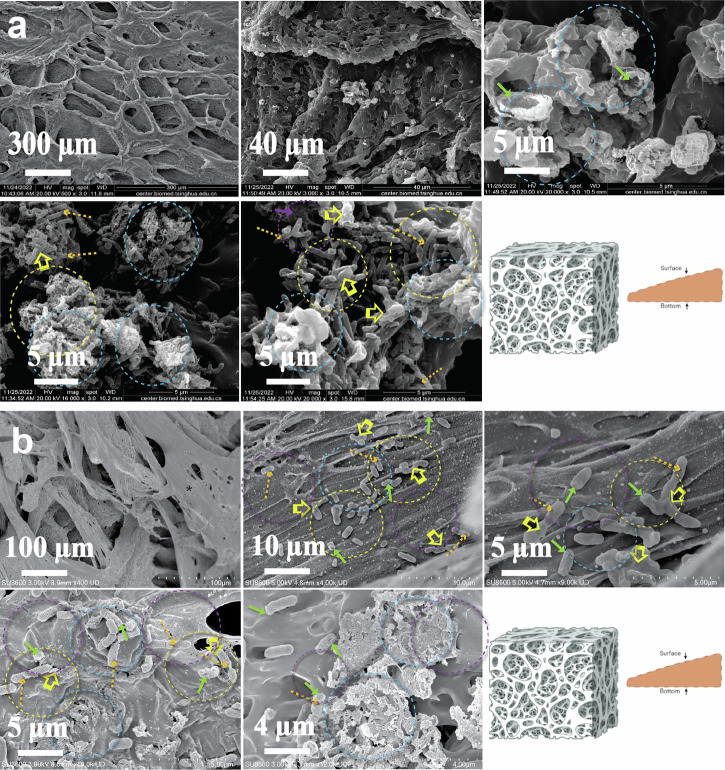
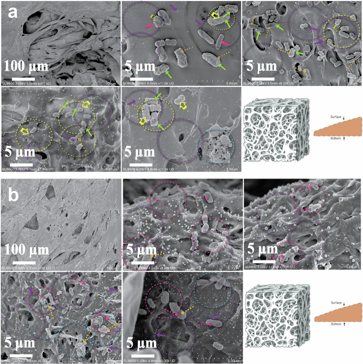
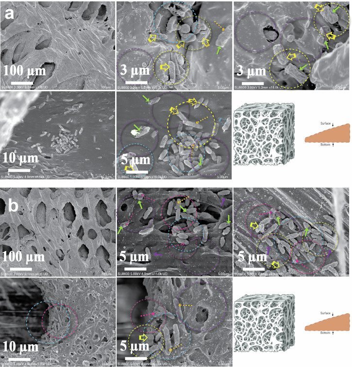
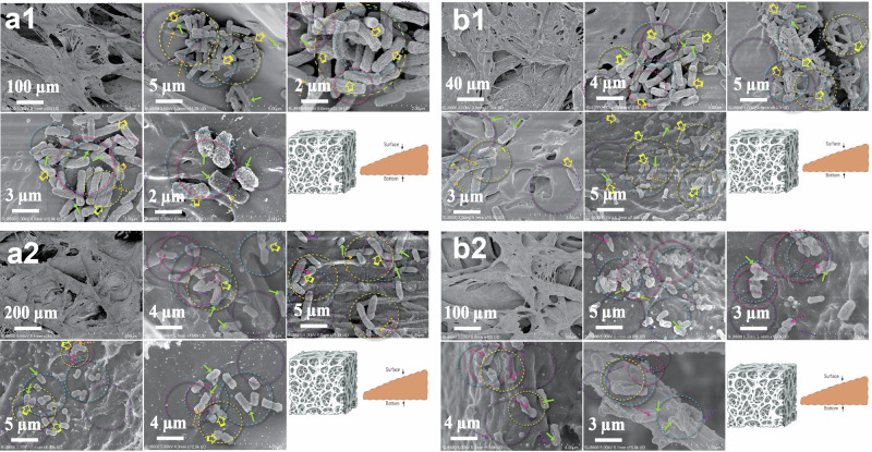
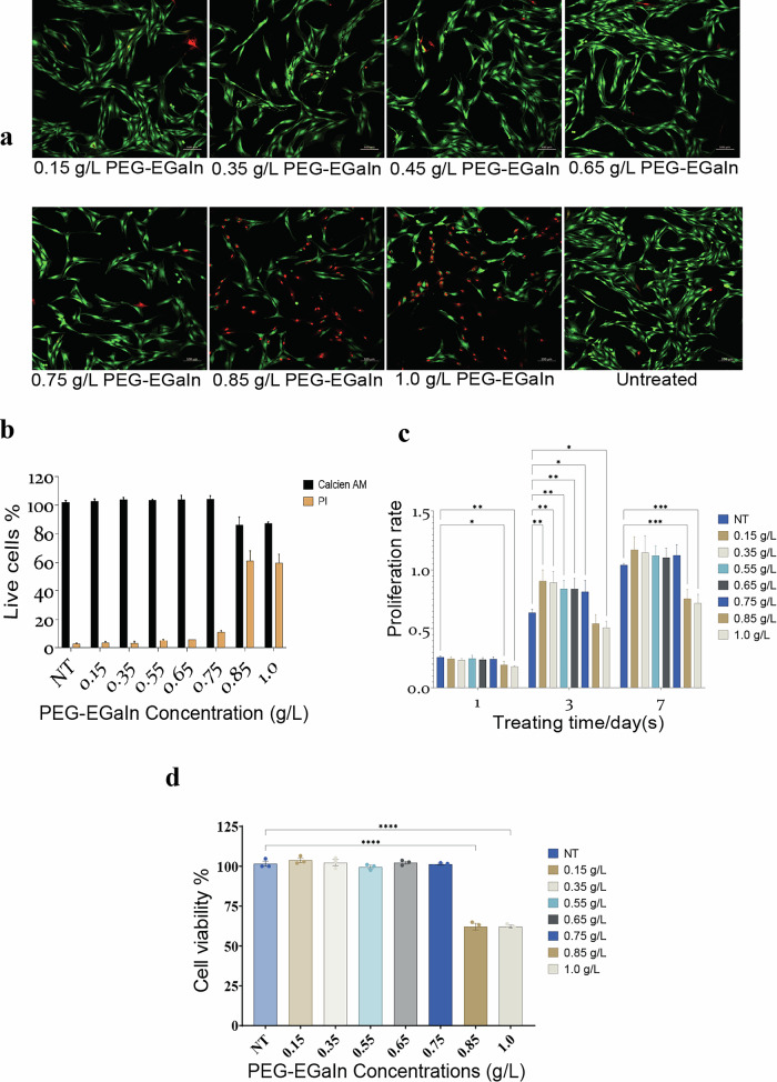
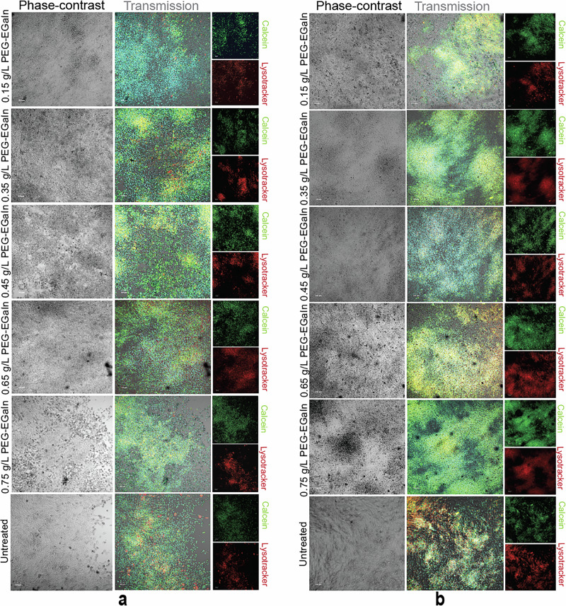
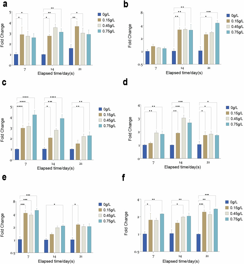
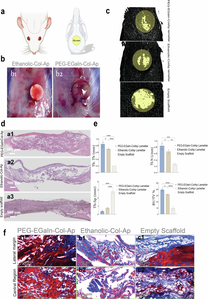
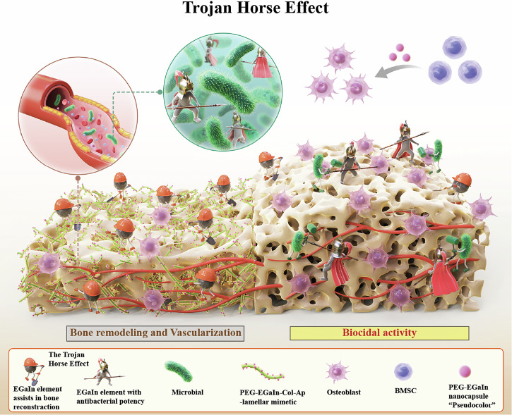
Critical bone defects, exacerbated by infections, pose significant challenges to bone healing and homeostasis, necessitating the development of dual-functional biomimetics that combine anti-infective and reparative capabilities. The EGaIn holds promise across various disciplines, though its interactions with microbial cells require further elucidation. This investigation evaluates the antimicrobial efficacy of PEG-EGaIn nanocapsules against a spectrum of bacterial, employing electron microscopy. Constructs containing 1.5% PEG-EGaIn hinder biofilm-producing bacteria, while 3% concentrations amplify the biocidal effect. Furthermore, the nanocapsules promoted osteogenic differentiation rBMSCs, evidenced by enhanced mineralization and upregulation of key osteogenic genes. In addressing large bone defects, PEG-EGaIn-Col-Ap-lamellar and ethanolic-mediated Col-Ap-lamellar constructs serve as potential solutions for bone resorption mitigation and osteo-angiogenesis. Bone-remodeling were validated through μ-CT and histomorphometry confirming no evidence of chronic inflammation or fibrosis. In this study, PEG-EGaIn nanocapsules emerge as potent biocide and bone repair, underscoring their potential in combating antibiotic resistance and enhancing bone healing.


Conflict of interest statement

Competing interests: The authors declare no competing interests.

Figures

Fig. 1
Fig. 1. Spatial-temporal interrogation of the E. coli-K12 to PEG-EGaIn-coated PCL construct as detected by FE-SEM.
a 1.5% PEG-EGaIn coated PCL, b 3% PEG-EGaIn coated PCL. Morphological Changes: (a, b). Asterix = conditioning film deposited onto the PCL-mimetics forming intersecting pore networks covering the entire mimetic framework; Purple arrows and dotted circles = PEG-EGaIn nanocapsules; green arrows= membrane distortion (i.e., telescoping or invaginations) close to the polar and septal regions; pale blue dotted circles = leakage of intracellular contents; Yellow open arrows and dotted circles = abnormal textures (i.e., membrane blebbing and clumping); Orange dotted arrows = Disruption in cell cycle regulation and division (abnormal amitosis), (n = 5).
Fig. 2
Fig. 2. Spatial-temporal interrogation of the S. aureus to PEG-EGaIn-coated PCL construct as detected by FE-SEM.
a 1.5% PEG-EGaIn coated PCL, b 3% PEG-EGaIn coated PCL.Morphological Changes: (a, b). Asterix = conditioning film deposited onto the PCL-mimetics forming intersecting pore networks covering the entire mimetic framework; Purple arrows and dotted circles = PEG-EGaIn nanocapsules; green arrows= membrane distortion (i.e., telescoping or invaginations); pink open arrows and dotted circles = membrane contours displaying blisters or cavitations); pale blue dotted circles = leakage of intracellular contents; Yellow open arrows and dotted circles = abnormal textures (i.e., membrane blebbing and clumping); Orange dotted arrows = Disruption in cell cycle regulation and division (abnormal amitosis) (n = 5).
Fig. 3
Fig. 3. Spatial-temporal interrogation of the P. aeruginosa (PAO1) to PEG-EGaIn-coated PCL construct as detected by FE-SEM.
a 1.5% PEG-EGaIn coated PCL, b 3% PEG-EGaIn coated PCL. Morphological Changes: (a, b). Asterix = conditioning film deposited onto the PCL-mimetics forming intersecting pore networks covering the entire mimetic framework; Purple arrows and dotted circles = PEG-EGaIn nanocapsules; green arrows= membrane distortion (i.e., telescoping or invaginations) close to the polar and septal regions;; pink open arrows and dotted circles = membrane contours displaying blisters or cavitations); pale blue dotted circles = leakage of intracellular contents; Yellow open arrows and dotted circles = abnormal textures (i.e., membrane blebbing and clumping); Orange dotted arrows = Disruption in cell cycle regulation and division (abnormal amitosis) (n = 5).
Fig. 4
Fig. 4. Spatial-temporal interrogation of the K. pneumoniae (ATCC and 226) to PEG-EGaIn-coated PCL construct as detected by FE-SEM.
a1, b1 1.5% PEG-EGaIn coated PCL, a2, b2 3% PEG-EGaIn coated PCL. Morphological Changes: (a12) K. pneumoniae ATCC and (b12) K. pneumoniae-226. Asterix = conditioning film deposited onto the PCL-mimetics forming intersecting pore networks covering the entire mimetic framework; Purple arrows and dotted circles = PEG-EGaIn nanocapsules; green arrows= membrane distortion (i.e., telescoping or invaginations) close to the polar and septal regions;; pink open arrows and dotted circles = membrane contours displaying blisters or cavitations; pale blue dotted circles = leakage of intracellular contents; Yellow open arrows and dotted circles = abnormal textures (i.e., membrane blebbing and clumping); Orange dotted arrows = Disruption in cell cycle regulation and division (abnormal amitosis), (n = 5).
Fig. 5
Fig. 5. PEG-EGaIn nanocapsules effects on rBMSCs.
a Representative merged fluorescent confocal imaging of the treated rBMSCs with PEG-EGaIn nanocapsules (0.15, 0.35, 0.45, 0.65, 0.75, 0.85, and 1.0 g/L). The cells were stained with Calcein AM and Propidium Iodine (PI) following treatment period (48 h) (n = 6). b Population of live/dead cells presented as a percentage of all counted cells. Data is represented as mean ± SD and values were compared by the repeated measures by one-way AVOVA (n = 4). c CCK-8 assay was performed to determine proliferation rate at different treating time points (1, 3, and 7 d). Data is presented as mean ± SD and significance were compared by the repeated measures by two-way ANOVA test denoted as *p < 0.05, **p < 0.01, ***p < 0.001, and ****p < 0.0001. (n = 6). d CCK-8 assay was performed prior to Live/Dead staining to determine cell viability of rBMSCs treated with gradient series of PEG-EGaIn nanocapsules (0.15, 0.35, 0.45, 0.65, 0.75, 0.85, and 1.0 g/L). Data is presented as mean ± SEM and significance is denoted as *p < 0.05, **p < 0.01, ***p < 0.001, ****p < 0.0001 by one-way ANOVA. Data were normalized with respect to untreated, (n = 4).
Fig. 6
Fig. 6. PEG-EGaIn nanocapsules (0.15-, 0.35-, 0.45-, 0.65- and 0.75 g/L) promotes the proliferation and osteogenic differentiation of rBMSCs.
a, b The cells were cultured with calcein, and stained with Hoechst 33342 and Lysotracker-red at the end of each induction period (days 7 and 16). a Representative confocal live-imaging of the treated cells following calcification induction with gradient series of PEG-EGaIn nanocapsules for day7 showed crystalline deposits which produced a particularly distinctive pattern that was easily seen under the phase-contrast as light sheets of nucleation points. b Representative confocal live-imaging following day 16 mineralizing rBMSCs precipitated significant apatite minerals appearing as dense nucleation points that expanded into a continuous matrix. Note the similar pattern of staining visualized by calcein labeling (top-right panel) and lysotracker-red (bottom-right panel); overlap between the phase-contrast (left panel) and merged fluorescent images in composite setting (right panel), scale bars representation 100 μm. Representative pictures are shown, (n = 3).
Fig. 7
Fig. 7. Real-time polymerase chain reaction (RT-qPCR) quantification of gene expression profiles of osteoblastic markers.
a ON/SPARC b RUNX2 c POSTN d BGLAP/OCN e ENPP1/ PC-1, and f ALPL on rBMSCs following induction with PEG-EGaIn treatments (0.15-, 0.35-, 0.45-, 0.65- and 0.75 g/L) for 7,14, and 21 days. Data are represented as mean ± SD (n = 6). Data is presented as mean ± SD and significance were compared by the repeated measures by two-way ANOVA test denoted as *p < 0.05, **p < 0.01, ***p < 0.001, ****p < 0.0001. Data were normalized with respect to gene expression of untreated, (n = 3).
Fig. 8
Fig. 8. Newly bone formation in a rat cranial bone defect model.
a, b In vivo rat calvarial regeneration within the rat calvaria critical-size defect. Critical-sized bone defects (ca. 5 mm in diameter) were generated in the calvarium of SD-rat. c Three-dimensional µ-CT images were also examined to determine the microarchitecture and distribution of the newly formed mineralized tissue. Representative scale bars 1 mm. e Quantity analysis of the bone formation (BV/TV, Tb. Th, Tb. N and Tb.Sp). Data are represented as mean ± SEM and significance were compared by the repeated measures by one-way ANOVA test denoted as *P < 0.05 between groups ****P < 0.0001 (BV/TV, Tb. Th and Tb.Sp), **P < 0.01, ***P < 0.001 (Tb. N) (n = 4). df Histological analysis with Hematoxylin & Eosin and Masson’s Trichrome staining of samples at 12 weeks. Representative scale bars 500 µm (d) and 100 µm (f) (n = 4).
Fig. 9
Fig. 9. A graphical summary of the diverse properties of the PEG-EGaIn nanomaterial, leveraging the trojan horse concept.
Microbial presence and its metabolites affecting the orthopedic implant. The PEG-EGaIn nanomaterial targeting to bone infection by destroying and disordering the bacterial membrane. The Col-Ap-lamellar construct containing PEG-EGaIn nanocapsules can enhance bone regeneration by activating skeletal interoception.

Similar articles

References

    1. De Long, W. G. Jr. et al. Bone grafts and bone graft substitutes in orthopaedic trauma surgery. A critical analysis. J. Bone Jt. Surg. Am.89, 649–658 (2007). - PubMed
    1. Amini, A. R., Laurencin, C. T. & Nukavarapu, S. P. Bone tissue engineering: recent advances and challenges. Crit. Rev. Biomed. Eng.40, 363–408 (2012). - PMC - PubMed
    1. Sheikh, Z., Brooks, P. J., Barzilay, O., Fine, N. & Glogauer, M. Macrophages, foreign body giant cells and their response to implantable biomaterials. Materials8, 5671–5701 (2015). - PMC - PubMed
    1. Witherel, C. E., Abebayehu, D., Barker, T. H. & Spiller, K. L. Macrophage and fibroblast interactions in biomaterial-mediated fibrosis. Adv. Healthcare Mater.8, e1801451 (2019). - PMC - PubMed
    1. Roope, L. S. J. et al. The challenge of antimicrobial resistance: what economics can contribute. Science364, eaau4679 (2019). - PubMed

MeSH terms