. 2025 Jul 1;16(1):5800.
doi: 10.1038/s41467-025-61243-w.

Self-reinforced piezoelectric chip for scaffold-free repair of critical-sized bone defects

Affiliations

Self-reinforced piezoelectric chip for scaffold-free repair of critical-sized bone defects

Wei Li et al. Nat Commun. .

Abstract

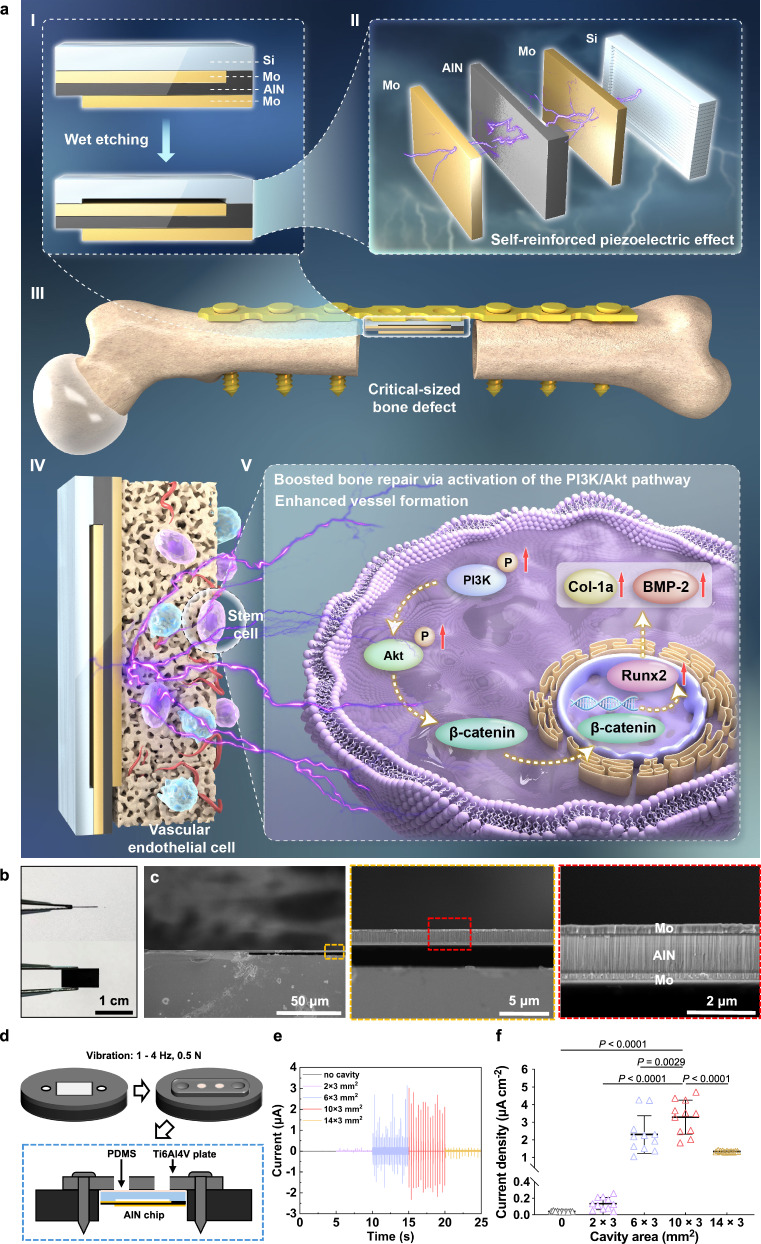
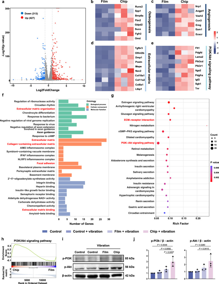
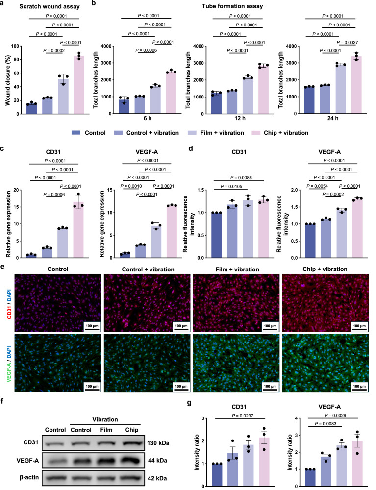
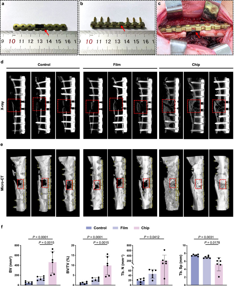
The use of piezoelectric materials to treat critical-sized bone defects typically requires additional stimulation to generate their piezoelectric properties and the implantation of scaffolds to promote bone repair. Here we present a self-reinforced piezoelectric chip and demonstrate its efficacy in the treatment of critical-sized bone defects. Specifically, the chip is comprised of the third-generation semiconductor aluminum nitride (AlN) as a piezoelectric layer, molybdenum (Mo) electrodes, and a silicon substrate with an optimized internal cavity structure. All these components are confirmed to be non-cytotoxic. This design enables the chip to provide self-sustained and long-term electrical signals in response to physiological vibrations. After being implanted into a rabbit critical-sized femoral defect model, the chip creates a localized bioelectric microenvironment, thereby promoting vascularized bone repair within 4 weeks without using any scaffolds and additional tools. Moreover, the chip can be fixed onto the clinically used orthopedic plate system, representing a universal plug-and-play strategy.


Conflict of interest statement

Competing interests: The authors declare no competing interests.

Figures

Fig. 1
Fig. 1. Design and characterization of the self-reinforced piezoelectric chip for scaffold-free repair of critical-sized bone defects.
a The smart chip is a multilayer structure, including a Si substrate, Mo electrodes, and an AlN piezoelectric layer (II). An internal cavity was etched into the Si substrate using a wet etching method to enhance sensitivity to physiological vibrations (I). The chip generates alternating electrical signals under vibration, converting mechanical energy into a localized bioelectric microenvironment. After being integrated with a clinically used plate and implanted at the critical-sized bone defect site (III), the chip synergistically activates the PI3K/Akt signaling pathway (V), thereby promoting the proliferation and osteogenic differentiation of stem cells (IV). Meanwhile, the chip enhances angiogenesis by upregulating VEGF-A and CD31 expression. Hence, the chip provides a promising strategy for critical-sized bone defect repair through robust electrical stimulation and vascularized bone regeneration, achieving an efficient scaffold-free bone repair. b Photos of the chip. c Cross-sectional SEM images of the chip with a 10 × 3 mm2 cavity. d Schematic diagram of the current testing system for the chips. A testing disk with a central cavity (10 × 5 mm2) is used to simulate a bone defect. The chips are adhered to a Ti6Al4V plate using Polydimethylsiloxane (PDMS), and then the plate is fixed across the defect with screws. A vertical cyclic force (0.5 N at 1−4 Hz) is applied perpendicular to the disk to mimic physiological vibration. e Electrical signal of the chips integrated with Ti6Al4V plates under vibration. f Statistical analysis of current density derived from panel (e). Data are presented as mean ± SD (n = 11). Two-sided ANOVA with a Tukey’s post hoc test for multiple comparisons. Source data are provided as a Source Data file.
Fig. 2
Fig. 2. In vitro osteogenic effect of the chip.
ALP staining (a) and ALP activity (c) of BMSCs cultured in the control, control + vibration, film + vibration, and chip + vibration groups for 7 days. Alizarin Red staining (b) and its qualitative analysis (d) of the extracellular matrix of BMSCs cultured in the control, control + vibration, film + vibration, and chip + vibration groups for 21 days. e Expression levels of osteogenic-related genes, including Col-1a, BMP-2, and Runx2 of BMSCs, were measured and normalized to the GAPDH. f Representative images of immunofluorescence staining of BMP-2 (Red) and Runx2 (Green). g Semi-qualitative analysis of BMP-2 and Runx2 expression derived from panel (f). h Western blot results of the expression of Runx2, Col-1a, and BMP-2 after 7 days of culture. i Quantitative study of the grayscale of protein expression derived from panel (h). Data are presented as mean ± SD (n = 3 biological replicates). Two-sided ANOVA with a Tukey’s post hoc test for multiple comparisons. Source data are provided as a Source Data file.
Fig. 3
Fig. 3. Transcriptome analysis of osteogenic differentiation of BMSCs.
a Volcano plots of the chip + vibration versus the film + vibration. Heatmap of osteogenesis (b), angiogenesis (c), extracellular matrix (d), and PI3K/Akt signaling pathway (e) related genes. GO (f) and KEGG (g) pathway enrichment analysis. h GSEA for the PI3K/Akt signaling pathway. i Western blotting analysis for the expression of p-PI3K and p-Akt of BMSCs in the control, control + vibration, film + vibration, and chip + vibration groups for 7 days of incubation. j Quantitative analysis of the grayscale of protein expression derived from panel (f). Data are presented as mean ± SD (n = 3 biological replicates). Two-sided ANOVA with a Tukey’s post hoc test for multiple comparisons. Source data are provided as a Source Data file.
Fig. 4
Fig. 4. In vitro angiogenesis.
a Quantification of wound healing assay. b Quantitative evaluation of total branches length in tube formation assay. c Expression levels of angiogenic genes CD31 and VEGF-A of HUVECs cultured for 3 days. Semi-qualitative analysis (d) and representative immunofluorescence staining images (e) of CD31 (Red) and VEGF-A (Green) of HUVECs in different groups. Western blot results (f) and quantitative analysis (g) of protein expression of CD31 and VEGF-A. Data are presented as mean ± SD (n = 3 biological replicates). Two-sided ANOVA with a Tukey’s post hoc test for multiple comparisons. Source data are provided as a Source Data file.
Fig. 5
Fig. 5. The chip promotes bone formation in rabbit critical-sized femoral defects.
Photos of the chip on the Ti6Al4V plate (a, b) and the implantation of the chip (c). The chip is highlighted by the red arrow. Representative 2D X-ray images (d) and 3D reconstruction images (e) of bone defect in the control, film, and chip groups at 4 weeks post-operation. f Quantitative analysis of BV, BV/TV, Tb. N and Tb. Sp derived for panel (e). Data are presented as mean ± SD (n = 6 biological replicates). Two-sided ANOVA with a Tukey’s post hoc test for multiple comparisons. Source data are provided as a Source Data file.
Fig. 6
Fig. 6. Histological and immunohistochemistry staining.
Representative images of H&E staining (a), Masson’s trichrome staining (b) and Goldner’s trichrome staining (c) in different groups. Immunohistochemistry staining of BMP-2 (d) and Runx2 (e) at 4 weeks post-operation. The newly formed bone tissues were highlight by black arrows. F represent fibrous tissues, O represents old bone tissues, Ti represents Ti6Al4V plate, and S represents screws. All experiments were repeated independently at least three times with similar results.

Similar articles

References

    1. Schemitsch, E. H. Size matters: Defining critical in bone defect size!. J. Orthop. Trauma31, S20–S22 (2017). - PubMed
    1. Dimitriou, R., Jones, E., McGonagle, D. & Giannoudis, P. V. Bone regeneration: current concepts and future directions. BMC Med.9, 66 (2011). - PMC - PubMed
    1. Yuan, X. et al. Recent advances in 3D printing of smart scaffolds for bone tissue engineering and regeneration. Adv. Mater.36, e2403641 (2024). - PubMed
    1. Lu, J., Gao, Z., He, W. & Lu, Y. Harnessing the potential of hyaluronic acid methacrylate (HAMA) hydrogel for clinical applications in orthopaedic diseases. J. Orthop. Transl.50, 111–128 (2025). - PMC - PubMed
    1. Nain, A. et al. Progress in the development of piezoelectric biomaterials for tissue remodeling. Biomaterials307, 122528 (2024). - PubMed

LinkOut - more resources