. 2025 Jul 1;16(1):5782.
doi: 10.1038/s41467-025-61261-8.

A skin-interfaced three-dimensional closed-loop sensing and therapeutic electronic wound bandage

Affiliations

A skin-interfaced three-dimensional closed-loop sensing and therapeutic electronic wound bandage

Xingcan Huang et al. Nat Commun. .

Abstract

Chronic wound healing is a complex and long-standing problem, that has been a major and critical clinical concern around the world for years. Recent advances in digital wound dressings open new possibilities for solving the problem. Here, we report a battery-free, fully permeable, skin-adhesive, stretchable electronic wound bandage (iSAFE) for intelligent wound management. This electronic bandage exhibits superior properties in multiple features and can be conformally adhered to the skin wound. In addition, the iSAFE can accurately assess the wound conditions in-situ and thus adaptively perform localized drug release. The results from both in vitro and in vivo studies on animals prove the validity of wound monitoring, wound healing boosting and intelligent closed-loop wound management. Clinical trials on patients across age 18 to 95 with various types of wounds are performed. These results all indicate the unique and universality of the reported technology for wound monitoring and management.


Conflict of interest statement

Competing interests: The authors declare no competing interests.

Figures
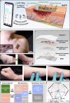
Fig. 1
Fig. 1. Skin adhesive, battery-free intelligent, stretchable, permeable and multi-functional electronics (iSAFE) system for wound management.
a Schematic illustration depicting iSAFE as a multi-functional wound electronic capable of closed-loop sensing and treatment for infected chronic wounds. Highly-integrated configuration endows iSAFE with superior breathability, waterproofness, tissue adhesiveness, anti-bacterial activity, and on-demand therapeutic ability. b Optical images of the iSAFE attached onto the chest. The enlarged image demonstrates the robust adhesion of the iSAFE to skin. c Exploded-view illustration of iSAFE highlighting key layers. d Optical images showing the comparison between conventional wound dressing (left) and our iSAFE (right) for ankle wound treatment. e Optical images of the iSAFE system adhering to wrist and arm and its deformation with skin. f Block diagram of the key components of the iSAFE system. g A radar map comparing the critical performances (i.e., permeability, anti-bacterial ability, skin adhesion, stretchability, waterproofness, sensing, and drug delivery) of our iSAFE against other published systems.
Fig. 2
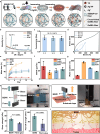
Fig. 2. Preparation and characterization of the bioadhesive, multi-functional BEWI.
a Schematic illustration of the fabrication process of the BEWI. b XRD patterns of the electrospun SEBS, GelMA, SEBS/GelMA and BEWI films. c Conductivities of BEWIs with different ratios of SEBS and GelMA (w/w) (n = 3 independent replicates, one-way two-sided analysis of variance test, NS denotes not significant, mean value ± SD). d Stress-strain curves of different electrospun films. e Mass loss of the electrospun SEBS, GelMA, SEBS/GelMA and BEWI films after incubation in PBS containing collagenase (3 μ/mL) for 14 days (n = 3 independent replicates, mean value ± SD). f Water vapor transmission rate and (g) water sorption of different electrospun films (n = 3 independent replicates, mean value ± SD). Schematics and optical images and of the (h) lap shear and (i) peel-off tests. Comparison of (j) shear strength and (k) peel-off strength between BEWI and commercial fibrin glue (n = 3 independent replicates, Two-sided Student’s t test, *P  <  0.05, mean value ± SD). l Schematic illustration showing the mechanism of tissue adhesiveness of BEWI. SEBS mentioned in this work represents the hydrophilic SEBS/F127.
Fig. 3
Fig. 3. In vitro evaluation of biocompatibility, pro-healing, and anti-bacterial performances of BEWI.
a Live/dead staining of NIH/3T3 cells cultured with different materials for 24, 48, and 72 h. b Cell viability calculated based on live/dead staining images (n = 3 independent replicates, mean value ± SD). c Cell proliferation evaluation via CCK-8 test (n = 3 independent replicates, mean value ± SD). d Scratch assay to analyze the migration ability of NIH/3T3 cells pretreated with BEWI. Control is the group without BEWI treatment. e Quantification of the scratch closure rate (n = 3 independent replicates, one-way two-sided analysis of variance test, **P  <  0.01, ***P  <  0.001, mean value ± SD). f Images of cultivated E. coli and S. aureus colonies onto Luria-Bertani (LB) agar plates after being treated with different materials. g Quantitative assessment of anti-bacterial rate of various materials against E. coli and S. aureus (n = 3 independent replicates, one-way two-sided analysis of variance test, mean value ± SD). For the E. coli group, ****P  <  0.0001, ***P  =  0.0003 and, *P  = 0.0211. For the S. aureus group, ****P  <  0.0001, ***P  =  0.0001, **P  =  0.0055 and, *P  = 0.0313.
Fig. 4
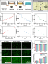
Fig. 4. Characterization of stretchable biosensors based on the electrospun SEBS film.
a Schematic illustration of the fabrication process of the stretchable SEBS electrode. b Schematic illustration showing the electrode layout of working electrode (i.e., thermal, pH and glucose sensors), counter electrode (CE) and reference electrode (RE) onto BEWI. ce Output responses of the glucose, pH and Tem sensors as a function of time. fh Corresponding calibration curves of the glucose, pH and Tem sensors. i Biocompatibility evaluation of pH, glucose and Tem sensors by live/dead staining. Quantitative analysis of (j) cell viability and (k) cell proliferation co-cultured with different specimens (n = 3 independent replicates, mean value ± SD).
Fig. 5
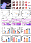
Fig. 5. In vivo investigation of iSAFE for in-situ wound monitoring and accelerated wound healing.
a Optical image of iSAFE adhered to the rat with a chronic wound on its back for in-situ wound monitoring and therapy. b Representative images and schematic illustrations showing the change of diabetic infected wounds with time after different treatments. In-situ measurement of dynamic changes of (c) glucose concentration, (d) pH and (e) Tem in the infected diabetic wound with different treatments (n = 3 independent replicates, one-way two-sided analysis of variance test, *P  <  0.05, **P  <  0.01, ***P  <  0.001 and ****P  <  0.0001, mean value ± SD). f Representative histologic images of wound tissues harvested from diabetic rats with different treatments for 14 days. Top and bottom left, hematoxylin and eosin (H&E); bottom right, Masson’s trichrome (MT). Quantitative analysis of (g) epidermal thickness, (h) length of wound area, (i) re-epithelialization, (j) appendage count, (k) scar elevation index and (l) collagen density of skin tissues after different treatments (n = 3 independent replicates, one-way two-sided analysis of variance test, *P  <  0.05, **P  <  0.01, ***P  <  0.001 and ****P  <  0.0001, mean value ± SD).
Fig. 6
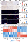
Fig. 6. Mechanism analysis of the wound healed through iSAFE treatment.
Representative immunostaining images for (a) cluster of differentiation 68 (CD68), interleukin 6 (IL-6), alpha smooth muscle actin (α-SMA) and Ki-67 (red) (Cell nuclei were stained with 4′, 6-diamidino-2-phenylindole (DAPI, blue)) from wound tissues after different treatments. Quantity analysis of the IHC staining (b) CD 68 positive rate, (c) average optical density (AOD) of IL-6, (d) α-SMA+ cells density, and (e) Ki-67 positive density of wounds with three different wound dressings after 6 days treatment (n = 3 independent replicates, one-way two-sided analysis of variance test, *P  <  0.05, **P  <  0.01, ***P  <  0.001 and ****P  <  0.0001, mean value ± SD). f Heatmap showing the comparison of differentially expressed genes (DEGs) in the wound tissues between the control group and the iSAFE treatment group (P < 0.05).
Fig. 7
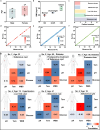
Fig. 7. Clinical assessment of the in situ wound monitoring glucose, pH and Tem.
a Box plot showing the age and gender distribution of 10 enrolled patients. b Box plot showing the BMI distribution. Here, the normal number is 4 (n = 4), the overweight number is 3 (n = 3), and the obese number is 3 (n = 3). Minima, maxima, center, bounds of the box and whiskers and percentiles are shown in the box plots of (a and b). c Column plot displaying the diseases distribution in these 10 patients. Analysis of the accuracy rate of (d) glucose, (e) pH and (f) Tem sensors in the iSAFE system by comparing with data collected from commercial instruments. g Patient-specific correlation matrices of parameters assessed by sensors and the wound bed score (WBS) for the 6 tracked patients. The scale bar represents Pearson’s correlation coefficient (rp). Part elements in this figure were Created in BioRender. H, K. (2025) https://BioRender.com/nd4kln3.

Similar articles

References

    1. Yu, X. et al. Needle-shaped ultrathin piezoelectric microsystem for guided tissue targeting via mechanical sensing. Nat. Biomed. Eng.2, 165–172 (2018). - PubMed
    1. Song, E. et al. Miniaturized electromechanical devices for the characterization of the biomechanics of deep tissue. Nat. Biomed. Eng.5, 759–771 (2021). - PubMed
    1. Wang, C. et al. Monitoring of the central blood pressure waveform via a conformal ultrasonic device. Nat. Biomed. Eng.2, 687–695 (2018). - PMC - PubMed
    1. Zhang, B. et al. A three-dimensional liquid diode for soft, integrated permeable electronics. Nature628, 84–92 (2024). - PubMed
    1. Li, J. et al. Thin, soft, wearable system for continuous wireless monitoring of artery blood pressure. Nat. Commun.14, 5009 (2023). - PMC - PubMed

LinkOut - more resources