. 2025 Jul 1;15(1):22067.
doi: 10.1038/s41598-025-03419-4.

The function of PCSK9 in doxorubicin-induced cardiotoxicity and its underlying mechanism

Affiliations

The function of PCSK9 in doxorubicin-induced cardiotoxicity and its underlying mechanism

Shuai Shi et al. Sci Rep. .

Abstract

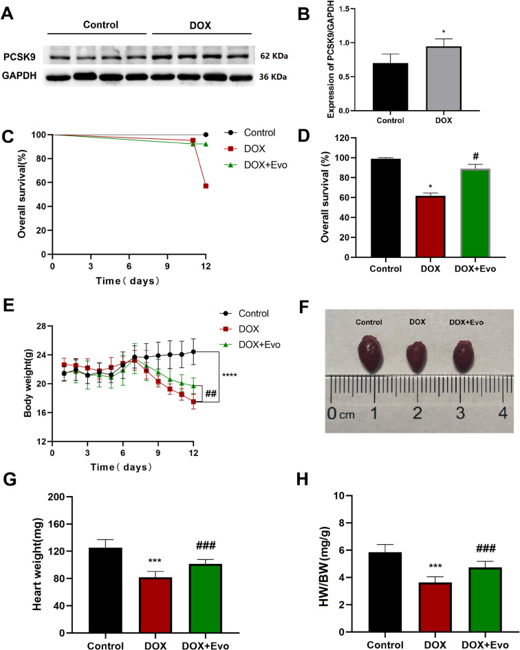
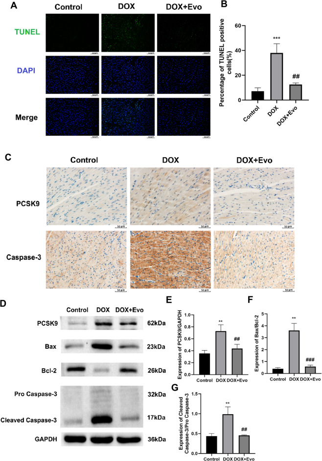
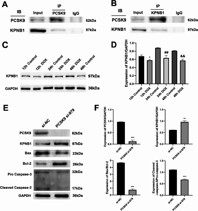
Doxorubicin (DOX) is an anthracycline class of chemotherapy drug, the application of which is limited due to its cardiotoxic effects. Recombinant Proprotein Convertase Subtilisin/Kexin Type 9 (PCSK9), is a serine protease pivotal in lipid metabolism and has a profound correlation with the onset of cardiovascular diseases. This study uncovers a connection between PCSK9 and DOX-induced cardiotoxicity (DIC). This research found that injection of DOX in mice caused cardiac toxicity. DOX treatment up-regulated the expression of PCSK9 protein in myocardial tissue. Evolocumab (PCSK9 inhibitors) improved cardiac function, myocardial injury, and fibrosis in DOX-treated mice, indicating a protective effect against DIC. The mechanism involved modulation of cardiomyocyte apoptosis and regulation of apoptosis-related proteins, including Bax/Bcl-2 ratio and Cleaved Caspase-3/Pro Caspase-3 ratio. DOX exhibited concentration- and time-dependent cytotoxic effects on H9C2 cardiomyocytes, promoting apoptosis. PCSK9 nuclear aggregation occurred in H9C2 cardiomyocytes after DOX treatment, and PCSK9 interacted with the Importin subunit beta-1 (KPNB1) protein. Interference with PCSK9 up-regulated KPNB1 expression, affecting apoptosis-related proteins and improving DOX-induced H9C2 cardiomyocyte apoptosis. In short, the elucidation of this mechanism is helpful involve that PCSK9 inhibitor may be a potential drug for improving DIC.

Keywords: Apoptosis; Cardiotoxicity; Doxorubicin; KPNB1; PCSK9.


Conflict of interest statement

Declarations. Competing interests: The authors declare no competing interests. Ethics approval and consent to participate: Not applicable. Consent for publication: Not applicable.

Figures

Fig. 1
Fig. 1
DIC in mice. A Show the survival rate over 5 days (n = 10). B Bar graph of survival rate (n = 10). C Body weight of mice in both groups over 5 days (n = 10). D Images of the heart size of both groups of mice (n = 10). E Representative echocardiography images in the both groups of mice (n = 7). FM The Statistical results are IVSd, IVSs, LVIDd, LVIDs, LVPWd, LVPWs, LVEF and LVFS respectively (n = 7). N HE staining of myocardial tissue from both groups of mice (n = 6), scale bar = 50 μm. Data are presented as mean ± SD, *P < 0.05, **P < 0.01, ***P < 0.001, ****P < 0.0001, ns. No significant vs. Control group.
Fig. 2
Fig. 2
The expression of PCSK9 protein in myocardial tissue of mice was up-regulated after DOX treatment, and PCSK9 inhibitor improved DIC. A WB images of PCSK9 protein expression in cardiac tissue of both groups of mice (n = 8). B Gray scale scanning analysis of PCSK9/GAPDH protein expression (n = 8). C Show the survival rate over 12 days (n = 10). D Bar graph of survival rate (n = 10). E Body weight of the three groups of mice over 12 days (n = 10). F Images of the heart size of three groups of mice (n = 10). G Heart weight (n = 10). H Heart weight to body weight ratio (n = 10). Data are presented as mean ± SD, *P < 0.05, ***P < 0.001, vs. Control group, #P < 0.05, ##P < 0.01, ###P < 0.001 vs. DOX group.
Fig. 3
Fig. 3
PCSK9 inhibitor improved DOX-induced cardiac dysfunction, myocardial injury and myocardial fibrosis in mice. A Representative echocardiography images in the three groups of mice (n = 7). BI The Statistical results are IVSd, IVSs, LVIDd, LVIDs, LVPWd, LVPWs, LVEF and LVFS respectively (n = 7). J HE staining and Masson staining of myocardial tissue from the three groups of mice (n = 6), scale bar = 50 μm, myocardial fibers are shown in red, collagen fibers are shown in blue. Data are presented as mean ± SD, *P < 0.05, **P < 0.01, ***P < 0.001 vs. Control group, #P < 0.05, ##P < 0.01, ###P < 0.001 vs. DOX group.
Fig. 4
Fig. 4
PCSK9 inhibitor improved DOX-induced myocardial apoptosis. A TUNEL staining images of myocardial tissues of the three groups of mice (n = 6), scale bar = 100 μm. B The percentage of TUNEL positive in the three groups of mice (n = 6). C Immumohis tochemical staining images of PCSK9 and Caspase-3 (n = 8), scale bar = 50 μm. D WB images of PCSK9 and apoptosis-related protein expression in myocardial tissue of the three groups of mice (n = 5). EG Results of gray-scale scanning analysis of relative protein expression of PCSK9/GAPDH, Bax/Bcl-2 and Cleaved Caspase-3/Pro Caspase-3  (n = 5). Data are presented as mean ± SD, **P < 0.01, ***P < 0.001 vs. Control group, ##P < 0.01, ###P < 0.001 vs. DOX group.
Fig. 5
Fig. 5
The changes of PCSK9 gene and protein and the localization of PCSK9 in H9C2 cardiomyocytes after DOX treatment. A The cells were treated with DOX for 12 h, 24 h and 48 h (n = 6). B Changes in relative expression of PCSK9 mRNA after DOX treatment (n = 6). C WB images of PCSK9 protein expression after DOX treatment (n = 6). D Results of gray-scale scanning analysis of relative protein expression of PCSK9/GAPDH after DOX treatment (n = 6). E Ultra-high resolution images of changes in PCSK9 localization after 24 h and 48 h of DOX treatment (n = 5), scale = 10 μm. Data are presented as mean ± SD, **P < 0.01, ***P < 0.001 vs. 0µM group, #P < 0.05, ##P < 0.01 vs. 24 h control group, &&P < 0.01, &&&P < 0.001 vs. 48 h control group.
Fig. 6
Fig. 6
Interfering with PCSK9 improved DOX-induced apoptosis of H9C2 cardiomyocytes. A WB images of apoptosis-related protein expression after DOX treatment (n = 6). B Results of gray-scale scanning analysis of relative protein expression of Cleaved Caspase-3/Pro Caspase-3 after DOX treatment (n = 6). C Results of gray-scale scanning analysis of relative protein expression of Bax/Bcl-2 after DOX treatment (n = 6). D WB images of PCSK9 gene interference effect after DOX treatment (n = 6). E Results of gray-scale scanning analysis of relative protein expression interference PCSK9 gene after DOX treatment (n = 6). F TUNEL staining images of cells treated with DOX at different times (n = 6), scale bar = 100 μm. G Percentage of TUNEL positive cells treated with DOX at different times (n = 6). H TUNEL staining showed that PCSK9 interference improved DOX-induced apoptosis in H9C2 cardiomyocytes (n = 6), scale bar = 50 μm. I Percentage statistics of apoptosis after PCSK9 interference (n = 6). Data are presented as mean ± SD, *P < 0.05 vs. 12 h control group, **P < 0.01, ***P < 0.001 vs. si-NC group, ##P < 0.01, ###P < 0.001 vs. 24 h control group, &&&P < 0.001 vs. 48 h control group.
Fig. 7
Fig. 7
The mechanism of PCSK9 in DOX-induced apoptosis of H9C2 cardiomyocytes. A CO-IP forward verification (n = 5). B CO-IP reverse verification (n = 5), whole protein sample of H9C2 cardiomyocytes in Input group (positive control), Nonspecific IgG-binding proteins in IgG group (negative control). C WB images of KPNB1 protein expression after DOX treatment (n = 6). D Results of gray-scale scanning analysis of relative protein expression of KPNB1 after DOX treatment (n = 6). E WB images of KPNB1 and apoptosis-related proteins after interfering with PCSK9 (n = 5). F Results of gray-scale scanning analysis of relative protein expression of PCSK9, KPNB1, Bax/Bcl-2 and Cleaved Caspase-3/Pro Caspase-3 after interfering PCSK9 gene (n = 5). Data are presented as mean ± SD, *P < 0.05 vs. 12 h control group, **P < 0.01, ***P < 0.001 vs. si-NC group, ##P < 0.01 vs. 24 h control group, &&P < 0.01 vs. 48 h control group.
Fig. 8
Fig. 8
Research mechanism diagram. PCSK9 may interact with KPNB1 to induce apoptosis in cardiomyocyte and PCSK9 inhibitor may be a potential drug for improving DOX-induced cardiotoxicity.

References

    1. Grumbach, I. M. Cardio-oncology at the beginning of a new decade. J. Am. Heart Assoc.9, e015890. 10.1161/JAHA.120.015890 (2020). - PMC - PubMed
    1. Deo, S. V., Al-Kindi, S. G. & Oliveira, G. H. Management of advanced heart failure due to cancer therapy: the present role of mechanical circulatory support and cardiac transplantation. Curr. Treat. Opt. Cardiovasc. Med.17, 388. 10.1007/s11936-015-0388-8 (2015). - PubMed
    1. Renu, K., Abilash, V. G. & Arunachalam, S. Molecular mechanism of doxorubicin-induced cardiomyopathy—an update. Eur. J. Pharmacol.818, 241–253. 10.1016/j.ejphar.2017.10.043 (2018). - PubMed
    1. Henriksen, P. A. Anthracycline cardiotoxicity: an update on mechanisms, monitoring and prevention. Heart104, 971–977. 10.1136/heartjnl-2017-312103 (2018). - PubMed
    1. Yarmohammadi, F., Rezaee, R. & Karimi, G. Natural compounds against doxorubicin-induced cardiotoxicity: a review on the involvement of Nrf2/AREsignaling pathway. Phytother. Res.35, 1163–1175. 10.1002/ptr.6882 (2021). - PubMed

MeSH terms

LinkOut - more resources