. 2025 Jul 1;15(1):21261.
doi: 10.1038/s41598-025-03738-6.

Tanshinone IIA suppresses the proliferation and fibrosis of mesangial cell in diabetic nephropathy though WTAP-mediated m6A methylation

Affiliations

Tanshinone IIA suppresses the proliferation and fibrosis of mesangial cell in diabetic nephropathy though WTAP-mediated m6A methylation

Hao Peng et al. Sci Rep. .

Abstract

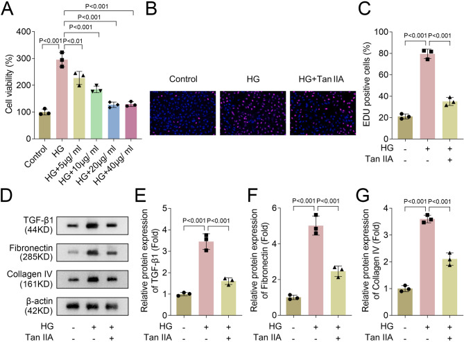
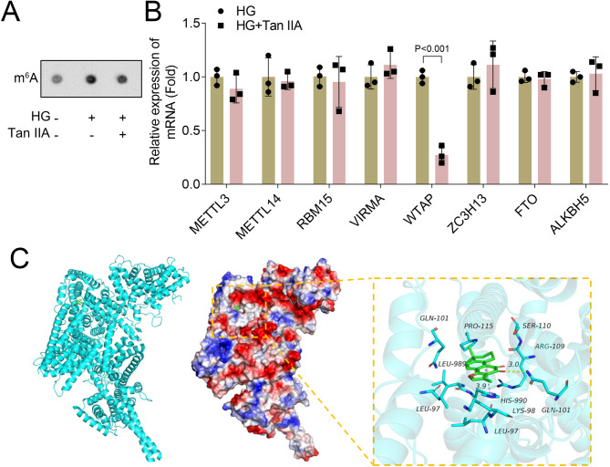
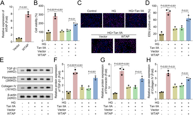
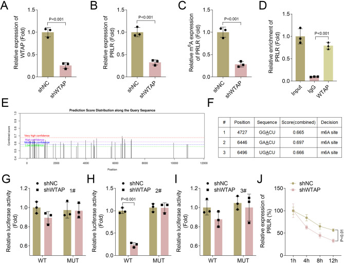
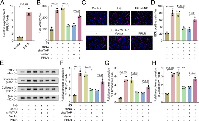
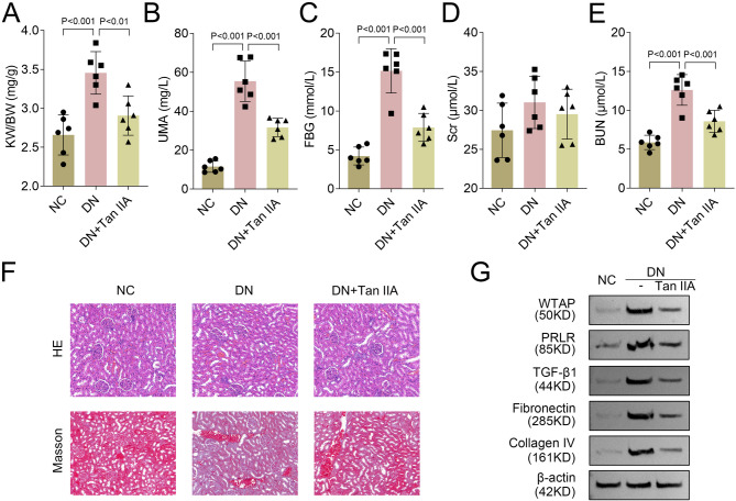
Diabetic nephropathy (DN) is often accompanied by mesangial cell proliferation and fibrosis. Tanshinone IIA (Tan-IIA) is the main fat-soluble component of Salvia miltiorrhiza. N6-Methyladenosine (m6A) modification is a widely studied epigenetic mechanism. This study aimed to investigate the role of Tan-IIA in DN and the underlying mechanism. Cell viability and proliferation were assessed via cell counting kit-8 and ethynyldeoxyuridine assays. Protein levels of fibrosis-related indicators were detected by Western blot. Reverse transcription-quantitative polymerase chain reaction was used to detect the levels of m6A-related enzymes. The interaction betweenWT1 associated protein (WTAP) and prolactin receptor (PRLR) was examined through RNA immunoprecipitation and dual-luciferase reporter assays. The animal DN models was established. Biochemical measurements of rat serum were performed using commercial kits. Hematoxylin&eosin and Masson trichrome staining were used for histopathological analysis. Results showed that Tan IIA treatment inhibited the cell proliferation and fibrosis of human renal mesangial cells (HRMCs). Besides, Tan IIA treatment regulated WTAP-mediated m6A modification. Overexpression of WTAP upregulated the cell proliferation and fibrosis of HRMCs. Mechanically, WTAP enhanced the stability of PRLR mRNA via m6A methylation. Subsequent rescue investigations revealed that overexpression of PRLR increased the cell proliferation and fibrosis of HRMCs. In the in vivo study, Tan IIA treatment reversed the renal injury in rats and decreased the protein levels of WTAP, PLRP, and fibrosis-related indicators in kidney tissues. Tan IIA suppressed the proliferation and fibrosis of HRMCs in DN though WTAP-mediated m6A methylation of PRLR.

Keywords: Diabetic nephropathy; Fibrosis; PRLR; Proliferation; Tanshinone IIA; WTAP; m6A.


Conflict of interest statement

Declarations. Competing interests: The authors declare no competing interests. Ethics approval and consent to participate: This study was approved by the Ethics Committee of Xiangyang Hospital of Traditional Chinese Medicine. All animal experiments should comply with the ARRIVE guidelines. All methods were carried out in accordance with relevant guidelines and regulations.

Figures

Fig. 1
Fig. 1
Tan IIA treatment inhibited the cell proliferation and fibrosis of HRMCs. HRMCs were cultured in commercial complete medium containing 10% fetal bovin serum and 1% penicillin/streptomycin. The atmosphere required for cell culture was 37 °C and 5% CO2. HRMCs were cultured with normal (5.5 mmol/L) or high (25 mmol/L) concentrations of D-glucose in serum-free medium. Besides, HRMCs were treated with different concentrations (5, 10, 20, and 40 μg/mL) of Tan IIA. (A) Cell viability in each group was detected by CCK-8 assay (n = 3); (B) EdU assay was performed to analyze cell proliferation (n = 3); (C) Quantification of EdU positive cells in each group (n = 3); (D) Western blot analysis of TGF-β1, fibronectin, and collagen IV protein levels in each group (n = 3); Quantification of (D) TGF-β1, (E) fibronectin, and (F) collagen IV protein levels in each group (n = 3). Data are expressed as the mean ± SD. Tan IIA, Tanshinone IIA; HRMCs, human renal mesangial cells; CCK-8, cell counting-kit 8; EdU, ethynyldeoxyuridine; TGF-β1, transforming growth factor-beta1.
Fig. 2
Fig. 2
Tan IIA treatment regulated WTAP-mediated m6A modification. (A) Dot blot assay was performed to assess the total m6A level in each group (n = 3); (B) RT-qPCR was performed to analyze the mRNA levels of METTL3, METTL14, RBM15, VIRMA, WTAP, ZC3H13, FTO, and ALKBH5 in each group (n = 3); (C) The 3D structure of the binding complex of WTAP with Tan IIA and the detail binding mode of the complex. The backbone of the protein was rendered in tube. Compound is rendered by green and the OGT is shown in blue sticks. Yellow and ray dash represents hydrogen bond. Data are expressed as the mean ± SD. Tan IIA, Tanshinone IIA; WTAP, Wilms tumor 1-associating protein; m6A, N6-methyladenosine; RT-qPCR, reverse transcription-polymerase chain reaction; METTL, methyltransferase; RBM15, RNA binding motif protein 15; VIRMA, vir-like m(6)A methyltransferase-associated protein; ZC3H13, zinc finger CCCH-type containing 13; FTO, alpha‐ketoglutarate dependent dioxygenase; ALKBH5, AlkB homolog 5.
Fig. 3
Fig. 3
Overexpression of WTAP upregulated the cell proliferation and fibrosis of HRMCs. Negative control pcDNA 3.1 and pcDNA 3.1-WTAP overexpression vectors were transfected into HRMCs using Lipo8000™ transfection reagent. (A) RT-qPCR was used to assess the expression of WTAP when WTAP was overexpressed in HRMCs (n = 3); (B) Cell viability in each group was detected by CCK-8 assay (n = 3); (C) EdU assay was performed to analyze cell proliferation (n = 3); (D) Quantification of EdU positive cells in each group (n = 3); (E) Western blot analysis of TGF-β1, fibronectin, and collagen IV protein levels in each group (n = 3); Quantification of (F) TGF-β1, (G) fibronectin, and (H) collagen IV protein levels in each group (n = 3). Data are expressed as the mean ± SD. HRMCs, human renal mesangial cells; RT-qPCR, reverse transcription-polymerase chain reaction; WTAP, Wilms tumor 1-associating protein; CCK-8, cell counting-kit 8; EdU, ethynyldeoxyuridine; TGF-β1, transforming growth factor-beta1.
Fig. 4
Fig. 4
WTAP enhanced the stability of PRLR mRNA via m6A demethylation. Short hairpin negative control (shNC) and shWTAP vectors were transfected into HRMCs using Lipo8000™ transfection reagent. (A) RT-qPCR was used to assess the expression of WTAP when WTAP was inhibited in HRMCs (n = 3); (B) RT-qPCR was used to assess the expression of PRLR when PRLR was suppressed in HRMCs (n = 3); (C) The m6A level of PRLR after WTAP inhibited was detected using MeRIP-qPCR assay (n = 3); (D) RIP assay was conducted to examine the interaction between WTAP and PRLR in HRMCs cells (n = 3); (E) Sequence-based RNA adenosine methylation site predictor database was used to predict m6A sites of PRLR; (F) The three predictive sites of PRLR; Dual-luciferase reporter assay was performed to evaluate the binding of WTAP and PRLR at sites (G) 1#, (H) 2#, and (I) 3# in HRMCs; (J) RNA stability assay was used to detect the existing PRLR expression when actinomycin D treated at different time points (1, 4, 8, and 12 h) in HRMCs (n = 3). Data are expressed as the mean ± SD. WTAP, Wilms tumor 1-associating protein; PRLR, prolactin receptor; m6A, N6-methyladenosine; RT-qPCR, reverse transcription-polymerase chain reaction; HRMCs, human renal mesangial cells; RIP, RNA immunoprecipitation; MeRIP, Methylated-RIP.
Fig. 5
Fig. 5
Overexpression of PRLR increased the cell proliferation and fibrosis of HRMCs. Negative control pcDNA 3.1, pcDNA 3.1-PRLP overexpression, shNC, and shWTAP vectors were transfected into HRMCs using Lipo8000™ transfection reagent. (A) RT-qPCR was used to assess the expression of PRLR when PRLR was overexpressed in HRMCs (n = 3); (B) Cell viability in each group was detected by CCK-8 assay (n = 3); (C) EdU assay was performed to analyze cell proliferation (n = 3); (D) Quantification of EdU positive cells in each group (n = 3); (E) Western blot analysis of TGF-β1, fibronectin, and collagen IV protein levels in each group (n = 3); Quantification of (F) TGF-β1, (G) fibronectin, and (H) collagen IV protein levels in each group (n = 3). Data are expressed as the mean ± SD. PRLR, prolactin receptor; RT-qPCR, reverse transcription-polymerase chain reaction; HRMCs, human renal mesangial cells; CCK-8, cell counting-kit 8; EdU, ethynyldeoxyuridine; TGF-β1, transforming growth factor-beta1.
Fig. 6
Fig. 6
Tan IIA treatment reversed the renal injury induced by DN. Creating a rat model mimicking diabetes in humans through a combination of HFD and STZ treatment. Twenty-two male Sprague–Dawley rats (6 weeks old) were randomly assigned to either negative controls (NC; n = 6) or HFD (n = 16) groups for 6 weeks at the start of the experiment. After that, the HFD group rats received intraperitoneal injections of STZ (35 mg/kg), while the NC group rats were injected with vehicle citrate buffer. Rats (n = 12) were randomly divided into DN group (n = 6) and Tan IIA-treatment group (DN + Tan IIA group, n = 6) after excluding four STZ-injected rats that did not meet the criteria for diabetes. (A) KW/BW, (B) UMA, (C) FBG, (D) Scr, and (E) BUN levels of the each group rats were shown (n = 6); F, Representative images of H&E and Masson trichrome staining of the each group kidney sections (magnification: × 200; n = 6); (G) Western blot was used to analyze the protein levels of WTAP, PRLR, TGF-β1, fibronectin, and collagen IV in each group rat kidneys (n = 6). Data are expressed as the mean ± SD. Tan IIA, Tanshinone IIA; DN, diabetic nephropathy; HFD, high-fat diet; STZ, streptozocin; KW/BW, kidney weight to body weight ratio; UMA, urinary microalbumin; FBG, fasting blood glucose; Scr, serum creatinine; BUN, blood urea nitrogen; H&E, hematoxylin&eosin; WTAP, Wilms tumor 1-associating protein; PRLR, prolactin receptor; TGF-β1, transforming growth factor-beta1.

Similar articles

References

    1. Alicic, R. Z., Rooney, M. T. & Tuttle, K. R. Diabetic kidney disease: Challenges, progress, and possibilities. Clin. J. Am. Soc. Nephrol.12, 2032–2045 (2017). - PMC - PubMed
    1. Gupta, S., Dominguez, M. & Golestaneh, L. Diabetic kidney disease: An update. Med. Clin. North. Am.107, 689–705 (2023). - PubMed
    1. Gross, J. L. et al. Diabetic nephropathy: Diagnosis, prevention, and treatment. Diabetes Care28, 164–176 (2005). - PubMed
    1. Samsu, N. Diabetic nephropathy: challenges in pathogenesis, diagnosis, and treatment. Biomed Res. Int.2021, 1497449 (2021). - PMC - PubMed
    1. Zou, M. et al. Safety and efficacy of hemodialysis and peritoneal dialysis in treating end-stage diabetic nephropathy: A meta-analysis of randomized controlled trials. Int. Urol. Nephrol.54, 2901–2909 (2022). - PubMed

MeSH terms

LinkOut - more resources