. 2025 Jul 1;15(1):21298.
doi: 10.1038/s41598-025-03722-0.

Surgical timing and approach for brainstem cavernous malformation warranting thorough preoperative evaluation

Affiliations

Surgical timing and approach for brainstem cavernous malformation warranting thorough preoperative evaluation

Xiao Qian et al. Sci Rep. .

Abstract

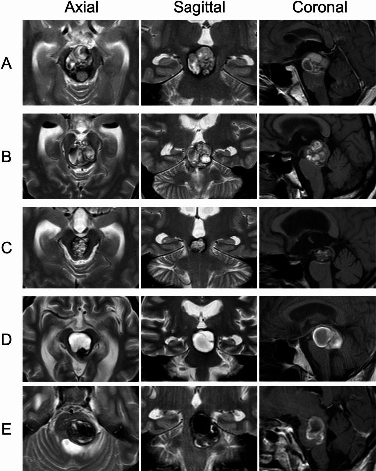
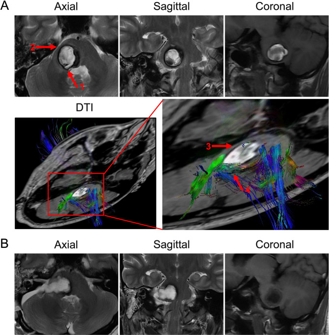
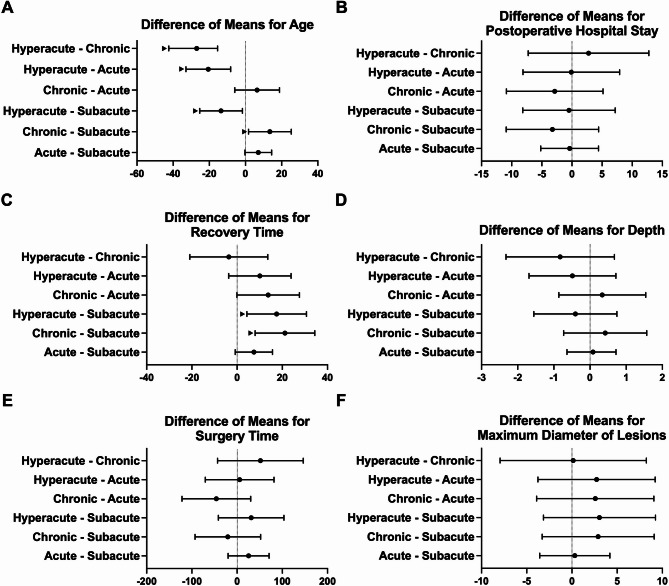
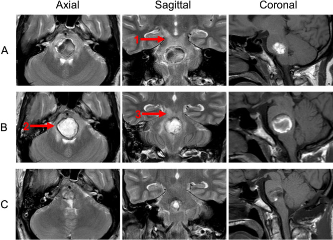
Hemorrhage from brainstem cavernous malformations (BSCMs) can cause severe neurological dysfunction. While surgery is effective, the optimal timing remains unclear. This retrospective study aims to identify the best timing for surgery after hemorrhage in BSCM patients. Data from patients who underwent surgery between 2012 and 2022 were analyzed. Patients were divided into groups with favorable and unfavorable outcomes based on their best post-treatment modified Rankin Scale (mRS) scores. They were further categorized into four groups according to the timing of surgery after hemorrhage: hyperacute (≤ 7 days), acute (8-20 days), subacute (21-56 days), and chronic (> 56 days). Clinical characteristics, imaging findings, prognosis, and outcomes were compared between the groups. A total of 135 BSCM patients were included, with most lesions located in the pons. Surgical timing distribution was as follows: hyperacute phase (12 cases), acute phase (42 cases), subacute phase (69 cases), and chronic phase (12 cases). Of these, 113 patients had favorable outcomes. Statistical analysis revealed a significant difference in mRS scores between the four groups, with the most pronounced differences observed between the acute/subacute phases and the hyperacute/chronic phases. In conclusion, performing surgery in the acute and subacute phases could improve neurological function in BSCM patients.

Keywords: Brainstem cavernous malformations; Outcome; Surgical resection; Surgical timing.


Conflict of interest statement

Declarations. Competing interests: The authors declare no competing interests.

Figures

Fig. 1
Fig. 1
This figure shows the different approaches to 5 lesions located in the midbrain. (A) The patient’s lesion occupied almost the entire midbrain, slightly to the right. The surgery was performed through the subtemporal approach. Analyzing from the sagittal position T1, the posterior median approach was also a selectable path. (B) Preoperative MRI images showed supratentorial hydrocephalus, part of the lesion protruded into the third ventricle, the patient’s lesion also occupied the entire midbrain, so the subtemporal approach and the transchoroidal approach were both feasible. Considering the patient’s hydrocephalus, the final choice was the transchoroidal approach + third ventricle fistula surgery. (C) The patient’s lesion was located inside the midbrain. Using the posterior median approach and the subtemporal approach will pass through a long distance of brain parenchyma and fiber, and the upper approach could more directly treat the lesion. The transcallosal interforniceal approach was finally adopted. (D) The lesion was located at the posterior midbrain, part of it protruded into the fourth ventricle. The poppen approach was finally chosen. (E) The lesion range invaded the upper part of the midbrain and pons, mainly located on the left side, close to the bottom of the fourth ventricle and the surface of the midbrain ventricular membrane. Both lateral and posterior approaches are feasible in this case, and finally subtemporal approach was adopted.
Fig. 2
Fig. 2
This figure presents a case violating two-point method. (A) This female patient was admitted for surgery 30 days after acute hemorrhage. Preoperative MRI indicated that the cavernous malformation was located in the right pons. According to the two-point method, the lesion was closer to the ependymal surface of the fourth ventricle (red arrow 1) than to the outer pia mater of the right pons (red arrow 2), suggesting a posterior approach should be adopted. However, preoperative diffuse tense images (DTI) indicated fewer fibers on the lateral side of the lesion (red arrow 3), and despite the shorter distance, there were more fibers gathered posteriorly (red arrow 4). (B) Therefore, a lateral approach was ultimately used to perform a total resection of the lesion. The patient had sensory and motor deficits before surgery, but these symptoms significantly improved three weeks post-surgery. In the final follow-up, no significant neurological deficits were left.
Fig. 3
Fig. 3
Use analysis of variance (ANOVA) to compare parameters pairwise among patients in different surgical timing groups. (• represents the mean, the bar represents the difference between means, and ▲ represents a P < 0.05 in the intergroup comparison).
Fig. 4
Fig. 4
This picture presents a case of waiting until the edema subsided and hemosiderin belt appeared before surgery. (A) The patient is a middle-aged male who suddenly experienced dizziness, hemiplegia, and sensory deficits. The patient was admitted to hospital on the 9th day after acute hemorrhage, and the MRI scan indicated a pontine cavernous malformation. On the T2 image, the boundary of the lesion was not clear, and there was severe surrounding edema, so surgery was postponed (red arrow 1). (B) Eventually, on the 22nd day after acute bleeding, another MRI indicated the formation of hemosiderin, the boundary was clear, and the edema was significantly reduced compared to before (red arrow 2, 3). (C) The patient then underwent surgery, with a median suboccipital approach, and the lesion was completely removed.

Similar articles

References

    1. Abla, A. A. et al. Advances in the treatment and outcome of brainstem cavernous malformation surgery: A single-center case series of 300 surgically treated patients. Neurosurgery68, 403–414 (2011). - PubMed
    1. <ISSVA-Classification-2018.pdf>.
    1. Gross, B. A., Batjer, H. H., Awad, I. A. & Bendok, B. R. Brainstem cavernous malformations. Neurosurgery64, E805-818 (2009). - PubMed
    1. Januszewski, J., Albert, L., Black, K. & Dehdashti, A. R. The usefulness of diffusion tensor imaging and tractography in surgery of brainstem cavernous malformations. World Neurosurg.93, 377–388 (2016). - PubMed
    1. Li, D. et al. Natural history of brainstem cavernous malformations: Prospective hemorrhage rate and adverse factors in a consecutive prospective cohort. J. Neurosurg.134, 917–928 (2020). - PubMed

LinkOut - more resources