. 2025 Jul 2;15(1):22807.
doi: 10.1038/s41598-025-04064-7.

Strictosamide and mitraphylline inhibit cancer cell motility by suppressing epithelial-mesenchymal transition via integrin α4-mediated signaling

Affiliations

Strictosamide and mitraphylline inhibit cancer cell motility by suppressing epithelial-mesenchymal transition via integrin α4-mediated signaling

Mücahit Varlı et al. Sci Rep. .

Abstract

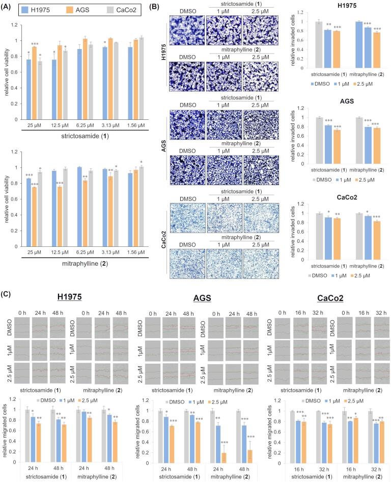
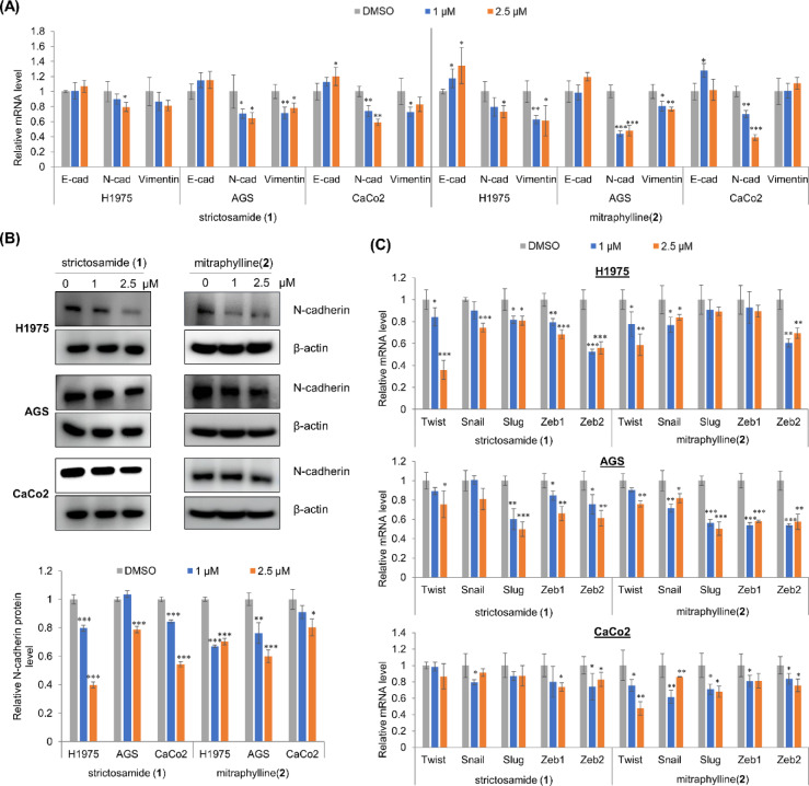
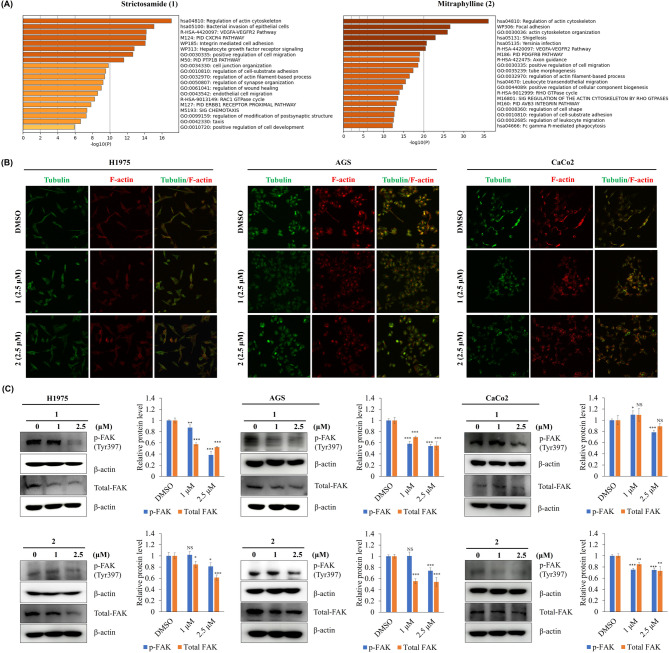
Epithelial-mesenchymal transition (EMT) is a critical process in cancer cell motility and metastasis. Monoterpene indole alkaloids (MIAs) have been widely investigated for biological activities, but rarely been explored for cell motility inhibition. This study aimed to discover natural products, especially focusing on MIAs, that inhibit EMT in cancer cells, based on our screening using multiple plant extracts. We found that an extract of the aerial parts of Uncaria scandens (Sm.) Wall. (Rubiaceae) decreased cancer cell motility. Targeted isolation exhibited eight MIAs. Among them, strictosamide and mitraphylline suppressed the invasion and migration of the cells. The alteration of the mRNA expression of the EMT effectors and transcription factors suggested that the EMT signaling pathway is related to the suppression of cancer cell motility by both compounds. RT-PCR gene array suggested inhibition of integrin α4 signaling as a potential mechanism of the EMT inhibition, which was supported by the quantitative analysis of the mRNA expressions of the related genes. Together, present study is the first report highlighting the cell motility-suppressive effects of strictosamide and mitraphylline. Our results suggested the potential applicability of strictosamide and mitraphylline for the prevention of metastasis.

Keywords: Bioactivity-guided fractionation; Integrin Α4β1 agonist; Metastasis suppressor; Spirocylic oxindole alkaloid; Vallesiachotaman-type alkaloid.


Conflict of interest statement

Declarations. Competing interests: The authors declare no competing interests.

Figures

Fig. 1
Fig. 1
Bioactivity screening and chemical investigation of U. scandens. (A) Representative images showing each insert in the invasion assay and the relative number of invaded cells. AGS cells were treated with fractions (5 µg/mL; with 0.1% DMSO) for 24 h. Data represent mean ± S.D. * p < 0.05; ** p < 0.01. (B) Chemical structures of the eight isolated compounds 18. (C) The LC-MS base peak ion (BPI) chromatogram of the alkaloid-rich fraction of U. scandens. The isolated compounds are denoted on the corresponding chromatographic peaks. Notably, the intensities of chromatographic peaks do not represent absolute quantity of each compound because the MS signal intensity is heavily affected by ionization efficiency of each molecule.
Fig. 2
Fig. 2
The effects of strictosamide (1) and mitraphylline (2) on cell viability and motility of H1975, AGS, and CaCo2 cells. (A) The cell viability of H1975, AGS, and CaCo2 cells treated with strictosamide (1) and mitraphylline (2) for 48 h. Data are represented as the mean ± S.D. of the differences between the cell viability of cells affected with strictosamide (1) and mitraphylline (2) compared with that of DMSO-treated controls. (B) Representative images of each insert and relative number of invaded cells in the Transwell invasion assay. Cells were treated with strictosamide (1) and mitraphylline (2) at concentrations of 1 and 2.5 µM for 24 h. (C) Representative images of each wound area and quantitative data of relative wound density in the scratch wounding migration assay using the IncuCyte system. Overlaid on phase contrast images, the green line indicates the initial wound line, while the red line indicates the migrated cell area at the specified time period. Data are represented as the mean ± S.D. *p < 0.05; ** p < 0.01; *** p < 0.001.
Fig. 3
Fig. 3
Strictosamide (1) and mitraphylline (2) downregulated the expression of EMT effectors and transcription factors. (A) The mRNA expression of EMT effectors (N-cadherin, vimentin and E-cadherin) were analyzed by qRT-PCR. The mRNA expressions were normalized against β-actin. (B) The protein levels of N-cadherin were measured. β-actin served as a loading control. (C) The mRNA expressions of EMT transcription factors (Twist, Snail, Slug, Zeb1, and Zeb2) were analyzed by qRT-PCR. The mRNA expressions were normalized against β-actin. Data are represented as the mean ± S.D. * p < 0.05; ** p < 0.01; *** p < 0.001.
Fig. 4
Fig. 4
Strictosamide (1) and mitraphylline (2) downregulated several cell motility-related factors. (A) Pathway-focused gene expression analysis using the Human Cell Motility RT2 Profiler™ PCR Array was performed to examine the mechanisms by which strictosamide and mitraphylline modulate cell motility. The scatter plot compares the gene on the PCR Array between the DMSO-strictosamide and DMSO-mitraphylline. The number of fold changes for ITGA4 arranged downwards is shown in the figure. (B) A network of interrelated genes affected by strictosamide (1) and mitraphylline (2). (C) Quantitative analysis of the mRNA expressions of CAV1, CAPN1, WASF1, WASL, RHO, RHOA, RHOB, ITGA4, FAK, SRC in H1975, AGS, and CaCo2 cells treated with 2.5 µM of strictosamide (1) and mitraphylline (2). Data are represented as the mean ± S.D. * p < 0.05; ** p < 0.01; *** p < 0.001.
Fig. 5
Fig. 5
Effect of strictosamide (1) and mitraphylline (2) on cell morphology and FAK protein level of cancer cells. (A) Biological processes linked to the pathways by strictosamide (1) and mitraphylline (2) found in Metascape. (B) The microtubule organization in the H1975, CaCo2, and AGS cells was examined using immunofluorescence microscopy following a 24 h treatment with strictosamide (1) and mitraphylline (2). Microtubules were stained with α-tubulin antibodies (green), while actin was stained with Alexa Fluor 568 phalloidin (red). (C) The protein levels of p-FAK and total FAK were measured. Cells were treated with 1 and 2.5 µM of strictosamide (1) and mitraphylline (2) for 48 h. β-actin served as a loading control. Data are represented as the mean ± S.D. * p < 0.05; ** p < 0.01; *** p < 0.001.
Fig. 6
Fig. 6
The responses to strictosamide (1) and mitraphylline (2) were evaluated in cells exposed α4β1 agonist. (A) Chemical structure of the α4β1 agonist molecule. (B) Cell viability in ITGA4 agonist treated in AGS cells. Cells were treated with α4β1 agonist for 48 h with indicated concentration. (C) Relative mRNA levels of N-cadherin, ITGA4 and FAK, after treatment with strictosamide (1) and mitraphylline (2) for 48 h in AGS cells. (D) Representative images of each insert and relative number of invaded cells in the Transwell invasion assay. AGS cells were treated with strictosamide (1) and mitraphylline (2) at concentrations of 1 and 2.5 µM for 24 h. The α4β1 agonist was treated at a concentration of 2.5 µM. Data are presented as mean ± S.D. Statistical significance was determined as follows: *p < 0.05, **p < 0.01, ***p < 0.001, NS: not significant (p > 0.05) compared to DMSO-treated cells; #p < 0.05, ##p < 0.01, ###p < 0.001, NS: not significant (p > 0.05) compared to α4β1 agonist-treated cells; @p < 0.05, @@p < 0.01, @@@p < 0.001, NS: not significant (p > 0.05) compared to strictosamide (1)- or mitraphylline (2)-treated cells.

Similar articles

References

    1. Ganesh, K. & Massagué, J. Targeting metastatic cancer. Nat. Med.27, 34–44 (2021). - PMC - PubMed
    1. Mittal, V. Epithelial mesenchymal transition in aggressive lung cancers. Adv. Exp. Med. Biol.890, 37–56 (2016). - PubMed
    1. Said, N. A. B. M. & Williams, E. D. Growth factors in induction of epithelial-mesenchymal transition and metastasis. Cells Tissues Organs.193, 85–97 (2011). - PubMed
    1. Lamouille, S., Xu, J. & Derynck, R. Molecular mechanisms of epithelial-mesenchymal transition. Nat. Rev. Mol. Cell. Biol.15, 178–196 (2014). - PMC - PubMed
    1. Santamaria, P. G., Moreno-Bueno, G., Portillo, F. & Cano, A. EMT: Present and future in clinical oncology. Mol. Oncol.11, 718–738 (2017). - PMC - PubMed

MeSH terms

LinkOut - more resources