

Fluorescent EGTA-derived carbon dots for turn on-off-on detection of Fe3+ and ascorbic acid via hydrothermal synthesis and cellular imaging
- PMID: 40594098
- PMCID: PMC12218165
- DOI: 10.1038/s41598-025-03845-4
Fluorescent EGTA-derived carbon dots for turn on-off-on detection of Fe3+ and ascorbic acid via hydrothermal synthesis and cellular imaging
Abstract
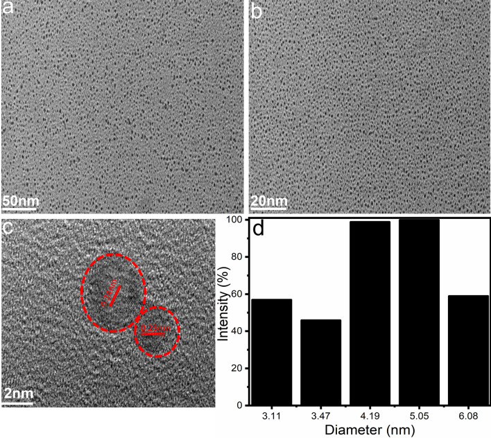
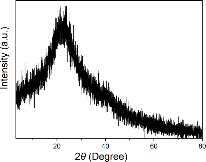
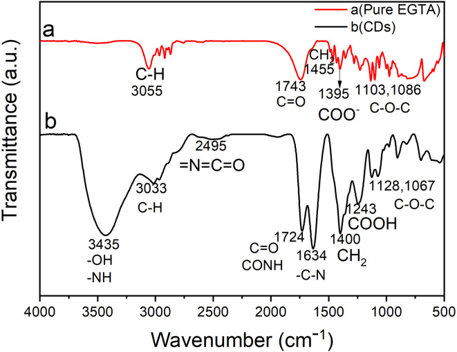
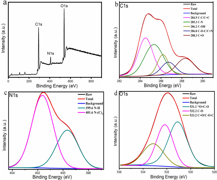
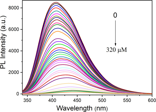
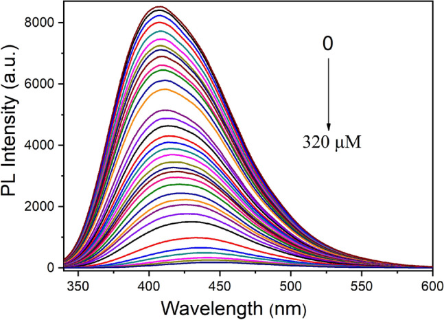
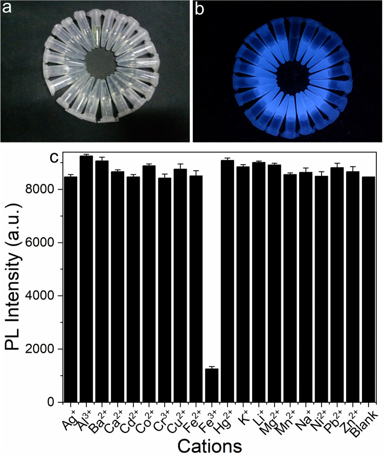
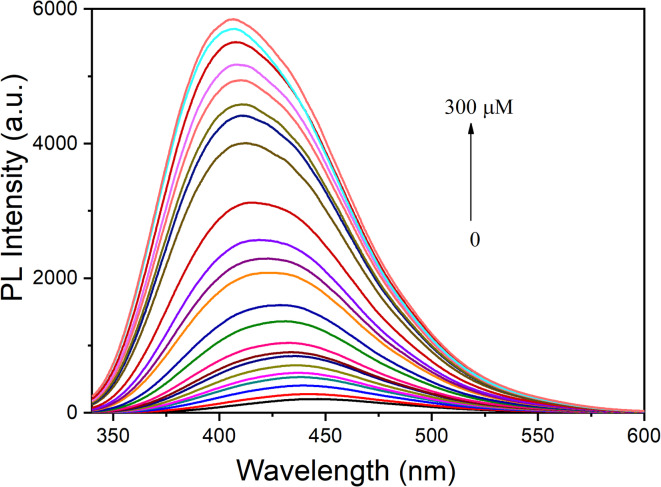
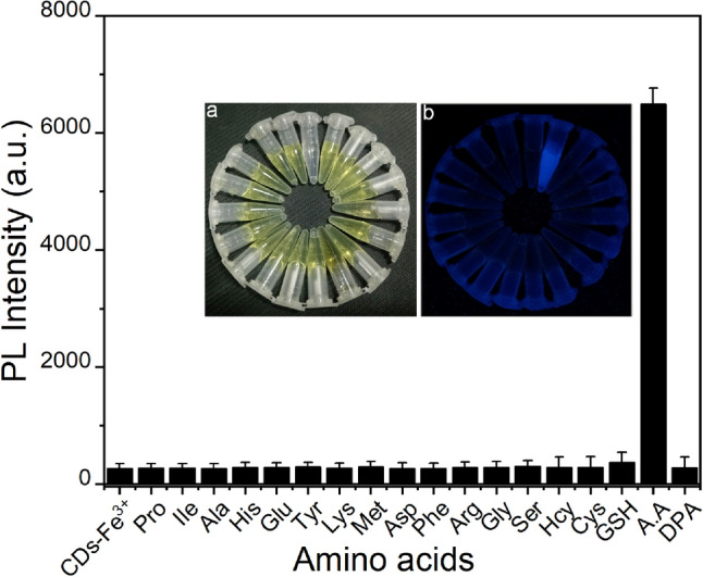
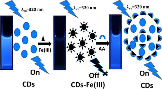
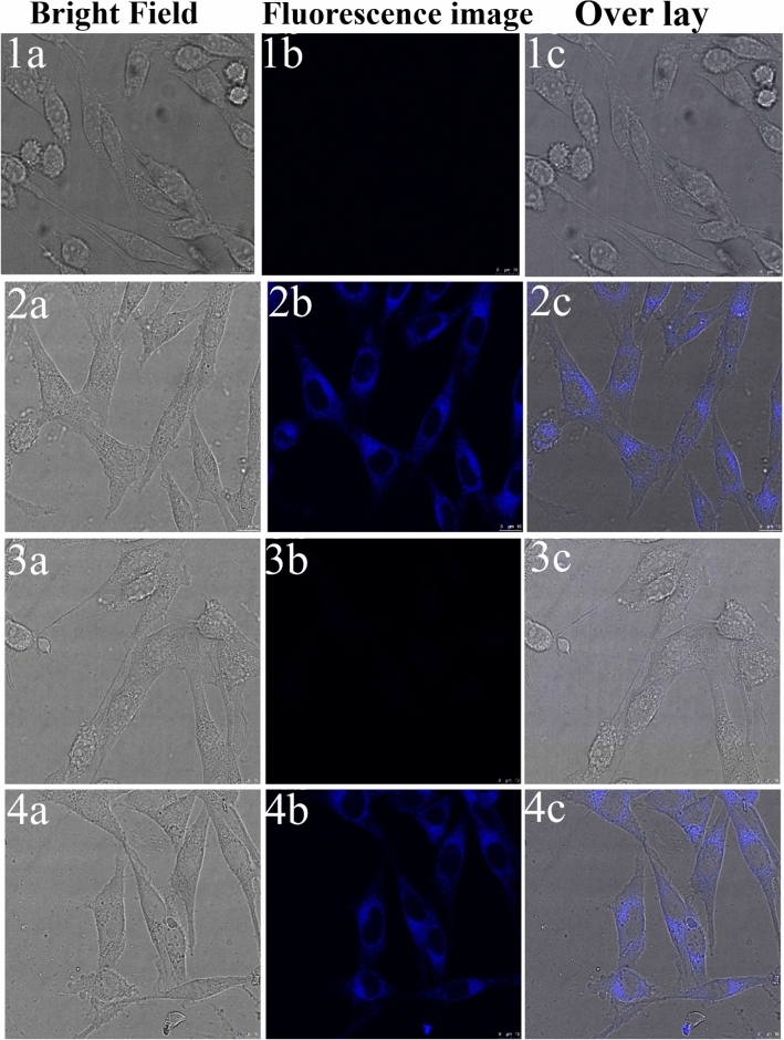
We present a novel, one-step hydrothermal synthesis of carbon dots (CDs) with intense blue fluorescence and a remarkably high quantum yield of 50%, using ethylene glycol tetraacetic acid (EGTA) as a single precursor eliminating the need for additional passivation agents. This streamlined strategy represents a significant advancement in the efficient production of functional CDs. The resulting CDs exhibit dual-mode fluorescence sensing, selectively detecting Fe3+ through a fluorescence quenching mechanism, with an ultra-low detection limit of 23 nM. Notably, the quenched fluorescence is fully restored upon the introduction of ascorbic acid (AA), enabling a highly sensitive "off-on" detection system with a detection limit of 21 nM. This approach demonstrates excellent selectivity for AA over dopamine and other amino acids, providing a reliable method for distinguishing AA in complex biological matrices. Furthermore, the CDs-based Fe3+/AA sensing system proves highly effective for bioimaging applications, allowing for clear visualization of Fe3+ and AA in living cells. Compared to conventional commercial assays, this method is cost-effective, simple, and scalable, offering a powerful tool for next-generation biosensing and bioimaging. The fluorescence-based "off-on" mechanism, combined with the versatility of CDs in real-sample analysis, highlights the innovation and practical value of this approach.
© 2025. The Author(s).
Conflict of interest statement
Declarations. Competing interests: The authors declare no competing interests.
Figures
Similar articles
-
"Signal-Switching" Fluorescence Sensing of Fe3+ and Ascorbic Acid by Songaria Cynomorium Polysaccharide-Doped Carbon Dots of the Tibetan Medicinal Herb.Luminescence. 2025 Jun;40(6):e70233. doi: 10.1002/bio.70233. Luminescence. 2025. PMID: 40555544
-
Ultrathin boron nanosheets: a novel fluorescent sensor for sensitive and selective detection of Fe3+ and ascorbic acid.Spectrochim Acta A Mol Biomol Spectrosc. 2025 Dec 15;343:126556. doi: 10.1016/j.saa.2025.126556. Epub 2025 Jun 11. Spectrochim Acta A Mol Biomol Spectrosc. 2025. PMID: 40513314
-
Fluorescent Nanozyme Based on Bimetallic Fe/Co-Doped Carbon Dots for Dual-Mode Detection of Ascorbic Acid and Hydrogen Peroxide.Anal Chem. 2025 Jul 22;97(28):15320-15328. doi: 10.1021/acs.analchem.5c02107. Epub 2025 Jul 9. Anal Chem. 2025. PMID: 40629988
-
High Luminescent Carbon Dots Derived from Fermented Beverages for Sensitive Sensing of Tartrazine in Foods.J Fluoresc. 2025 Jul;35(7):4989-4996. doi: 10.1007/s10895-024-03944-x. Epub 2024 Sep 25. J Fluoresc. 2025. PMID: 39320635 Review.
-
Effective Determination of Diosmin Using Nitrogen Doped Carbon Dots as Probe Based on Internal Filtering Effect.J Fluoresc. 2025 Jul;35(7):6097-6105. doi: 10.1007/s10895-024-03963-8. Epub 2024 Oct 17. J Fluoresc. 2025. PMID: 39419896 Review.
References
MeSH terms
Substances
Grants and funding
LinkOut - more resources
Full Text Sources
Medical
