. 2025 Jul 1;15(1):20426.
doi: 10.1038/s41598-025-06294-1.

ADCY4 inhibits cAMP-induced growth of breast cancer by inactivating FAK/AKT and ERK signaling but is frequently silenced by DNA methylation

Affiliations

ADCY4 inhibits cAMP-induced growth of breast cancer by inactivating FAK/AKT and ERK signaling but is frequently silenced by DNA methylation

Guangrui Pan et al. Sci Rep. .

Abstract

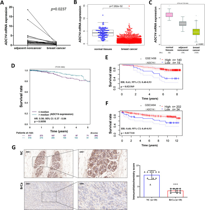
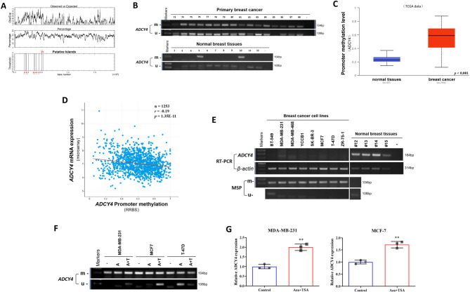
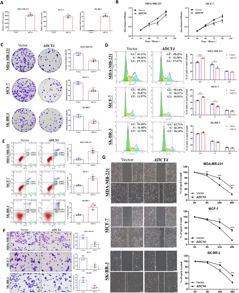
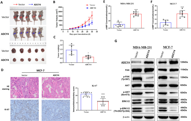
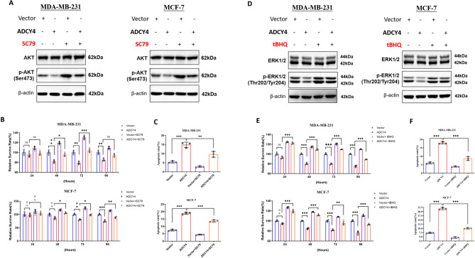
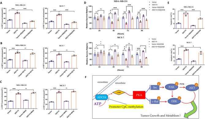
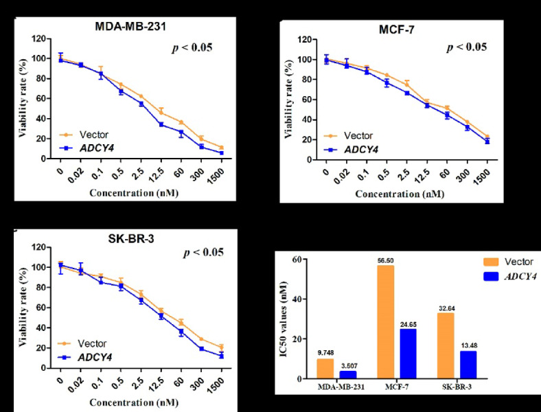
Local increases in cyclic adenosine monophosphate (cAMP) caused by specific adenylyl cyclases (ACs) can selectively modulate related proteins. AC-selective drugs have an advantage in side effect control, and the specific AC may finally be considered as a therapeutic target. We show that adenylyl cyclase 4 (ADCY4), which is silenced by DNA methylation and is critical for breast cancer (BC) patient survival, plays essential roles in anti-tumor effects in BC cells. DNA methyltransferase inhibitor and histone deacetylase inhibitor can restore ADCY4 mRNA expression in ADCY4-silenced BC cells. ADCY4 directly affects BC cell proliferation, apoptosis, invasion, and metastasis. Mechanistically, ADCY4 converts ATP to cAMP and activates cAMP/PKA signaling, leading to a decrease in the phosphorylation level of downstream FAK/AKT and ERK signaling and creating a suppression environment for cell survival. Ectopic ADCY4 inhibits BC growth, which is blocked by cAMP inhibition, activating AKT and ERK. The present study provides evidence that human BC relies upon this epigenetic silenced ADCY4-associated ATP-cAMP loop for phosphorylation and activation of FAK/AKT and ERK signaling. Also, ADCY4 increases BC cell chemosensitivity to paclitaxel. The observations demonstrates that ADCY4 is a significant tumor suppressor and that loss of ADCY4 functions by DNA methylation hampers cAMP signaling and triggers FAK/AKT and ERK signaling during breast tumorigenesis.

Keywords: ADCY4; Breast cancer; DNA methylation; FAK; cAMP.


Conflict of interest statement

Declarations. Competing interests: The authors declare no competing interests. Ethical approval and consent to participate: Ethics approval was received from the Affiliated Hospital of Chongqing Medical University. All participants provided written consent during their enrollment. Consent for publication: All patients provided written consent during enrollment for publication.

Figures

Fig. 1
Fig. 1
ADCY4 is downregulated in BC and associated with poor prognosis. (A) ADCY4 mRNA expression in primary BC (n = 20) and paired adjacent noncancer tissues (n = 20) by qRT-PCR with β-actin as a control (p = 0.0237, < 0.05). (B) ADCY4 mRNA expression in normal tissues and BC from TCGA datasets using R 3.6.3. package (p = 7.262E-52, < 0.001). (C) ADCY4 mRNA expression in normal tissues (n = 92), adjacent noncancer (n = 104), and BC (n = 1034) from GTEx and TCGA datasets (p < 0.001). (D) The OS rate in the group of high and low ADCY4 expression from TCGA datasets (p = 0.0056, < 0.01). Cutoff value: the median expression level. (E) The difference of OS rate for BC in high and low ADCY4 based on GSE1456 (p = 0.021569, < 0.05). (F) The difference of OS rate for BC in high and low ADCY4 based on GSE3494 (p = 0.017310, < 0.05). (G) Representative IHC staining images of ADCY4 protein expression in adjacent normal breast and BC tissues. The IHC score of ADCY4 in 10 BC specimens (IHC score: 3.10 ± 0.88) was lower than paired 10 adjacent normal breast samples (IHC score: 10.50 ± 2.12) (Mean ± S.D). NC, adjacent normal breast tissues; BrCa, BC tissues. ***p < 0.001.
Fig. 2
Fig. 2
Identification of ADCY4 silenced by promoter methylation in BC. (A) The CpG islands in the LTBP4 nucleotide sequence (EMBOSS-cpgplot). Each red arrows represents a single CpG island. (B) ADCY4 methylation in primary BC tissues (n = 171) and normal tissues (n = 11) measured by MSP. m-, methylated, u-, unmethylated. Gel images showed the representational graphs. (C) The differences in methylation levels of ADCY4 in primary BC (n = 793) and normal tissues (n = 97) from TCGA datasets. (D) The correlation between ADCY4 methylation and expression from TCGA-BRCA datasets was analyzed using cBioPortal online software (n = 1253, r=-0.19, p = 1.35E-11, < 0.001); (E) ADCY4 mRNA expression and methylation status in BC cell lines. m-, methylated, u-, unmethylated. (F) ADCY4 methylation status with 5-aza-2-deoxycytidine (A) and trichostatin A (T) treatments in BC cell lines. m-, methylated, u-, unmethylated. (G) ADCY4 expression with 5-aza-2-deoxycytidine (A) and trichostatin A (T) treatments in BC cell lines by qRT-PCR. **p < 0.01.
Fig. 3
Fig. 3
ADCY4 suppresses BC in vitro. (A) Ectopic expression of ADCY4 in BC cell lines was measured by RT-qPCR. (B) Cell viabilities evaluated at 24, 48, 72, and 96 h after ectopic expressing ADCY4 in MDA-MB-231 and MCF-7 cells. (C) Representative colony formation of vector- and of ADCY4-expressing BC cells (left panel). Rates are shown as mean ± SD (right panel) from three independent experiments. (D) Cell cycle distribution measured in vector- and ADCY4-expressing BC cells by flow cytometry. Representative distribution plots and histograms of alterations are shown. (E) Percentages of apoptotic cells with ADCY4 ectopic expression were evaluated. Cell apoptosis alterations were revealed by histograms. (F) The cellular invasion abilities of BC cells upon ectopic expression of ADCY4 were measured by transwell assays with matrigel. (G) Cell migration abilities of BC cells evaluated by wound healing assays. Photographs captured at 0, 6, 24 and 48 h. Representative images were photographed following fixation and staining. Scale bars: 50 μm. ***p < 0.001, **p < 0.01, *p < 0.05.
Fig. 4
Fig. 4
ADCY4 suppresses BC in vivo and is critical for cAMP/PKA and its downstream signaling pathways. (A) Representative photos showing tumor growth 30 days after subcutaneously injected MCF-7 cells with ADCY4-overexpression or not. (B) Tumors’ volume was measured on each three days. (C) The weight of vector-group and ADCY4-group tumors were measured respectively (n = 5). (D) Representative images of HE staining (original magnification: 800×), The protein levels of Ki-67 was determined by immunohistochemistry (original magnification: 400×). The histogram shows the intensity of immunostaining. All statistical data were shown as mean ± S.D. (E, F) ADCY4 promotes cAMP generation in MDA-MB-231 and MCF-7 cells. (G) WB analysis for phospho-FAK (Tyr397), FAK, phospho-AKT (Ser473), AKT, phospho-ERK1/2 (Thr202/Tyr204), ERK1/2 with ADCY4 ectopic expression, β-actin was used as a loading control. *p < 0. 05, ***p < 0.001.
Fig. 5
Fig. 5
Phosphorylation of AKT and ERK signaling is suppressed by ADCY4 ectopic expression. (A) MDA-MB-231 and MCF-7 cells with ADCY4 ectopic expression or control cells were treated with vehicle (DMSO) or SC79, WB analysis for phospho-AKT (Ser473) and AKT. β-actin was used as a loading control. (B) MDA-MB-231 and MCF-7 cells with ADCY4 ectopic expression or control cells were treated the same as in (A), the cell viabilities were measured by MTT assay at 24, 48, 72, and 96 h. (C) MDA-MB-231 and MCF-7 cells with ADCY4 ectopic expression or control cells were treated the same as in (A), the percentages of apoptotic cells were measured. (D) MDA-MB-231 and MCF-7 cells with ADCY4 ectopic expression or control cells were treated with vehicle (DMSO) or tBHQ, WB analysis for phospho-ERK1/2 (Thr202/Tyr204) and ERK. β-actin was used as a loading control. (E) MDA-MB-231 and MCF-7 cells with ADCY4 ectopic expression or control cells were treated the same as in (D), the cell viabilities were measured by MTT assay at 24, 48, 72, and 96 h. (F) MDA-MB-231 and MCF-7 cells with ADCY4 ectopic expression or control cells were treated the same as in (D), the percentages of apoptotic cells were measured. ***p < 0.001, **p < 0.01, *p < 0.05.
Fig. 6
Fig. 6
cAMP plays an important role in ADCY4-regulated signaling pathways. (A) MDA-MB-231 and MCF-7 cells with ADCY4 ectopic expression or control cells were treated with vehicle (DMSO) or SQ22536, cAMP concentration was measured by ELISA assay. (B) MDA-MB-231 and MCF-7 cells with ADCY4 ectopic expression or control cells were treated with vehicle (DMSO) or SC79, cAMP concentration was measured the same as in (A). (C) MDA-MB-231 and MCF-7 cells with ADCY4 ectopic expression or control cells were treated with vehicle (DMSO) or tBHQ, cAMP concentration was measured the same as in (A). (D) MDA-MB-231 and MCF-7 cells with ADCY4 ectopic expression or control cells were treated the same as in (A), the cell viabilities were measured by MTT assay at 24, 48, 72, and 96 h. (E) MDA-MB-231 and MCF-7 cells with ADCY4 ectopic expression or control cells were treated the same as in A), the percentages of apoptotic cells were measured. (F) Proposed model of alternative mechanism of conversing ATP to cAMP and suppression of downstream FAK/AKT and ERK signaling via ADCY4 during breast carcinogenesis. Loss of ADCY4 is detected in BC and tightly correlated with promoter CpG methylation. ADCY4 restoration inhibits breast carcinoma cell growth and metablism through cAMP/PKA and its downstream FAK/AKT and ERK signaling pathways. ***p < 0.001, **p < 0.01, *p < 0.05.
Fig. 7
Fig. 7
ADCY4 sensitized the respond of BC cells to paclitaxel. (AC) Viability rate of proliferation in ADCY4 ectopic expression or control BC cells (MDA-MB-231, MCF-7, and SK-BR-3) treated with gradient concentration of Paclitaxel. (D) IC50 values of paclitaxel for BC cells.

Similar articles

References

    1. Sung, H. et al. Global cancer statistics 2020: GLOBOCAN estimates of incidence and mortality worldwide for 36 cancers in 185 countries. Cancer J. Clin.71, 209–249. 10.3322/caac.21660 (2021). - PubMed
    1. Loibl, S., Poortmans, P., Morrow, M., Denkert, C. & Curigliano, G. Breast cancer. Lancet397, 1750–1769. 10.1016/S0140-6736(20)32381-3 (2021). - PubMed
    1. Holubekova, V. et al. Epigenetic regulation by DNA methylation and MiRNA molecules in cancer. Future Oncol.13, 2217–2222. 10.2217/fon-2017-0363 (2017). - PubMed
    1. Pierre, S., Eschenhagen, T., Geisslinger, G. & Scholich, K. Capturing adenylyl cyclases as potential drug targets. Nat. Rev. Drug Discov.8, 321–335. 10.1038/nrd2827 (2009). - PubMed
    1. Dong, H., Claffey, K. P., Brocke, S. & Epstein, P. M. Inhibition of breast cancer cell migration by activation of cAMP signaling. Breast Cancer Res. Treat.152, 17–28. 10.1007/s10549-015-3445-9 (2015). - PubMed