. 2025 Jul 2;15(1):23131.
doi: 10.1038/s41598-025-05074-1.

Combined treatment using bismuth sulfide nanoparticles loaded with NANOG decoy oligodeoxynucleotides under X-ray radiation for breast cancer cells

Affiliations

Combined treatment using bismuth sulfide nanoparticles loaded with NANOG decoy oligodeoxynucleotides under X-ray radiation for breast cancer cells

Sara Heidari et al. Sci Rep. .

Abstract

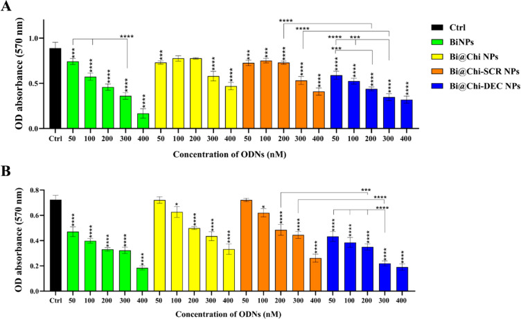
Our goal in this study was to develop bismuth sulfide nanoparticles (NPs) that were functionalized with chitosan and incorporated with decoy oligodeoxynucleotides (ODNs) specifically targeting the NANOG transcription factor (designated as Bi@Chi-DEC NPs) in triple-negative breast cancer cells. FT-IR, UV-vis, FESEM, EDX, TEM, DLS, release kinetics, and hemolysis assays were done to validate the successful synthesis of Bi@Chi-DEC NPs. The synthesized spherical particles exhibited a size distribution averaging 213.8 nm, with a zeta potential measured at -3.27 mV. The anticancer properties of the synthesized nanoparticles, along with X-ray irradiation (2Gy), were assessed through a series of cellular assays, including MTT, cellular uptake, apoptosis, cell cycle analysis, scratch, and tumorsphere formation assays on MDA-MB-231 breast cancer cells. Treatment with the synthesized nanoparticles and X-irradiation resulted in a significant reduction in cell viability, tumorsphere formation, and cellular migration, while concurrently enhancing the rate of apoptotic cells and inducing cell cycle arrest at the G2/M phase. It can be inferred that Bi@Chi-DEC NPs possess the potential to serve as a therapeutic modality for cancer treatment, particularly when utilized along with radiation therapy. Further, in vivo studies are warranted to substantiate the efficacy of this therapeutic approach.

Keywords: Bismuth nanoparticle; Breast cancer; Combinational therapy; Decoy oligodeoxynucleotides (ODNs); NANOG transcription factor; Radiotherapy.


Conflict of interest statement

Declarations. Competing interests: The authors declare no competing interests. Ethical approval: Informed consent was obtained from all individual participants included in the study. This study was approved by the Research Ethics Committees of Zanjan University of Medical Sciences, Zanjan, Iran (Ethical Code: IR.ZUMS.REC.1400.518). Moreover, all methods were performed in accordance with the relevant guidelines and regulations.

Figures

Fig. 1
Fig. 1
A schematic representation of Bi@Chi-DEC NPs drug delivery mechanism along with X-irradiation as a novel therapeutic strategy on LNCaP prostate cancer cell line.
Fig. 2
Fig. 2
(A, B, C) The FTIR spectrum and (D) UV–vis analysis of BiNPs, Bi@Chi NPs, Bi@Chi-SCR, and Bi@Chi-DEC showed the successful synthesis of different formulations.
Fig. 3
Fig. 3
(A, B) FESEM and EDX analysis of Bi@Chi-DEC NPs. (C, D) TEM and average size distribution of Bi@Chi-DEC NPs.
Fig. 4
Fig. 4
(A, B) Average size and (C, D) average zeta potential for groups of BiNPs, Bi@Chi NPs, Bi@Chi-SCR NPs, Bi@Chi-DEC NPs.
Fig. 5
Fig. 5
Investigating the release of ODNs at (A) pH = 7.4 and (B) pH = 5.8. (C) Hemolysis analysis on synthesized nanostructures. *p < 0.05, **p < 0.01, ***p < 0.001, and ****p < 0.0001.
Fig. 6
Fig. 6
(A) The cellular uptake rate of Bi@Chi NPs nanostructures (negative control) and Bi@Chi-labeled ODNs (ODNs labeled with Cy3) in the MDA-MB-231 cell line at concentrations of 25, 50, 100, 200, and 300 nM (B) Statistical graph showing uptake rate of nanostructured groups in different concentrations. **p < 0.01, ***p < 0.001, and ****p < 0.0001.
Fig. 7
Fig. 7
Cytotoxicity evaluation of different groups of nanostructured BiNPs, Bi@Chi, Bi@Chi-SCR, and Bi@Chi-DEC NPs at concentrations of 50, 100, 200, 300, and 400 nM on MDA-MB-231 cells in the condition of (A) without X-ray exposure and (B) with X-ray exposure. *p < 0.05, **p < 0. 01, *** p < 0.001, and ****, p < 0.0001.
Fig. 8
Fig. 8
The cell cycle evaluation in the treatment with different nanostructures (BiNPs, Bi@Chi, Bi@Chi-SCR, and Bi@Chi-DEC) with a concentration of 300 nM in conditions (A and C) without X-ray exposure and (B and D) with X-ray exposure. *p < 0.05, **p < 0.01, *** p < 0.001, and **** p < 0.0001.
Fig. 9
Fig. 9
Cell apoptosis evaluation in the treatment with different nanostructures (BiNPs, Bi@Chi, Bi@Chi-SCR, and Bi@Chi-DEC) with a concentration of 300 nM in conditions (A and C) without X-ray exposure and (B and D) with X-ray exposure. *p < 0.05, **p < 0.01, *** p < 0.001, and **** p < 0.0001.
Fig. 10
Fig. 10
Cell migration inhibition evaluation after treatment with 150 nM of BiNPs, Bi@Chi, Bi@Chi-SCR, and Bi@Chi-DEC NPs nanostructures on MDA-MB-231 cells at (A) 0h after treatment, (B) 72h later in without X-ray exposure conditions and (C) 72h later in with X-ray exposure conditions. (D) Statistical analysis of the data from the percentage of cell migration inhibition. **p < 0.01 and ****p < 0.0001.
Fig. 11
Fig. 11
Tumorsphere formation evaluation after treatment with 150 nM of BiNPs, Bi@Chi, Bi@Chi-SCR, and Bi@Chi-DEC NPs nanostructures on MDA-MB-231 cells in conditions (A) without X-ray exposure and (B) with X-ray exposure. (C) Statistical analysis of the data obtained from the examination of tumorsphere forming ability. * p < 01 and **p < 0.01.

Similar articles

References

    1. Lu, X., Mazur, S. J., Lin, T., Appella, E. & Xu, Y. The pluripotency factor nanog promotes breast cancer tumorigenesis and metastasis. Oncogene33(20), 2655–2664 (2014). - PMC - PubMed
    1. Han, J. et al. RNA interference-mediated silencing of NANOG reduces cell proliferation and induces G0/G1 cell cycle arrest in breast cancer cells. Cancer Lett.321(1), 80–88 (2012). - PubMed
    1. Choi, S. C. et al. Nanog regulates molecules involved in stemness and cell cycle-signaling pathway for maintenance of pluripotency of P19 embryonal carcinoma stem cells. J. Cell. Physiol.227(11), 3678–3692 (2012). - PubMed
    1. Sun, Y.-S. et al. Risk factors and preventions of breast cancer. Int. J. Biol. Sci.13(11), 1387 (2017). - PMC - PubMed
    1. Badrigilan, S. et al. Bismuth-based nanomaterials: Recent advances in tumor targeting and synergistic cancer therapy techniques. Adv. Healthc. Mater.9(7), 1901695 (2020). - PubMed