. 2025 Jul 1;15(1):22157.
doi: 10.1038/s41598-025-06849-2.

Acetaminophen, a new tool for the refinement of the experimental infection of toxoplasmosis in mice

Affiliations

Acetaminophen, a new tool for the refinement of the experimental infection of toxoplasmosis in mice

Nathalie Moiré et al. Sci Rep. .

Abstract

In recent years, animal welfare gained increasing importance in society and especially in scientific research. It has become necessary to refine the experimental procedures as much as possible in infectiology as in our reference model, toxoplasmosis, in accordance with the 3Rs rule and various ethical concerns. Thus, the establishment of a treatment using analgesics would provide relief to animals acutely infected with Toxoplasma gondii. However, the use of analgesics should in no way alter the pathophysiology of the disease and the host immune response, so as not to interfere with the initial scientific study. Little is currently known about the use of acetaminophen (APAP) in an infectious model. In the present work, we studied the impact of APAP at a reference dose of 30 mg/kg/day in a mouse model of acute toxoplasmosis. Zoonotic, telemetric, behavioral, histological and immune parameters were analyzed to better characterize the consequences of treatment with APAP either by gavage or by self-medication in Gel Water. APAP administered by gavage did not induce cellular or tissue toxicity or alter the physiological development of the mice. In addition, the nature of Gel Water itself, independent of APAP, had an effect on the immune response. APAP improved overall well-being and slowed the onset of clinical signs without altering the physiopathology or the immune responses induced by T. gondii. These first results in mice confirmed our initial hypothesis that APAP appears to be a pharmacological tool to refine and improve animal welfare during the acute phase of toxoplasmosis. Therefore, our project has highlighted the combination of specific markers to contribute to animal welfare in mice. In the long term, the use of APAP could be extended to other infectious models with other target animal species.

Keywords: Acetaminophen; Behavior; Immune response; Mice; Toxoplasmosis; Welfare.


Conflict of interest statement

Declarations. Competing interests: The authors declare no competing interests. Ethics statement: Experiments were carried out according to EU directives and French regulations (Directive 2010/63 / EU, 2010; Rural Code, 2018; Decree n ° 2013–118, 2013, https://www.legifrance.gouv.fr/loda/id/JORFTEXT000027038013/ ). All experimental procedures have been evaluated and approved by the Ministry of Higher Education and Research (APAFIS # 2018021917268751.V3—13634). The procedures involving mice were evaluated by the Val de Loire ethics committee (CEEA VdL, committee number 19) and took place at the INRAE Platform for Experimental Infection PFIE (UE-1277 PFIE, INRAE Centre-Val-de Loire) Valley research, Nouzilly, France, https://doi.org/ https://doi.org/10.15454/1.5535888072272498e12 .

Figures

Fig. 1
Fig. 1
Experimental timelines. (A) Evaluation of APAP cytotoxicity in naïve CBA/J mice. (B) Effect of APAP on Toxoplasma gondii pathophysiology (acute and chronic phases) and impact on the immune response and behavior in CBA/J mice. MB Marble burying test.
Fig. 2
Fig. 2
Histological analyses of target tissues (spleen, liver, kidney and stomach) following 5 consecutive days of APAP treatment. Observation after Haemalun-Eosin staining, scale bar: 100 µm. Images (AE) section of spleen (white circle: macrophage; square: white pulp; asterisk: red pulp); (FJ) section of liver (HA hepatic artery, BD Bile Duct); (KO) stomach section (GF Fundic Gland, M Muscle); (PT) section of right kidney (Gl tubular Glomerulus, Tub collecting tube). (Representative of 4 animals).
Fig. 3
Fig. 3
Spleen inflammation after APAP treatment. (A) Spleen weight (n = 7, except control n = 2) and (B) number of splenocytes (n = 4) after 5 days of consecutive treatment with APAP. Data were analyzed by Kruskal–Wallis test followed by Dunn’s multiple comparisons test and expressed as median ± interquartile range, * indicates P < 0.05.
Fig. 4
Fig. 4
Zootechnical parameters during infection. Monitoring of temperature (A and C) and weight (B and D) of mice during the acute phase following infection with 120 cysts (A and B) or 15 cysts (C and D), with or without APAP treatment from D7 to D11 (data expressed as mean ± SEM, * indicates P < 0.05, ** P < 0.01, Two-way ANOVA followed by Tukey’s multiple comparisons test, n = 8).
Fig. 5
Fig. 5
Clinical follow-up. Clinical score during the acute phase following infection with 120 cysts (A) or 15 cysts (B) with or without APAP treatment from D7 to D11 (data expressed as mean ± SEM, * P < 0.05, ** P < 0.01, *** P < 0.001, Two-way ANOVA followed by Tukey’s multiple comparisons test, n = 8).
Fig. 6
Fig. 6
Analysis of behavior during infection. Marble Burying test on D0, D10 after infection with 120 cysts (A, B) and on D0, D10, D38 after infection with 15 cysts (C, D, E) Data were analyzed by Kruskal–Wallis test followed by Dunn’s multiple comparisons test and expressed as median ± interquartile range, * indicates P < 0.05, ** P < 0.01, *** P < 0.001, n = 8, except control at day 38 n = 4).
Fig. 7
Fig. 7
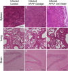
Histological analyses of target tissues during acute phase. Haemalun-Eosin staining of organs from mice of different groups infected with a lethal dose (120 cysts) for 12 days and treated with APAP from D7 to D11, scale bar: 100 µm. Images (AC) spleen section; (DF) lung section; (GI) brain section. Pictures representative of several sections from 3 different animals.
Fig. 8
Fig. 8
Histological analyses of target tissues during chronic phase. Haemalun-Eosin staining of organs from mice of different groups infected with a chronic dose (15 cysts) for 12 days and treated with APAP from D7 to D11, Scale: 100 µm. Images (AC) spleen section; (DF) lung section; (GI) brain section. Pictures representative of several sections from 3 different animals.
Fig. 9
Fig. 9
Splenocyte cytokine response. Production of cytokines in supernatants of splenocytes activated in vitro by specific antigens of T. gondii from mice after 12 days of infection with 120 cysts (A, B, C) or 42 days of infection with 15 cysts and 5 days of APAP-treatment or without treatment. A, B, C infected with 120 cysts and D, E, F infected with 15 cysts. (A) and (D) IL-6; (B) and (E) IL-12; and (C) and (F) IFN-γ. Data were analyzed by Kruskal–Wallis test followed by Dunn’s multiple comparisons test and are expressed as median ± interquartile range, * P < 0.05, n = 5.
Fig. 10
Fig. 10
Post-infection distribution of immune splenic populations. Percentage of splenocyte populations after 12 days of infection with 120 cysts (A, C, E and G) or 42 days of infection with 15 cysts (B, D, F, H) and 5 days of APAP-treatment. A and B: F4/80+ (macrophages), C and D: CD4+ T cells; E and F: CD8+ T cells; G and H: CD19+ B cells. Data were analyzed by Kruskal–Wallis test followed by Dunn’s multiple comparisons test and are expressed as median ± interquartile range, * P < 0.05, ** P < 0.01, n = 5.
Fig. 11
Fig. 11
Humoral response after infection. Humoral response in mice infected with 120 cysts (A) or 15 cysts (B) and APAP-treated (data are expressed as mean ± SD, n = 4). Data were analyzed by two-way ANOVA followed by Tukey’s multiple comparisons test. a **** P < 0.0001 in comparison to day 0, b **** P < 0.0001 in comparison to control group of the same day, c **** P < 0.0001 in comparison to the same group of day 11.
Fig. 12
Fig. 12
Parasite load during infection with T. gondii. A to C T. gondii parasite number in 200 ng of total tissue DNA at day 12 after infection with 120 cysts in lung (A) and brain (C) and at day 42 after infection with 15 cysts in lung (B). (D) brain cyst counts. Data were analyzed by Kruskal–Wallis test followed by Dunn’s multiple comparisons test and expressed as median ± interquartile rang, n = 5. (E) Illustration of cyst in brain.

Similar articles

References

    1. Russell, W.M.S., and Burch, R.L. The principles of humane experimental technique. Methuen, Wheathampstead (UK): Universities Federation for Animal Welfare (as reprinted 1992) (1959).
    1. Tannenbaum, J. & Bennett, B. T. Russell and Burch’s 3Rs then and now: the need for clarity in definition and purpose. J. Am. Assoc. Lab. Anim. Sci.54(2), 120–132 (2015). - PMC - PubMed
    1. Layton, R. et al. The impact of stress and anesthesia on animal models of infectious disease. Front. Vet. Sci.10, 1086003 (2023). - PMC - PubMed
    1. Bayne, K. Environmental enrichment and mouse models: Current perspectives. Animal Model Exp. Med.1(2), 82–90 (2018). - PMC - PubMed
    1. Beauge, C. & Riou, M. Etude de l’impact et choix de l’enrichissement du milieu sur l’élevage de lignées de souris transgéniques exempts d’organismes pathogènes spécifiques (EOPS) et opportunistes. STAL Sci. Tech. Anim. Lab.48, 42–52 (2020).

LinkOut - more resources