

Automatic melanoma detection using an optimized five-stream convolutional neural network
- PMID: 40594676
- PMCID: PMC12215865
- DOI: 10.1038/s41598-025-05675-w
Automatic melanoma detection using an optimized five-stream convolutional neural network
Abstract
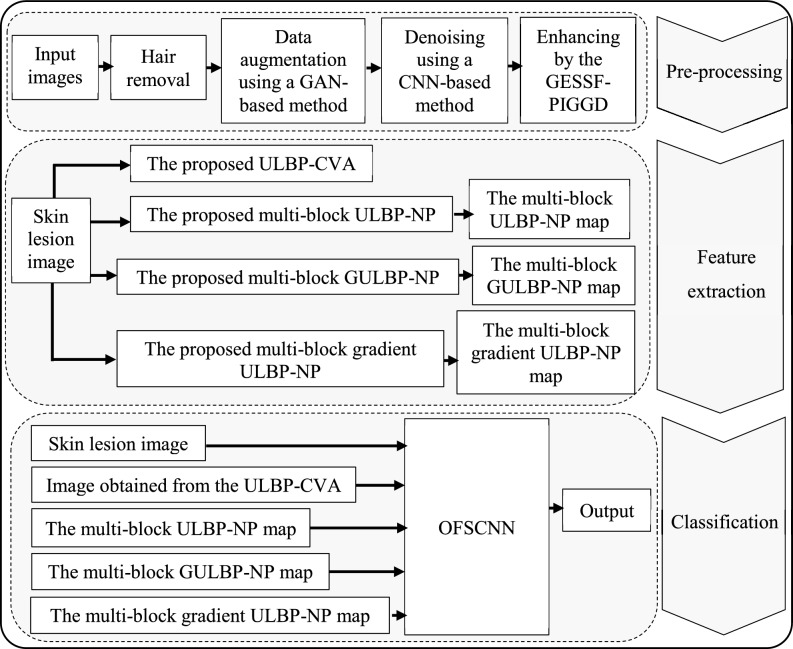
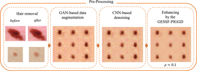
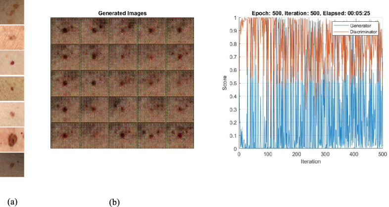
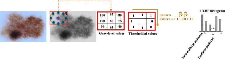
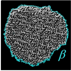
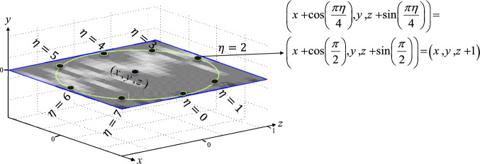
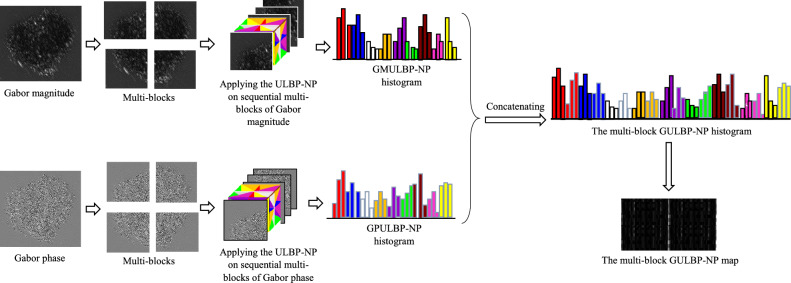
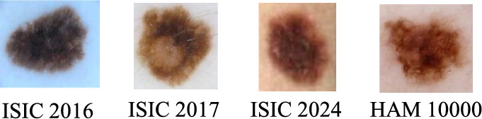
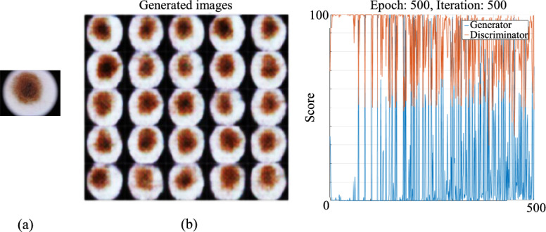
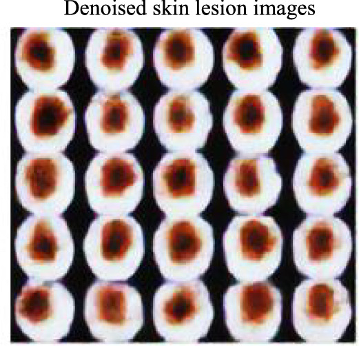
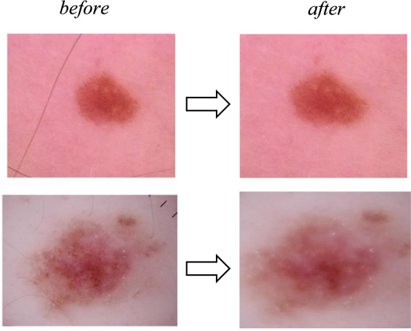
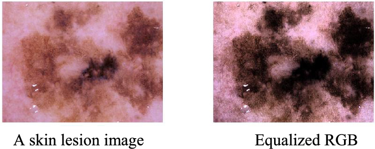
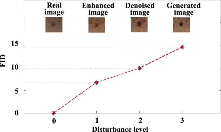
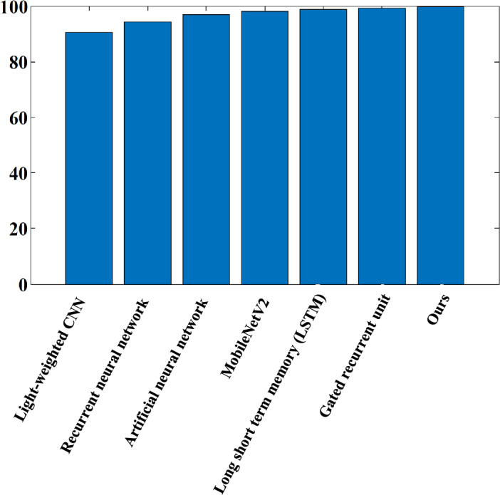
Melanoma is among the deadliest forms of malignant skin cancer, with the number of cases increasing dramatically worldwide. Its early and accurate diagnosis is crucial for effective treatment. However, automatic melanoma detection has several significant challenges. These challenges include the lack of a balanced dataset, high variability within melanoma lesions, differences in the locations of skin lesions in images, the similarity between different skin lesions, and the presence of various artifacts. In addition, the previous deep-learning techniques for diagnosing melanoma cannot recognize the unique relations between samples. For this reason, these convolutional neural networks (CNNs) cannot perceive the changed or rotated image samples as similar. To address these issues in this paper, we have done pre-processing such as hair removal, balancing the skin lesion images using a generative adversarial network (GAN)-based method, denoising using a CNN-based method, and image enhancement. In addition, we propose four new methods to extract key features: the hybrid ULBP and Chan-Vese algorithm (ULBP-CVA), multi-block ULBP on the nine suggested planes (multi-block ULBP-NP), a combination of multi-block Gabor magnitude and phase with ULBP-NP (multi-block GULBP-NP), and combining multi-block gradient magnitude and orientation with ULBP-NP (multi-block gradient ULBP-NP). We suggest nine planes to grab the most vital information about skin lesions in any direction for accurate coding. These introduced planes can capture synchronous spatial and local variations. Hence, very similar lesions can be differentiated by revealing small changes in these small planes. Finally, we propose an optimized four-stream CNN (OFSCNN) for classification. It can simultaneously classify the lesion color, lesion edges, texture features, local-spatial frequency features, and multi-oriented gradient features. The simulation results of our proposed method are promising compared to the most relevant state-of-the-art methods for melanoma detection in dermoscopy images. Our proposed method has automatically detected melanoma in 99.8%, 99.9%, 99.62%, and 99.6% of the HAM 10000, ISIC 2024, ISIC 2017, and ISIC 2016 datasets, respectively.
Keywords: Classification; Convolutional neural network; Denoising; Melanoma detection; Skin cancer.
© 2025. The Author(s).
Conflict of interest statement
Competing interests: The authors declare no competing interests. Ethics approval: This manuscript, or a large part of it, has not been published, was not, and is not being submitted to any other journal. All text and graphics, except for those marked with sources, are original works of the authors. All authors each made a significant contribution to the research reported and have read and approved the submitted manuscript.
Figures


Similar articles
-
Skin-CAD: Explainable deep learning classification of skin cancer from dermoscopic images by feature selection of dual high-level CNNs features and transfer learning.Comput Biol Med. 2024 Aug;178:108798. doi: 10.1016/j.compbiomed.2024.108798. Epub 2024 Jun 25. Comput Biol Med. 2024. PMID: 38925085
-
Advancing dermoscopy through a synthetic hair benchmark dataset and deep learning-based hair removal.J Biomed Opt. 2024 Nov;29(11):116003. doi: 10.1117/1.JBO.29.11.116003. Epub 2024 Nov 19. J Biomed Opt. 2024. PMID: 39564076 Free PMC article.
-
Enhanced melanoma and non-melanoma skin cancer classification using a hybrid LSTM-CNN model.Sci Rep. 2025 Jul 10;15(1):24994. doi: 10.1038/s41598-025-08954-8. Sci Rep. 2025. PMID: 40640352 Free PMC article.
-
Integrating Patient Data Into Skin Cancer Classification Using Convolutional Neural Networks: Systematic Review.J Med Internet Res. 2021 Jul 2;23(7):e20708. doi: 10.2196/20708. J Med Internet Res. 2021. PMID: 34255646 Free PMC article.
-
Drugs for preventing postoperative nausea and vomiting in adults after general anaesthesia: a network meta-analysis.Cochrane Database Syst Rev. 2020 Oct 19;10(10):CD012859. doi: 10.1002/14651858.CD012859.pub2. Cochrane Database Syst Rev. 2020. PMID: 33075160 Free PMC article.
References
-
- Bhatt, H., Shah, V., Shah, K., Shah, R. & Shah, M. State-of-the-art machine learning techniques for melanoma skin cancer detection and classification: a comprehensive review. Intell. Med.3, 180–190 (2023).
-
- Pereira, P. M. et al. Dermoscopic skin lesion image segmentation based on local binary pattern clustering: comparative study. Biomed. Signal Process. Control59, 101924 (2020).
MeSH terms
LinkOut - more resources
Full Text Sources
Medical
Miscellaneous
