

Independent component analysis of oddball EEG recordings to detect Parkinson's disease
- PMID: 40594683
- PMCID: PMC12214538
- DOI: 10.1038/s41598-025-07645-8
Independent component analysis of oddball EEG recordings to detect Parkinson's disease
Abstract
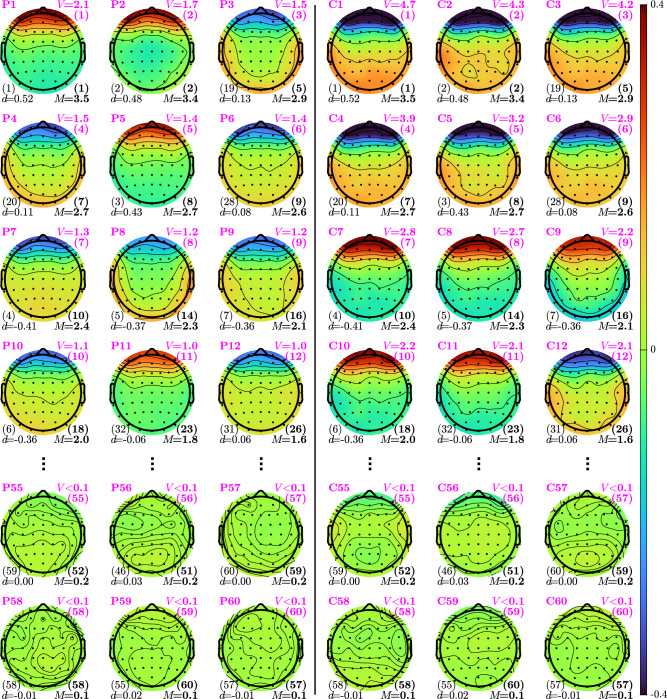
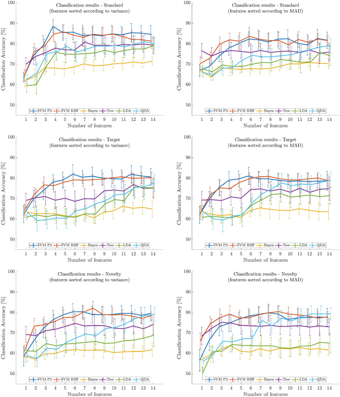
Parkinson's Disease (PD) is one of the most common diseases affecting the human brain, thus approaches are needed to help diagnose it. Since the changes caused by PD are visible in electroencephalograms (EEG), analysis of EEG represents one such approach. In this study, we used 25 EEG recordings of PD patients and 25 of healthy controls, subjected to auditory tasks, available in the Parkinson's Oddball database. The mean age of the PD patients was 69.7 years (std. 8.7) and 69.3 years (std. 9.6) of the control subjects. We employed the Independent Component Analysis (ICA) method to characterize the PD and control EEG recordings, to represent the changes in habituation as a response to different auditory events via the ICA components in the form of topological distributions, and to classify the EEG recordings of the two groups. Characterization of the frontal and central electrodes of the topological distribution showed high separation power to differentiate EEG recordings of the PD patients and healthy subjects. The average classification results using 5-fold cross-validation over 50 trials and the first four features ranked according to the variance of the ICA components, while the features were logarithm of the variance of the ICA components, yielded the following performances: classification accuracy of 88.56%, sensitivity of 89.36%, and specificity of 87.76%. The use of the ICA method appears to be a promising approach for characterizing and classifying auditory EEG recordings.
Keywords: Classification; Electroencephalogram; Independent component analysis; Parkinson’s disease.
© 2025. The Author(s).
Conflict of interest statement
Declarations. Competing interests: The authors declare that they have no competing interests.
Figures

Similar articles
-
Use of β-adrenoreceptor drugs and Parkinson's disease incidence in women from the French E3N cohort study.J Parkinsons Dis. 2025 Jun;15(4):789-804. doi: 10.1177/1877718X251330993. Epub 2025 Apr 29. J Parkinsons Dis. 2025. PMID: 40302366
-
Tissue Factor and Its Cerebrospinal Fluid Protein Profiles in Parkinson's Disease.J Parkinsons Dis. 2024;14(7):1405-1416. doi: 10.3233/JPD-240115. J Parkinsons Dis. 2024. PMID: 39240648 Free PMC article.
-
Modulation of Cerebellar Oscillations with Subthalamic Stimulation in Patients with Parkinson's Disease.J Parkinsons Dis. 2024;14(7):1417-1426. doi: 10.3233/JPD-240065. J Parkinsons Dis. 2024. PMID: 39331106 Free PMC article.
-
Interventions for preventing falls in Parkinson's disease.Cochrane Database Syst Rev. 2022 Jun 6;6(6):CD011574. doi: 10.1002/14651858.CD011574.pub2. Cochrane Database Syst Rev. 2022. PMID: 35665915 Free PMC article.
-
Magnetic resonance perfusion for differentiating low-grade from high-grade gliomas at first presentation.Cochrane Database Syst Rev. 2018 Jan 22;1(1):CD011551. doi: 10.1002/14651858.CD011551.pub2. Cochrane Database Syst Rev. 2018. PMID: 29357120 Free PMC article.
References
MeSH terms
LinkOut - more resources
Full Text Sources
Medical
Research Materials
Miscellaneous
