. 2025 Jul 1;15(1):21112.
doi: 10.1038/s41598-025-05781-9.

An intricate role of Ang II/AT1 in the modulation of monosodium glutamate-induced pulmonary fibrosis by TGF-β/Smad through quercetin

Affiliations

An intricate role of Ang II/AT1 in the modulation of monosodium glutamate-induced pulmonary fibrosis by TGF-β/Smad through quercetin

Arnab Banerjee et al. Sci Rep. .

Abstract

To investigate the protective actions of the natural flavonoid quercetin against monosodium glutamate (MSG)-induced pulmonary fibrosis in rats, the present study targets the modulation of the TGF-β/Smad signaling pathway and the involvement of Ang II/AT1. The experimental model involved the treatment of rats with MSG (0.6 g/kg body weight) for 4 weeks and quercetin dosages of 25 mg, 50 mg, and 100 mg/kg body weight. The study applied the combination of biochemical, molecular, and histopathological evaluation to identify the role of quercetin in impacting major cytokines (IL-17, IL-19, TGF-β, VEGF), oxidative stress markers (TBARS, NO, SOD, CAT, GSH), extracellular matrix components (collagen-I, α-SMA, fibronectin), and fibrosis gene expression (TGF-β1, Smad2/3/4, CTGF, Snail, Slug). MSG treatment increased pro-fibrotic cytokines, oxidative stress, and deposition of collagen in a significant amount, while administration of quercetin dose-dependently reversed the alterations. Quercetin also reversed the activity of antioxidant enzymes, reduced inflammatory cytokines, and inhibited TGF-β/Smad signaling as indicated by lowered TGF-β receptor II activation and following Smad phosphorylation. Molecular docking demonstrated that quercetin competitively binds to TGF-β receptor II to inhibit MSG-induced fibrotic signaling. Quercetin inhibits MSG-induced lung fibrosis by inhibiting collagen accumulation and inflammatory cell invasion and has the potential to produce therapeutic effects by modulating TGF-β/Smad signaling and restoring lung tissue homeostasis.

Keywords: Monosodium glutamate; Oxidative stress; Pro-fibrotic cytokines; Pulmonary fibrosis; Quercetin; TGF-β/Smad.


Conflict of interest statement

Declarations. Competing interests: The authors declare no competing interests.

Figures

Fig. 1
Fig. 1
Experimental design (BioRender was used to make the figure: https://biorender.com).
Fig. 2
Fig. 2
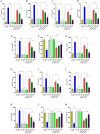
MSG-induced lung fibrosis: role of key cytokines, growth factors, and quercetin in modulating dysregulated signaling pathways (A-G). Quercetin regulates the equilibrium of pro and anti-fibrotic factors in MSG-fed rats (H-M). Data were expressed as Mean ± SEM. Significance level based on one-way ANOVA, P ≤ 0.05. Significance level based on Tukey’s post hoc test (n = 5): NC vs. MSG: a, NC vs. QL/QM/QH: b, QL vs. QM vs. QH: c, QL/QM/QH vs. MSG + QL/MSG + QM/MSG + QH: d, MSG vs. MSG + QL: e, MSG vs. MSG + QM: f, MSG vs. MSG + QH: g, MSG + QL vs. MSG + QM vs. MSG + QH: h [*P ≤ 0.05, **P ≤ 0.01, ***P ≤ 0.001, NS: not significant].
Fig. 3
Fig. 3
MSG-induced pulmonary fibrosis and ECM remodeling were modulated by quercetin (A-M). Cross-talk between oxidative stress and pulmonary fibrosis: possible protection through quercetin against MSG-incited changes in the lung (H-L). Data were expressed as Mean ± SEM. Significance level based on one-way ANOVA, P ≤ 0.05. Significance level based on Tukey’s post hoc test (n = 5): NC vs. MSG: a, NC vs. QL/QM/QH: b, QL vs. QM vs. QH: c, QL/QM/QH vs. MSG + QL/MSG + QM/MSG + QH: d, MSG vs. MSG + QL: e, MSG vs. MSG + QM: f, MSG vs. MSG + QH: g, MSG + QL vs. MSG + QM vs. MSG + QH: h [*P ≤ 0.05, **P ≤ 0.01, ***P ≤ 0.001, NS: not significant].
Fig. 4
Fig. 4
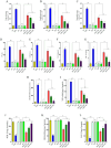
Quercetin as an anti-fibrotic molecule blunted the occurrence of MSG-induced lung fibrosis via suppressing TGF-β (A-G). Data were expressed as Mean ± SEM. Significance level based on one-way ANOVA, P ≤ 0.05. Significance level based on Tukey’s post hoc test (n = 5): NC vs. MSG: a, NC vs. QL/QM/QH: b, QL vs. QM vs. QH: c, QL/QM/QH vs. MSG + QL/MSG + QM/MSG + QH: d, MSG vs. MSG + QL: e, MSG vs. MSG + QM: f, MSG vs. MSG + QH: g, MSG + QL vs. MSG + QM vs. MSG + QH: h [*P ≤ 0.05, **P ≤ 0.01, ***P ≤ 0.001, NS: not significant].
Fig. 5
Fig. 5
TXNIP and inflammatory pathways: Role of MSG and quercetin in the development of pulmonary fibrosis through balance in redox and myofibroblast differentiation (A-D). Data were expressed as Mean ± SEM. Significance level based on one-way ANOVA, P ≤ 0.05. Significance level based on Tukey’s post hoc test (n = 5): NC vs. MSG: a, NC vs. QL/QM/QH: b, QL vs. QM vs. QH: c, QL/QM/QH vs. MSG + QL/MSG + QM/MSG + QH: d, MSG vs. MSG + QL: e, MSG vs. MSG + QM: f, MSG vs. MSG + QH: g, MSG + QL vs. MSG + QM vs. MSG + QH: h [*P ≤ 0.05, **P ≤ 0.01, ***P ≤ 0.001, NS: not significant]. The cytoarchitectonic view of lung tissues was also depicted in this figure, which shows the restorative impact of quercetin in the MSG-induced altered cytomorphology by HE (HE), Picrosirius red (PSR), and Masson’s trichrome (MT) staining. Histological examination of lung tissue in NC, MSG, and two graded concentrations of quercetin with or without MSG showed the variants in terms of structure integrity and fibrotic responses. Normal alveolar structures with minimal inflammatory infiltration were seen in the NC group by hematoxylin and eosin staining (HE). On the other hand, the MSG group presented intense pulmonary fibrosis, whose histological appearance was disturbed due to higher collagen deposition and inflammatory cell infiltration in all of the used staining methods. Interstitial edema and inflammation were mainly emphasized under HE staining (HE), whereas Picrosirius red staining (PSR) indicated the existence of thick fibers that represent fibrotic remodeling. Areas stained with Masson’s trichrome revealed an increase compared with controls (MT). The treatment at two concentrations of quercetin has not shown any alterations of the lung histology compared with the NC group, indicating that this compound has limited or no significant influence on the lung independently. However, quercetin + MSG offered protective and significantly reduced the level of fibrotic changes with all the stainings ; thus, there is evidence that although quercetin itself does not produce marked remodeling of lung tissue, it effectively rescues from the pathological effects of MSG and saves the structure and attenuates inflammatory responses.
Fig. 6
Fig. 6
Role of quercetin in MSG-induced changes in pro-inflammatory and pro-fibrotic gene expression (gene expression: A-U; heatmap: V) by regulating AT1 and TGF-β/Smad pathway. Data were expressed as Mean ± SEM. Significance level based on one-way ANOVA, P ≤ 0.05. Significance level based on Tukey’s post hoc test (n = 5): NC vs. MSG: a, NC vs. QL/QM/QH: b, MSG vs. MSG + QH: g [**P ≤ 0.01, NS: not significant].
Fig. 7
Fig. 7
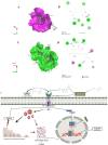
Molecular docking analysis of MSG and quercetin with TβR-II (AutoDock Vina and Discovery Studio were used to make the figure). (A) The 3D interaction of MSG with the TβR-II, as shown by Discovery Studio Visualizer, is described in terms of surface and major interacting residues. (B) A 2D diagram for classic hydrogen and carbon-hydrogen bonds between MSG and other residues of TGF-β RII such as Asn40, Gln41, and Trp65. (C) Representation of the 3D illustration of the binding interaction of quercetin with TβR-II. In this illustration, the receptor is shown to be surficial towards the involvement of residues important in the interaction. (D) 2D map of quercetin interaction, with hydrogen bonds, Pi-Pi T-shaped and Amide-Pi stacking interaction modes towards the key residues involved: Asn40, Gln41, Trp65, Phe126. The interaction distances are given in Å. (E) The mechanistic overview of MSG on lung to cause fibrotic responses by activating TGF-β/Smad and the role of quercetin in such anomalous situation (BioRender was used to make the figure: https://biorender.com).
Fig. 8
Fig. 8
The hypothetical target pathway by which quercetin inhibits TβR and Ang II/AT1 to modulate TGF-β/Smad in MSG-induced pulmonary fibrosis (BioRender was used to make the figure: https://biorender.com).

Similar articles

References

    1. Wilson, M. S. & Wynn, T. A. Pulmonary fibrosis: pathogenesis, etiology and regulation. Mucosal Immunol.2 (2), 103–121. 10.1038/mi.2008.85 (2009). - PMC - PubMed
    1. Ye, Z., Hu, Y. & TGFβ1 Gentlemanly orchestrator in idiopathic pulmonary fibrosis (Review). Int. J. Mol. Med.48 (1), 132. 10.3892/ijmm.2021.4965 (2021). - PMC - PubMed
    1. Mallikarjuna, P., Zhou, Y. & Landström, M. The synergistic Cooperation between TGF-β and hypoxia in Cancer and fibrosis. Biomolecules12 (5), 635. 10.3390/biom12050635 (2022). - PMC - PubMed
    1. Park, M. J. et al. IL-1-IL-17 signaling axis contributes to fibrosis and inflammation in two different murine models of systemic sclerosis. Front. Immunol.9, 1611. 10.3389/fimmu.2018.01611 (2018). - PMC - PubMed
    1. Lazar, M. et al. The role of cytokines and molecular pathways in lung fibrosis following SARS-CoV-2 infection: A physiopathologic (Re)view. Biomedicines12 (3), 639. 10.3390/biomedicines12030639 (2024). - PMC - PubMed

MeSH terms