. 2025 Jul 1;9(1):209.
doi: 10.1038/s41698-025-01006-4.

Dual treatment with Val-083 and AZD1775 shows potent anti-tumor activity in diffuse midline glioma models

Affiliations

Dual treatment with Val-083 and AZD1775 shows potent anti-tumor activity in diffuse midline glioma models

Bavani Subramaniam et al. NPJ Precis Oncol. .

Abstract

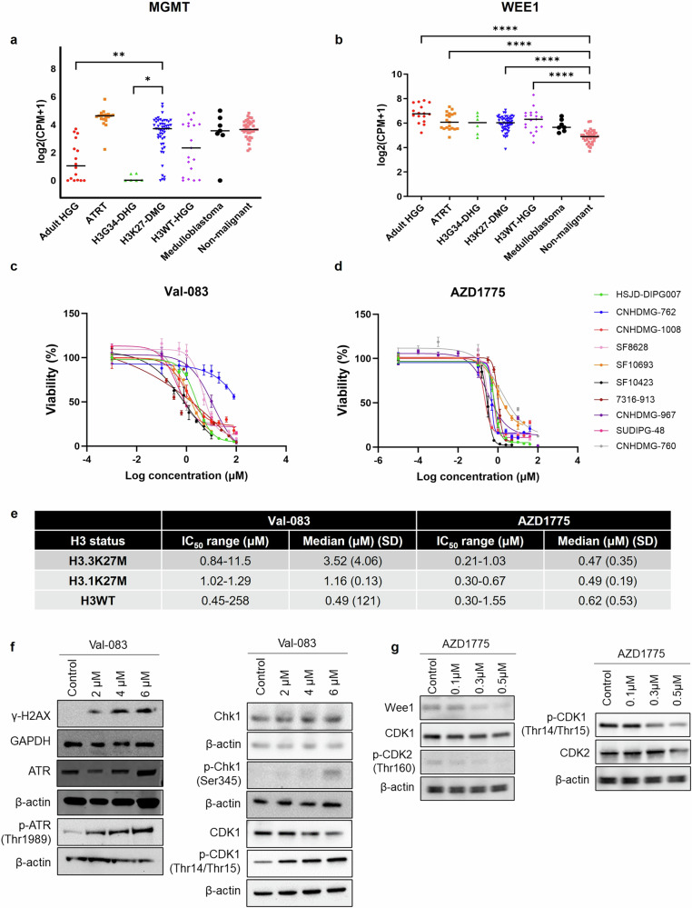
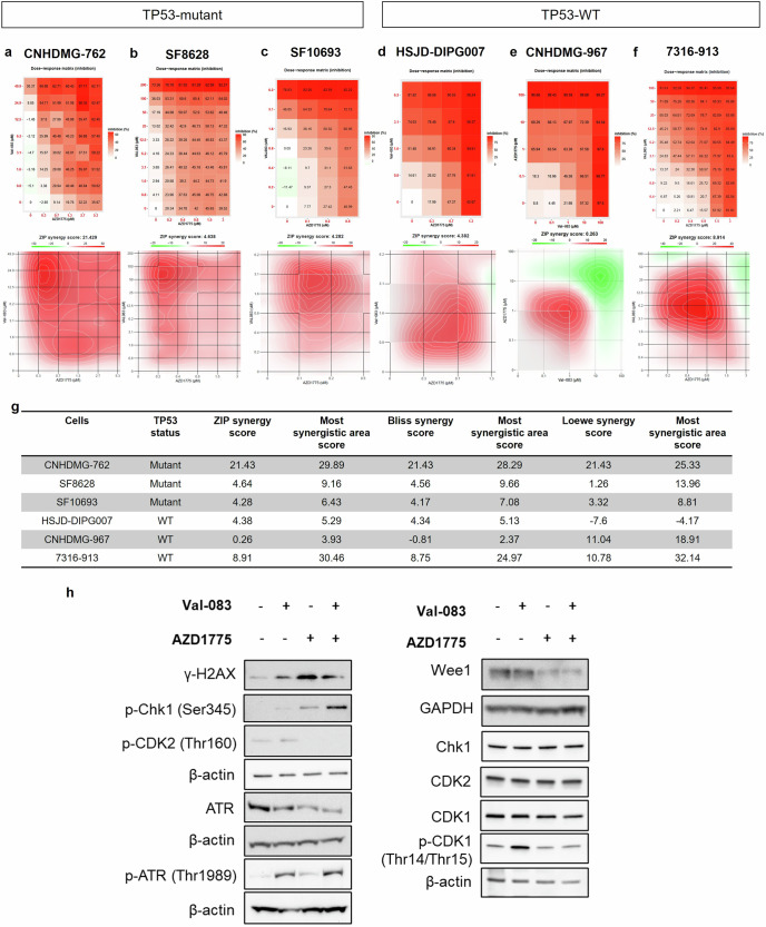
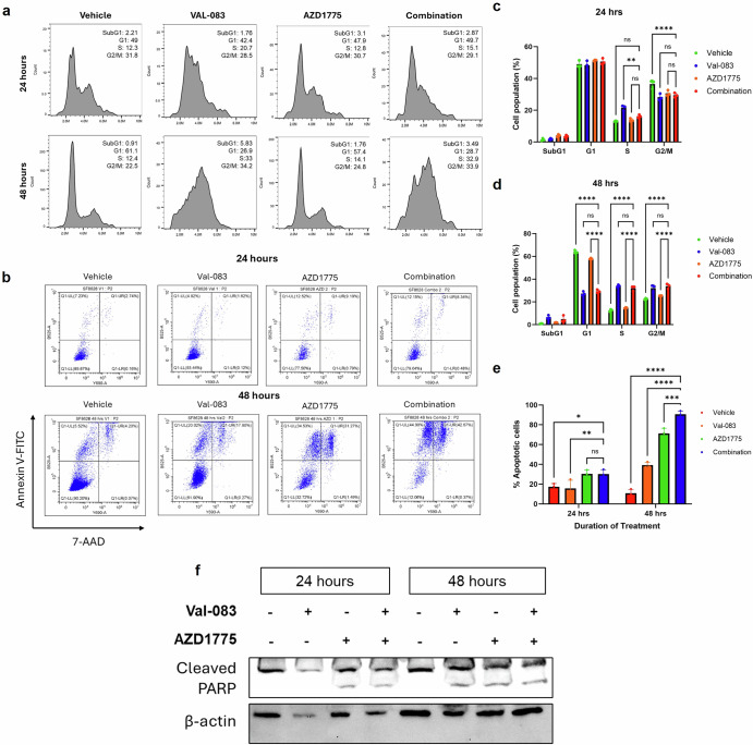
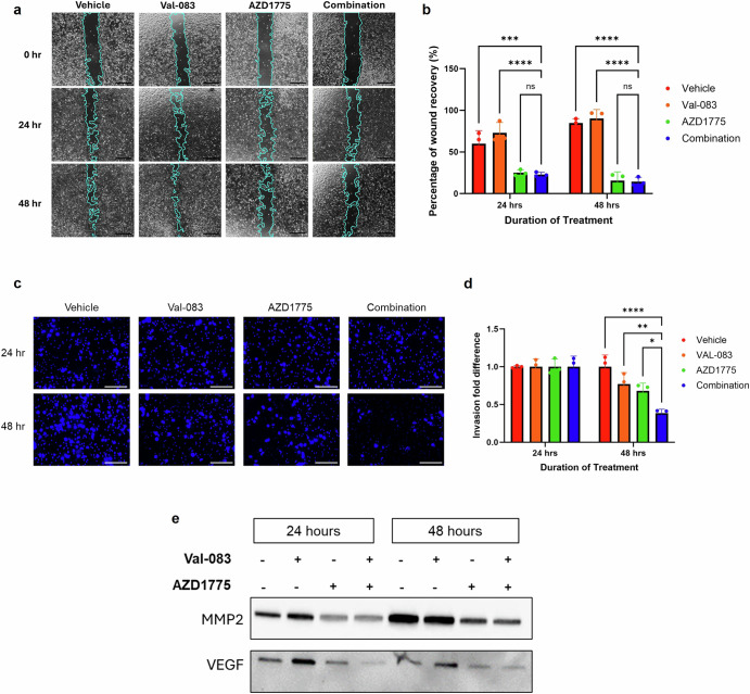
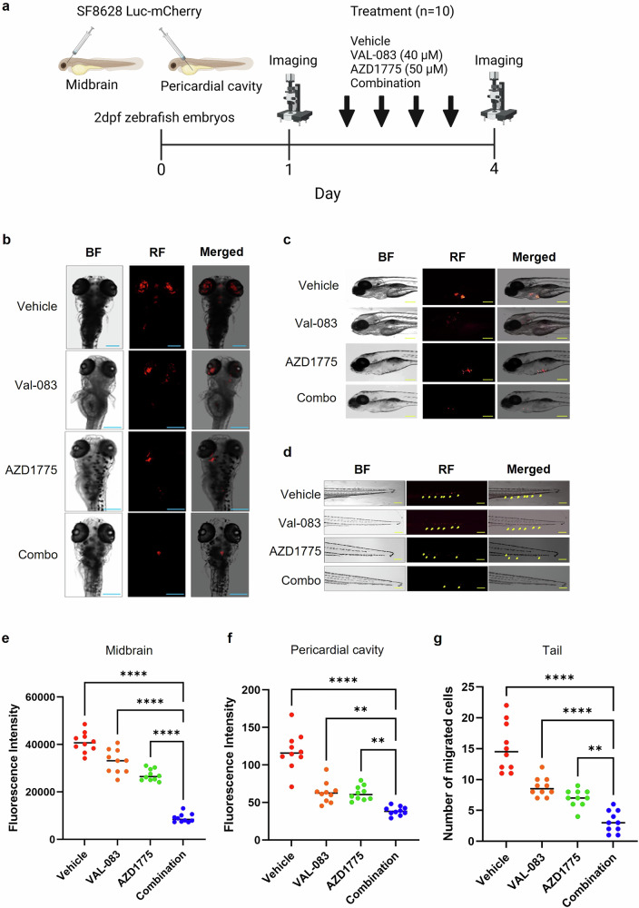
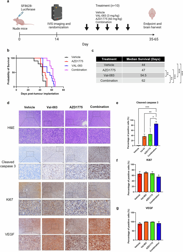
H3K27M diffuse midline gliomas (DMG) are characterized by p53 mutations and hypomethylation of MGMT, a DNA-repair enzyme, leading to resistance towards chemotherapeutic agents such as temozolomide (TMZ). As an alternative, we investigated the efficacy of a functionally different DNA-damaging agent, Val-083, on our DMG models. Val-083 is a blood-brain barrier penetrant DNA targeting agent that induces DNA N7-guanine interstrand crosslinks, which is unrepairable by MGMT. As Val-083 also triggers S/G2 phase cell cycle arrest for DNA repair, we evaluated its combined efficacy with Wee1 inhibitor, AZD1775. AZD1775 functions by inhibiting Wee1, at G2/M checkpoint to prevent phosphorylation of CDK1 and propel cells into the M phase. This subsequently overrides cell cycle arrest and drives cells with DNA damage into premature mitosis and apoptosis. Our results showed that Val-083 and AZD1775 work additively on a range of p53 mutant and p53 wildtype DMG models to inhibit cell growth, induce DNA damage and alter cell cycle. In addition, the combination drugs led to significant increase in the number of cells undergoing apoptosis, and a decrease in the migration and invasion activity of the cells. In vivo, the combination of both drugs led to significant reduction in tumor growth in zebrafish xenograft models and prolongation of survival in mice xenograft models. Our findings indicate that Val-083 and AZD1775 in combination demonstrate promising efficacy in DMGs, providing a clinical rationale for positioning these arms in future therapies.


Conflict of interest statement

Competing interests: The authors declare no competing interests.

Figures

Fig. 1
Fig. 1. Efficacy and mechanism of action of Val-083 and AZD1775 on DMG cells.
Gene expression analysis of CCMA datasets reveals significantly higher (a) MGMT expression in H3K27-DMG cell lines compared to adult HGG and H3G34-DHG cell lines, and (b) significantly higher WEE1 expression in adult HGG, ATRT, H3K27-DMG and H3WT-HGG cell lines compared to non-malignant cells. Kruskal-Wallis test with Dunn’s multiple comparisons test was conducted; *p < 0.05; **p < 0.01; ***p < 0.001; ****p < 0.0001. c, d Dose response curve of Val-083 and AZD1775 on ten DMG/HGG cell lines. e IC50 range and median IC50 of Val-083 and AZD1775 on responsive cell lines based on their H3 status. f Val-083 induces DNA damage and DNA repair response through ATR-mediated signaling pathway. g AZD1775 inhibits Wee1 kinase to reduce inhibitory phosphorylation of CDK1 and CDK2 and induce cell cycle checkpoint impairment.
Fig. 2
Fig. 2. Combination of Val-083 and AZD1775 is additive or synergistic on multiple DMG/HGG cells.
Dose response curve and ZIP synergy scores reveal that Val-083 and AZD1775 combination is synergistic on (a) CNHDMG-762 and additive on (b) SF8628, (c) SF10693 (d) HSJD-DIPG-007, e CNHDMG-967 and (f) 7316-913. g ZIP, Bliss and Loewe synergy scores with the most synergistic area scores on the six DMG cell lines. h Western blot assay demonstrates that Val-083 and AZD1775 combination induces DNA damage (upregulated γ-H2AX), activates DNA damage response via ATR and Chk1, and reduces inhibitory phosphorylation of CDK1 and CDK2 initiated by Val-083.
Fig. 3
Fig. 3. Val-083 and AZD1775 combination leads to cell cycle perturbation and apoptosis.
a 24 h and 48 h cell cycle assay reveal increased cell population at S and G2/M phases which is modified with the addition of AZD1775. b 24 h and 48 h apoptosis assay reveal increased cells in the early and late apoptotic phase upon Val-083 and AZD1775 single and combination treatment. c Percentage of cell population at SubG1, G1, S and G2/M phases after 24 h of treatment with vehicle, Val-083, AZD1775 and combination drugs. d Percentage of cell population at SubG1, G1, S and G2/M phases after 48 h of treatment with vehicle, Val-083, AZD1775 and combination drugs. e Percentage of total apoptotic cells is significantly increased upon AZD1775 and combination treatment at 24- and 48 h post-treatment, while Val-083 treatment is significant only after 48 h of treatments. f Western blot analysis of cleaved PARP shows that the combination treatment significantly increases cleaved PARP activation. All data are shown as mean ± standard deviation (n = 3). Statistically significant difference (Two-way ANOVA, Tukey’s post-hoc test) is shown as *p < 0.05; **p < 0.01; ***p < 0.001; ****p < 0.0001 or ns (not significant).
Fig. 4
Fig. 4. Val-083 and AZD1775 combination inhibits migration and invasion of DMG cells.
a 24 h and 48 h scratch assay image captured using Olympus EP50 microscope. Scale bar = 300 µm. b Plot shows that percentage of wound recovery in the combination group is significantly reduced in comparison to vehicle and Val-083 groups after 24 and 48 h of treatment. c Transwell inserts captured using EVOS M5000 after 24 h and 48 h invasion assay. Scale bar = 150 µm. d Combination treatment significantly inhibits invasion of cells when compared to vehicle, Val-083 and AZD1775 treated groups after 48 h of treatment. e Western blot analysis of MMP2 and VEGF shows that AZD1775 and combination treatment significantly reduces expression of these proteins. All plots are shown as mean ± standard deviation (n = 3). Statistically significant difference (One-way ANOVA or two-way ANOVA, Tukey’s post-hoc test) is shown as *p < 0.05; **p < 0.01; ***p < 0.001; ****p < 0.0001 or ns (not significant).
Fig. 5
Fig. 5. Val-083 and AZD1775 reduces tumor growth and migration in zebrafish models.
a Schematic figure of treatment plans for SF8628 zebrafish xenograft models. b SF8628 zebrafish xenograft (midbrain) models at 4 days post injection (DPI) treated with vehicle, Val-083, AZD1775 and combination (combo) treatments. Scale bar = 750 µm. c SF8628 pericardial cavity tumor models of zebrafish at 4 DPI, treated with vehicle, Val-083, AZD1775 and combo treatments. Scale bar = 750 µm. d Migration of SF8628 cells to the tail region of zebrafish models. Scale bar = 750 µm. e Val-083 and AZD1775 as a single drug or as a combination shows significantly reduced tumor growth in the midbrain region, f pericardial cavity and (g) reduced migration of tumor cells towards the tail region after 3 days of treatment. All plots are shown as mean ± standard deviation (n = 10 for midbrain; n = 10 for pericardial cavity). Statistically significant difference (One-way ANOVA, Tukey’s post-hoc test) is shown as **p < 0.01; ****p < 0.0001.
Fig. 6
Fig. 6. Val-083 and AZD1775 lead to apoptosis of tumor cells and prolonged survival of mouse xenograft models.
a Schematic figure of SF8628 mice xenograft model treatment plans. b Survival curve of SF8628 mouse orthotopic tumor models treated with vehicle control, AZD1775, Val-083 and combination of both drugs. c Table indicates median survival of animals in different treatment groups. The combination treatment significantly extended survival of the animals in comparison to vehicle control and single treatments. d Images of H&E and IHC staining of tumor regions from harvested mouse brain of different treatment groups, captured using Olympus VS200. Scale bar = 100 µm. e IHC staining of cleaved caspase 3 shows that the combination treatment leads to significantly increased number of cells undergoing apoptosis in the Val-083, AZD1775 and combination groups. IHC staining of (f) Ki67 and (g) VEGF) shows no significant difference between treatment groups. All plots are shown as mean ± standard deviation (n = 10 for survival curve; n = 3 for IHC). Statistically significant difference (One-way ANOVA, Tukey’s post-hoc test) is shown as *p < 0.05; **p < 0.01; ***p < 0.001; and ****p < 0.0001.

Similar articles

References

    1. Louis, D. N. et al. The 2016 World health organization classification of tumors of the central nervous system: a summary. Acta Neuropathol.131, 803–20 (2016). - PubMed
    1. Ostrom, Q. T. et al. CBTRUS statistical report: primary brain and central nervous system tumors diagnosed in the United States in 2006−2010. Neuro Oncol.15, ii1–56 (2013). - PMC - PubMed
    1. Hoffman, L. M. et al. Clinical, radiologic, pathologic, and molecular characteristics of long-term survivors of diffuse intrinsic pontine glioma (DIPG): a collaborative report from the international and european society for pediatric oncology dipg registries. J. Clin. Oncol.36, 1963–72 (2018). - PMC - PubMed
    1. Vanan, M. I. & Eisenstat, D. D. DIPG in children - what can we learn from the past?. Front Oncol.5, 237 (2015). - PMC - PubMed
    1. Buczkowicz, P. et al. Genomic analysis of diffuse intrinsic pontine gliomas identifies three molecular subgroups and recurrent activating ACVR1 mutations. Nat. Genet.46, 451–6 (2014). - PMC - PubMed

LinkOut - more resources