. 2025 Jul 1;15(1):21920.
doi: 10.1038/s41598-025-06876-z.

Use of bovine serum albumin might impair immunofluorescence signal in thick tissue samples

Affiliations

Use of bovine serum albumin might impair immunofluorescence signal in thick tissue samples

Anna Chwastowicz et al. Sci Rep. .

Abstract

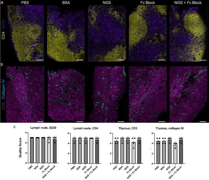
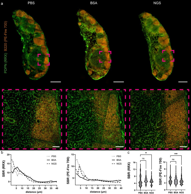
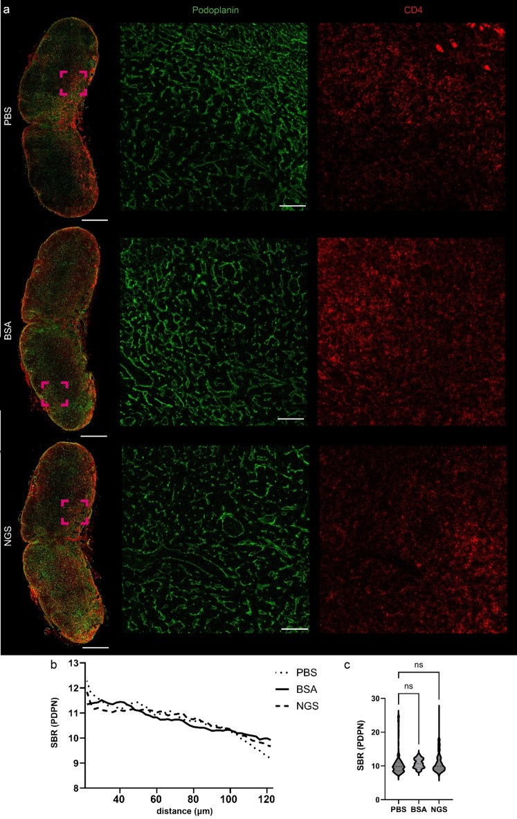
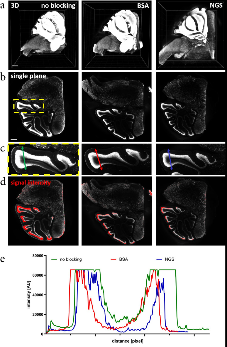
Significant progress in microscopic imaging techniques allowed transition from predominantly qualitative methods to a powerful tool for quantitative research, driven by improved instrumentation and computational power. Furthermore, previously limited to thin, laser-permeable tissue sections, imaging techniques have been revolutionized by the advent of tissue optical clearing. This innovation enables the visualization and quantitative analysis of entire organs and even whole bodies at cellular resolution. However, achieving high-quality imaging depends not only on the transparency of the tissue preparation but also on precise immunofluorescence labeling to ensure accurate signal detection and reliable study outcomes. In this study, we evaluated whether various reagents that are typically applied during the tissue blocking step prior to immunofluorescence staining affect the quality of the obtained image in thick and optically cleared samples. We demonstrate that the commonly employed tissue blocking step does not improve imaging conditions and even can substantially degrade fluorescence signal quality, particularly in large, optically cleared tissues such as whole mouse brain hemispheres.

Keywords: BSA; Bovine serum albumin; Confocal microscopy; Immunofluorescence; Tissue clearing.


Conflict of interest statement

Declarations. Competing interests: The authors declare no competing interests.

Figures

Fig. 1
Fig. 1
Immunofluorescence staining of murine lymphoid tissue samples processed with and without the protein blocking step prior to incubation with antibodies. (a) immunolabeling of murine lymph node against B220 (marker of B-lymphocytes) and CD4 (marker of T-helper lymphocytes) with secondary antibody conjugated with AF-488 and AF-647, respectively. (b) immunolabeling of murine thymus against collagen IV with secondary antibody conjugated with AF-568 and against CD3 (marker of T lymphocytes) with AF-647 directly conjugated antibody. (c) Four pathologists independently and blindly evaluated the quality of immunofluorescence staining in the panels (a) and (b), using a subjective scoring scale from 1 to 5, where 5 represents optimal conditions. No significant differences in image quality were observed between the PBS (no blocking agent) group and the other groups. BSA, bovine serum albumin, NGS, normal goat serum. Scale bar, 100 μm.
Fig. 2
Fig. 2
Protein blocking step with BSA diminishes signal-to-background ratio of commonly used fluorophores. (a) immunolabeling of murine lymph node against B220 (marker of B-lymphocytes) with secondary antibodies conjugated with AF-488, −555 and AF-647, ROI is zoomed in lower panels. (b) mean SBR values of three commonly used fluorophores across 40 μm imaging range, n = 7–8 per group and (c) SBR values pooled from 15 to 40 μm imaging range, one-way ANOVA with Dunn’s multiple comparison test, n = 7–8 per group; ****P < 0.0001, **P < 0.01. Scale bar, 300 μm and 30 μm in zoomed areas.
Fig. 3
Fig. 3
Immunofluorescence staining of murine lymph node tissue samples with and without the protein blocking step. (a) immunolabeling of murine lymph node against B220 (marker of B-lymphocytes) with primary antibody conjugated with Pe-Fire 700 (orange) and against podoplanin with secondary antibody conjugated with rhodamine red X (RRX, green), ROI is zoomed in lower panels. (b) mean SBR values of used fluorophores across 40 μm imaging range, n = 13–14 per group and (c) SBR values pooled from 15 to 40 μm imaging range, one-way ANOVA with Dunn’s multiple comparison test, n = 13–14 per group; **P < 0.01, *P < 0.05. Scale bar, 500 μm and 50 μm in zoomed areas.
Fig. 4
Fig. 4
Immunofluorescence staining of thick murine lymph node tissue samples processed with and without the protein blocking step prior to incubation with primary Abs. (a) immunolabeling of murine lymph node against podoplanin (marker of fibroblastic reticular cells and lymphatic endothelial cells) and CD4 followed by secondary antibodies conjugated with AF-555 and AF-647, respectively. ROI is zoomed in lower panels. (b) mean SBR values of used fluorophores across 20–125 μm imaging range, n = 11–12 per group and (c) SBR values pooled from 20 to 120 μm imaging range, one-way ANOVA with Dunn’s multiple comparison test, n = 11–12 per group. BSA, bovine serum albumin, NGS, normal goat serum. Scale bar, 400 μm and 50 μm in zoomed areas.
Fig. 5
Fig. 5
NeuN in cerebellum imaged using light-sheet microscope. (a) 3D reconstruction of images acquired using light-sheet microscope. (b) single plane taken from the middle of 3D Z-stack. Yellow ROI indicates zoom in (c). (c) Zoomed fragment of the cerebellum. Colored arrows indicate axis selected for NeuN signal intensity analysis (d). Red color indicates signal saturation. Note higher NeuN signal intensity when no blocking was used. (e) Signal intensity of NeuN antibody penetration. Scale bar in (a) 800 μm, (b) 400 μm.
Fig. 6
Fig. 6
No blocking enhances antibody penetration into deeper layers in immunofluorescence staining combined with iDISCO clearing technique. a1-a2 iDISCO clearing combined with immunostaining for c-Fos after behavioral appetitive training in mouse brain imaged using light-sheet microscope. a1 – single planes in XYZ, standard iDISCO (with blocking), a2 single planes in XYZ – iDISCO performed without blocking step. Note increased penetration of antibody and signal with no blocking. c-Fos positive cell is marked with an arrowhead. The 3D panel depicts a reconstruction of the Z-stack covering a piece of cortex and the hippocampus. Scale bar in a1-a2 500 μm, in 3D panel 1000 μm.

Similar articles

References

    1. Richardson, D. S. & Lichtman, J. W. Clarifying Tissue Clearing Cell162, 246–257 (2015). - PMC - PubMed
    1. Yu, T., Zhu, J., Li, D. & Zhu, D. Physical and chemical mechanisms of tissue optical clearing. iScience24, 102178 (2021). - PMC - PubMed
    1. Costantini, I. et al. In-vivo and ex-vivo optical clearing methods for biological tissues: review. Biomed. Opt. Express BOE. 10, 5251–5267 (2019). - PMC - PubMed
    1. Susaki, E. A. et al. Advanced CUBIC protocols for whole-brain and whole-body clearing and imaging. Nat. Protoc.10, 1709–1727 (2015). - PubMed
    1. Zhu, J. et al. MACS: rapid aqueous clearing system for 3D mapping of intact organs. Adv. Sci.7, 1903185 (2020). - PMC - PubMed

Substances

LinkOut - more resources