. 2025 Jul 2;15(1):22961.
doi: 10.1038/s41598-025-07242-9.

Unraveling chain specific ubiquitination in cells using tandem ubiquitin binding entities

Affiliations

Unraveling chain specific ubiquitination in cells using tandem ubiquitin binding entities

Muhammad Shahzad Ali et al. Sci Rep. .

Abstract

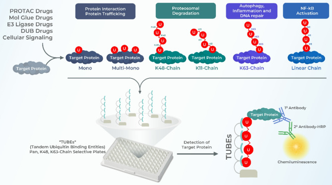
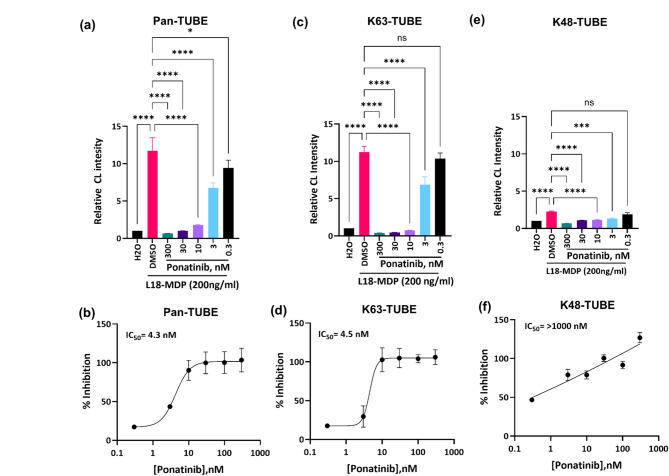
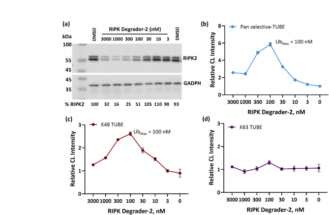
Polyubiquitination of proteins serves distinct functions that are governed by the nature of polyubiquitin chains built on target proteins. Among the eight distinct type of ubiquitin chains, lysine 48 (K48)-linked chains are specifically associated with proteasomal degradation, while lysine 63 (K63)-linked chains are primarily involved in regulating signal transduction and protein trafficking. The ubiquitin-proteasome system (UPS) has recently been exploited in drug discovery and introduced PROTACs (Proteolysis Targeting Chimeras), or molecular glues (MGs), to hijack ubiquitin E3 ligases, to facilitate the targeted degradation of specific proteins. However, assessment of PROTAC or MG mediated endogenous target protein ubiquitination in a linkage-specific manner in high throughput format remains a challenge. In this study, we applied chain-specific TUBEs (Tandem Ubiquitin Binding Entities) with nanomolar affinities for polyubiquitin chains in HTS assays to investigate the ubiquitination dynamics of RIPK2, a key regulator of inflammatory signaling. Using L18-MDP to induce K63 ubiquitination of RIPK2 and RIPK degrader-2, a RIPK2 PROTAC to induce K48 ubiquitination, we demonstrate that chain-selective TUBEs can differentiate and unravel context dependent linkage specific ubiquitination of endogenous RIPK2. Potential application of this technology to other target proteins and cellular contexts will be discussed.

Keywords: E3 ligases; Molecular glues; PROTACs; Proteasome; TUBEs; Ubiquitin binding domains.


Conflict of interest statement

Declarations. Competing interests: M.S.A.,C.R., D.E.S., H.W., K.S., and T.B. are employees of Progenra Inc. These authors declare no competing interests. J.P. was an employee of LifeSensors Inc. which is the commercial owner of TUBE technology and their applications along with chain-selective TUBE-based assay plates.

Figures

Fig. 1
Fig. 1
Graphical representation of ubiquitin chain-specific TUBE based assay platform. Cellular signaling or exogenous ligands such as PROTACs and MGs can induce ubiquitination of target proteins that are diverse in linkage configuration. Functionally, monoubiquitination and multimonoubiquitination are important for protein trafficking, K48 and K11 chains are associated with proteasomal degradation and K63-chains are involved in autophagy, inflammation and DNA repair and Linear chains mediate NF-κB activation. Polyubiquitin binding TUBEs facilitate selective capture and detection of context-dependent polyubiquitination of endogenous proteins in a high throughput manner.
Fig. 2
Fig. 2
L18-MDP induces RIPK2 polyubiquitination and Ponatinib inhibits RIPK2 Activation in THP1 Cells. (a) RIPK2 expression and ubiquitination were analyzed following L18-MDP (200 ng/ml and 500 ng/ml) treatment for 30 and 60 min. Cell lysates were probed with anti-RIPK2 antibody. GAPDH was used as a loading control. Original western blot images are presented in Supplementary Fig. 2. (b) Cells were pre-treated with 100 nM Ponatinib followed by stimulation with L18-MDP (200 ng/ml) for 30 min and 60 min. Cell lysates were subjected to Pan-selective TUBE pull down. Pull down samples were separated on SDS-PAGE and probed with anti-RIPK2 antibody.
Fig. 3
Fig. 3
High throughput ELISA assay to differentiate linkage-specific ubiquitination of RIPK2. THP1 cells were pre-treated with DMSO (vehicle control) or indicated doses of Ponatinib for 30 min followed by treatment with water (control) or 200 ng/ml L18-MDP for 30 min. Cell lysates were incubated on pan-selective TUBE (a and b), K63-selective TUBE (c and d) or K48-TUBE (e and f) coated plates. Ubiquitinated RIPK2 was detected using an anti-RIPK2 antibody and an HRP-conjugated secondary antibody followed by enhanced chemiluminescence (CL) signal generation. Relative increase in ubiquitination (top panel, relative CL intensity) and percent inhibition and IC50s of RIPK2 ubiquitination by Ponatinib (lower panel) were plotted using GraphPad Prism (n = 3). Error bars represent standard deviation. One-way ANOVA, ****p < 0.0001, ***p = 0.0004, ns = non-significant.
Fig. 4
Fig. 4
Analysis of K63 polyubiquitination of endogenous RIPK2. THP1 cells were pre-treated with DMSO (vehicle control) or indicated doses Ponatinib for 30 min followed by treatment with water (control) or 200 ng/ml L18-MDP for 30 min. Cell lysates were subjected to pull downs using pan-selective TUBE (a), K63-selective TUBE (b) or K48-TUBE (c). 30 µg whole cell lysate (d) was used detect RIPK2 expression. Pull down samples and whole cell lysates were immunoblotted with anti-RIPK2 antibody. GAPDH was used as loading control for whole cell lysate. Original western blot images are presented in Supplementary Fig. 4.
Fig. 5
Fig. 5
RIPK-Degrader 2 Mediates K48 Ubiquitination and Degradation of RIPK2. K562 cells (1.5 × 106 cells/ml) were treated with DMSO (vehicle control) or RIPK-degrader-2, PROTAC at indicated doses for 45 min. Cell lysates (30 µg) were separated on SDS-PAGE and probed with anti-RIPK2 antibody to detect RIPK2 degradation. Immunoblotting with anti-GAPDH antibody was used for loading control. Band intensities of RIPK2 normalized to GAPDH were used to quantitate RIPK2 degradation (%) and are shown below the western blot. Original western blot images are presented in Supplementary Fig. 5 (a) Lysates (15 µg/well) from RIPK degrader-2 treated cells were captured on Pan-selective TUBE. (b), K48-TUBE (c) and K63-TUBE (d) followed by detection with anti-RIPK2 antibody. Signal were developed using enhanced chemiluminescence (CL) substrate and luminescence was read using BMG LabTech Clariostar plate reader. Relative CL intensities were calculated by dividing raw CL signals from PROTAC treatment to the DMSO control and plotted using GraphPad Prism. Error bars represent standard deviation (n = 3).
Fig. 6
Fig. 6
UbiTest analysis of RIPK Degrader-2 mediated ubiquitination of RIPK2. K562 cells were pre-treated with MG-132 (1.0 µM) for 60 min followed by treatment with DMSO (vehicle control) or indicated doses of RIPK degrader-2 for 45 min. Cell lysates (1.5 mg from each treatment condition) were incubated with pan-selective TUBE magnetic beads for overnight at 4 °C. Polyubiquitinated proteins were eluted from the beads, neutralized and incubated in the absence or presence of indicated DUBs (LifeSensors). (a) Treatment of samples with a cocktail of DUBs, (b) treatment with K48-selective DUB and (c) treatment with K63-selective DUB. After incubation with the DUBs, samples were separated on SDS-PAGE and probed with anti-RIPK2 antibody (n = 2). Original western blot images are presented in Supplementary Fig. 6.

Similar articles

References

    1. Hershko, A. & Ciechanover, A. The ubiquitin system. Annu. Rev. Biochem.67, 425–479 (1998). - PubMed
    1. Popovic, D., Vucic, D. & Dikic, I. Ubiquitination in disease pathogenesis and treatment. Nat. Med.20, 1242–1253 10.1038/nm.3739 (2014). - PubMed
    1. Ali, M. S. et al. The giant HECT E3 ubiquitin ligase HERC1 is aberrantly expressed in myeloid related disorders and it is a novel BCR-ABL1 binding partner. Cancers (Basel). 13, 1–14 (2021). - PMC - PubMed
    1. Ali, M. S. et al. The downregulation of both giant hercs, HERC1 and HERC2, is an unambiguous feature of chronic myeloid leukemia, and HERC1 levels are associated with leukemic cell differentiation. J. Clin. Med.11, 324 (2022). - PMC - PubMed
    1. Chaugule, V. K. & Walden, H. Specificity and disease in the ubiquitin system. Biochem. Soc. Trans.44, 212–227 (2016). - PMC - PubMed

LinkOut - more resources