. 2025 Jul 1;15(1):22379.
doi: 10.1038/s41598-025-07977-5.

CaM promotes cardiomyocyte mitophagy in myocardial ischemia-reperfusion injury involving in the regulation of the IP3R3-GRP75-VDAC1 complex

Affiliations

CaM promotes cardiomyocyte mitophagy in myocardial ischemia-reperfusion injury involving in the regulation of the IP3R3-GRP75-VDAC1 complex

Bi-Ying Liu et al. Sci Rep. .

Abstract

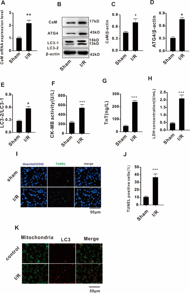
The pathogenesis of myocardial ischemia-reperfusion injury (MIRI) is not fully clear. This study aims to investigate the role of mitochondrial-associated endoplasmic reticulum membrane (MAM)-related calcium overload in mitophagy. In vitro and in vivo models were established to simulate MIRI. Cellular injury, apoptosis and mitophagy were measured and gene expression was analysized. The expression levels of glucose-regulated protein 75 (GRP75), receptor for inositol 1,4,5-trisphosphate (IP3R3), voltage-dependent anion-selective channel 1 (VDAC1), and calmodulin (CaM) and the mitochondrial calcium content, mitophagy and apoptosis were significantly increased in MIRI or hypoxia/reoxygenation (H/R) cells when compared to controls, but the mitochondrial membrane potential and ATP significantly decreased. GRP75 knockdown significantly inhibited CaM expression, mitochondrial calcium overload and mitophagy of H9C2 cells, whereas had no significant effect on IP3R3 and VDAC1 expression. CaM knockdown had no significant effect on the expression of GRP75, IP3R3 and VDAC1, and on mitochondrial calcium concentration, ATP levels and mitochondrial membrane potential of H9C2 cells, but significantly inhibited mitophagy and apoptosis. Collectively, these data suggest that the IP3R3-GRP75-VDAC1/CaM axis plays an important role in mitochondrial autophagy injury during myocardial ischemia-reperfusion and that it is a potential target for MIRI treatment.

Keywords: CaM; IP3R3-GRP75-VDAC1 complex; Mitochondrial calcium overload; Mitophagy; Myocardial ischemia reperfusion injury (MIRI).


Conflict of interest statement

Declarations. Competing interests: The authors declare no competing interests. Ethics approval: This study was approved by the Ethics Committee of the Affiliated Changsha Hospital of Hunan Normal University.

Figures

Fig. 1
Fig. 1
MIRI induces upregulation of the IP3R3-GRP75-VDAC1 complex proteins and mitochondrial Ca2+ overload in cardiomyocytes. (A). IP3R3, GRP75 and VDAC1 protein expression; (B, C and D). The relative ratio; (E).Rhod-2 fluorescence; (F).Mitochondrial calcium content; (G) ATP level; (H). Mitochondrial membrane potential; (I) Fold-change. A post-hoc test was used following the one-way ANOVA.*P < 0.05 vs. sham; **P < 0.01 vs. sham. IP3R3 receptor for inositol 1,4,5-trisphosphate, GRP75 glucose-regulated protein 75,, VDAC1 voltage-dependent anion-selective channel 1, ANOVA analysis of variance.
Fig. 2
Fig. 2
MIRI induces mitophagy and cell injury in cardiomyocytes. (A). CaM mRNA expression levels; (B). CaM, ATG4, LC3-2, and LC3-1 protein expression; (C, D, and E). The corresponding quantification analysis of protein expression; (F). CK activity; (G) TnT levels; (H). LDH levels; (I). TUNEL/Hoechst double staining. (J). TUNEL positive cell counting. (K). Co-localization of mitochondria and LC3. A post-hoc test was used following the one-way ANOVA analysis. *P < 0.05 vs. sham;**P < 0.01 vs. sham;***P < 0.001 vs. sham. MIRI myocardial ischemia-reperfusion injury, CaM calmodulin, ATG4 autophagy-regulating protease 4, LC3 microtubule-associated protein 1 A/1B-light chain 3, CK creatine kinase, TnT troponin-T, LDH lactate dehydrogenase, TUNEL terminal deoxynucleotidyl transferase dUTP nick end labeling, ANOVA analysis of variance.
Fig. 3
Fig. 3
GRP75 knockdown attenuates H/R-induced mitochondrial calcium overload in H9C2 cells. (A) and (B). GRP75 mRNA expression levels; (C) IP3R3, GRP75, and VDAC1 protein expression; (D, E, and F). The corresponding quantification analysis of protein expression; (G). Rhod-2 fluoresence; (H).Mitochondrial calcium content; (I). ATP levels; (J). Mitochondrial membrane potential; (K). Fold-change. A post-hoc test was used following the one-way ANOVA analysis. **P < 0.01 vs. control; ***P < 0.01 vs. NC siRNA;##P < 0.01 vs. H/R; ###P < 0.001 vs. H/R. GRP75 glucose-regulated protein 75, H/R hypoxia/reoxygenation, IP3R3 receptor for inositol 1,4,5-trisphosphate, VDAC1 voltage-dependent anion-selective channel 1, ANOVA analysis of variance.
Fig. 4
Fig. 4
GRP75 knockdown attenuates H/R-induced mitophagy in H9C2 cells. (A). CaM, ATG4, LC3-2, and LC3-1 protein expression; (B). The corresponding quantification analysis of CaM protein expression; (C) ATG4 mRNA expression; (D). The corresponding quantification analysis of ATG4 protein expression; (E) ATG4 mRNA expression; (F). The corresponding quantification analysis of the ratio of LC3-2 to LC3-1; (G). LDH levels; (H). TUNEL/Hoechst double staining. (I). TUNEL positive cell counting. (J). Co-localization of mitochondria and LC3. Post-hoc test was used following the one-way ANOVA analysis. *P < 0.05 vs. control;**P < 0.01 vs. control; #P < 0.05 vs. H/R. GRP75 glucose-regulated protein 75, H/R hypoxia/reoxygenation, CaM calmodulin, ATG4 autophagy-regulating protease 4, LC3 microtubule-associated protein 1 A/1B-light chain 3, LDH lactate dehydrogenase, TUNEL terminal deoxynucleotidyl transferase dUTP nick end labeling, ANOVA analysis of variance.
Fig. 5
Fig. 5
CaM knockdown exhibits no apparent effect on H/R-induced mitochondrial calcium overload in H9C2 cells. (A) and (B). CaM mRNA expression levels; (C). CaM, IP3R3, GRP75, and VDAC1 protein expression levels; (D, E, F, and G). The corresponding quantification analysis of protein expression; (H). Rhod-2 fluoresence; (I). Mitochondrial calcium content; (J). ATP levels; (K): mitochondrial membrane potential; (L). fold-change. A post-hoc test was used following the one-way ANOVA analysis.*P < 0.05 vs. control; **P < 0.01 vs. control; ***P < 0.01 vs. NC siRNA;##P < 0.01 vs. H/R; ###P < 0.001 vs. H/R. CaM calmodulin,H/R hypoxia/reoxygenation, IP3R3 receptor for inositol 1,4,5-trisphosphate, GRP75 glucose-regulated protein 75, VDAC1 voltage-dependent anion-selective channel 1, ANOVA analysis of variance.
Fig. 6
Fig. 6
Effects of CaM knockdown on H/R-induced mitophagy in H9C2 cells. (A). ATG4, LC3-2, and LC3-1 protein expression; (B and C). The corresponding quantification analysis of protein expression; (D). LDH levels; (E). TUNEL/Hoechst double staining; (F). TUNEL positive cell counting. (G). Co-localization of mitochondria and LC3. A post-hoc test was used following the one-way ANOVA test.*P < 0.05 vs. control; **P < 0.01 vs. control; #P < 0.05 vs. H/R. ##P < 0.01 vs. H/R. CaM, calmodulin,H/R, hypoxia/reoxygenation, ATG4, autophagy-regulating protease 4,LC3, microtubule-associated protein 1 A/1B-light chain 3, LDH, lactate dehydrogenase, TUNEL, terminal deoxynucleotidyl transferase dUTP nick end labeling, ANOVA, analysis of variance.
Fig. 7
Fig. 7
Schematic diagram of MIRI. MIRI led to an upregulated expression of the IP3R3-GRP75-VDAC1 complex proteins in cardiomyocytes, which resulted in mitochondrial calcium overload. In addition, MIRI led to an upregulated in the expression levels of CaM. The Ca2+ bind to CaM forming the Ca2+-CaM complex, which induces mitophagy-related gene (LC3 and ATG4) expression. This in turn leads to induction of mitophagy. Excessive mitophagy results in cell damage. MIRI myocardial ischemia-reperfusion injury, IP3R3 receptor for inositol 1,4,5-trisphosphate, GRP75 glucose-regulated protein 75, VDAC1 voltage-dependent anion-selective channel 1, CaM calmodulin; Ca2+, calcium ions, LC3 microtubule-associated protein 1 A/1B-light chain 3, ATG4 autophagy-regulating protease 4.

Similar articles

References

    1. Thiara, B. Cardiovascular disease. Nurs. Stand.29 (33), 60. 10.7748/ns.29.33.60 (2015). s44. - PubMed
    1. Lu, L., Liu, M., Sun, R., Zheng, Y. & Zhang, P. Myocardial infarction: symptoms and treatments. Cell. BiochemBiophys. 72 (3), 865–867. 10.1007/s12013-015-0553-4 (2015). - PubMed
    1. Neri, M., Riezzo, I., Pascale, N., Pomara, C. & Turillazzi, E. Ischemia/Reperfusion injury following acute myocardial infarction: A critical issue for clinicians and forensic pathologists. Mediators Inflamm.2017, 7018393. 10.1155/2017/7018393 (2017). - PMC - PubMed
    1. Frank, A. et al. Myocardial ischemia reperfusion injury: from basic science to clinical bedside. SeminCardiothoracVascAnesth(2012)16(3):123–132 .10.1177/1089253211436350 - PMC - PubMed
    1. Bugger, H. & Pfeil, K. Mitochondrial ROS in myocardial ischemia reperfusion and remodeling. BiochimBiophys Acta Mol. Basis Dis.1866 (7), 165768. 10.1016/j.bbadis.2020.165768 (2020). - PubMed

MeSH terms

LinkOut - more resources