Comparative Study
. 2025 Jul 1;15(1):21285.
doi: 10.1038/s41598-025-09329-9.

Superior fulcrum reconstruction improve tendon-to-bone healing in irreparable massive rotator cuff tears compared with superior capsule reconstruction

Affiliations
Comparative Study

Superior fulcrum reconstruction improve tendon-to-bone healing in irreparable massive rotator cuff tears compared with superior capsule reconstruction

Cheng Peng et al. Sci Rep. .

Abstract

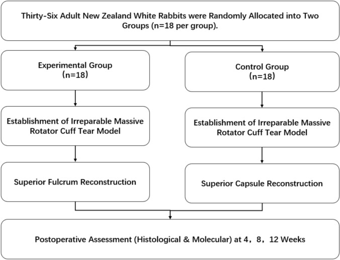
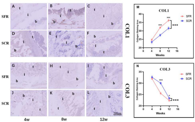
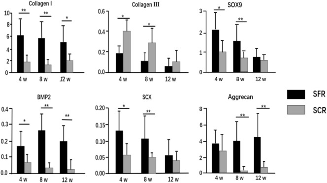
Recently, superior capsule reconstruction (SCR) has achieved some results in the treatment of irreparable massive rotator cuff tears (IMRCT), but the incidence of various postoperative complications is high. The study aims to establish a model of IMRCT in rabbits, and compare the histomorphology and molecular biology differences between superior fulcrum reconstruction (SFR) and SCR, so as to provide a new and effective treatment method for the clinical treatment of IMRCT. Thirty-six mature New Zealand white rabbits were required for the experiment to build the model of IMRCT. The supraspinatus and subscapular muscle of the thirty-six rabbits were cut off and randomly divided into two groups of eighteen rabbits in each group, with SFR and SCR. Six rabbits in each group were sacrificed at 4, 8, and 12 weeks after surgery for histological and molecular assessment. Macroscopically, no retear occurred in SFR group and SCR group after surgery. The results of molecular biological showed that the expression levels of COL1, BMP2, SCX and SOX9 in the SFR group were significantly higher than those in the SCR group at 4 and 8 weeks (P < 0.05), while the expression of COL3 was lower than that in the SCR group. There was no significant difference in the expression of Aggrecan between the two groups at 4 weeks (P > 0.05). At 12 weeks after surgery, the expression levels of COL1, BMP2 and Aggrecan in the SFR group were significantly higher than those in the SCR group (P < 0.05), while COL3, SCX and SOX9 were not significantly difference between the two groups (P > 0.05). Histologically, collagen fiber maturity and fibrocartilage regeneration in the SFR group were superior to those in the SCR group at 8 and 12 weeks (P < 0.05). However, at 4 weeks, there was no significant difference between the two groups (P > 0.05). In an IMRCT rabbit model, healing processes of SFR and SCR are different, but both repair techniques were effective. SFR outperformed SCR in collagen fiber maturity, fibrocartilage regeneration, and tendon regeneration.

Keywords: Histological; Irreparable massive rotator cuff tears; Molecularly; Rabbit model; Superior capsule reconstruction; Superior fulcrum reconstruction.


Conflict of interest statement

Declarations. Competing interests: The authors declare no competing interests. Ethical approval: All the methods and experiments of this study were carried out in accordance with the relevant guidelines and regulations of Anhui Medical University. Moreover, the present animal study was performed in accordance with the guidelines and approval of the Institutional Animal Care and Use Committees (IACUCs) of Anhui Medical University. This study was conducted in accordance with the ethical standards set forth in the Declaration of Helsinki and its later amendments or comparable ethical standards. Moreover, our study is reported in accordance with ARRIVE guidelines. All procedures involving animal participants were approved by the Ethics Committee of The First People’s Hospital of Hefei (LLSC-YY2019-54).

Figures

Fig. 1
Fig. 1
Allocation of experimental animals in this study. Experimental group: Underwent SFR following IMRCT induction. Control group: Underwent SCR following IMRCT induction. SFR superior fulcrum reconstruction, SCR superior capsule reconstruction, IMRCT irreparable massive rotator cuff tears.
Fig. 2
Fig. 2
Surgical procedure. (A) The harvested tissue of autologous peroneus longus tendon was braided. (B) The bone tunnels were in the humeral head. (C)The bone tunnel was in the superior glenoid. (D) The braided autologous peroneus longus tendon passed the bone tunnels and was tied on the proximal lateral humerus. (E) The harvested tissue of autologous fascia lata. (F) The bone tunnel was in the humeral head. (G) The bone tunnel was in the superior glenoid. (H) The fixed fascia lata.
Fig. 3
Fig. 3
Macroscopic view of the harvested specimens. (A) Gross specimen from the SFR group at 4 weeks. (B) Gross specimen from the SFR group at 8 weeks. (C) Gross specimen from the SFR group at 12 weeks. (D) Gross specimen from the SCR group at 4 weeks. (E) Gross specimen from the SCR group at 8 weeks. (F) Gross specimen from the SCR group at 12 weeks. SFR, superior fulcrum reconstruction; SCR, superior capsule reconstruction.
Fig. 4
Fig. 4
Comparison of healing interface maturity between SFR group and SCR group using hematoxylin and eosin–staining. (A) Tissue section of SFR group at 4 weeks. (B) Tissue section of SFR group at 8 weeks. (C) Tissue section of SFR group at 12 weeks. (D) Tissue section of SCR group at 4 weeks. (E) Tissue section of SCR group at 8 weeks. (F) Tissue section of SCR group at 12 weeks. (G) The graph shows the difference of the tendon maturing scores between groups. b, bone; i, interface; t, tendon; SFR, superior fulcrum reconstruction; SCR, superior capsule reconstruction. (100 × magnification, N = 6, *P < 0.05; **P < 0.01; ***P < 0.001).
Fig. 5
Fig. 5
Evaluation of fibrocartilage repair via Safranin O/Fast Green at the tendon-bone junction. (A) Tissue section of SFR group at 4 weeks. (B) Tissue section of SFR group at 8 weeks. (C) Tissue section of SFR group at 12 weeks. (D) Tissue section of SCR group at 4 weeks. (E) Tissue section of SCR group at 8 weeks. (F) Tissue section of SCR group at 12 weeks. (G) The graph shows the difference of the cartilage staining area between groups. b bone, i interface, t tendon, SFR superior fulcrum reconstruction, SCR superior capsule reconstruction. (100 × magnification, N = 6, *P < 0.05; **P < 0.01; ***P < 0.001).
Fig. 6
Fig. 6
Comparison of collagen expression between SFR group and SCR group using immunohistochemical staining, including COL1 (AF) and COL3 (GL). (AC) Tissue sections of COL1 from the SFR group at 4, 8, and 12 weeks postoperatively. (DF) Tissue sections of COL1 from the SCR group at 4, 8, and 12 weeks postoperatively. (GI) Tissue sections of COL3 from the SFR group at 4, 8, and 12 weeks postoperatively. (JL) Tissue sections of COL3 from the SCR group at 4, 8, and 12 weeks postoperatively. (M) The graph shows the difference of the collagen staining area of COL1 between groups. (N) The graph shows the difference of the collagen staining area of COL3 between groups. b bone, i interface, t tendon, SFR superior fulcrum reconstruction, SCR superior capsule reconstruction. (100 × magnification, N = 6, *P < 0.05; **P < 0.01; ***P < 0.001).
Fig. 7
Fig. 7
Gene expression analysis of COL1, COL3, BMP2, SCX, SOX9, and Aggracan at the tendon-bone junction in the SFR and SCR groups. Gene expression is reported relative to β-Actin reference. SFR superior fulcrum reconstruction, SCR superior capsule reconstruction. (*P < 0.05; **P < 0.01; ***P < 0.001).
Fig. 8
Fig. 8
Protein expression analysis of COL1, COL3, BMP2, SCX, SOX9, and Aggrecan at the tendon-bone junction in the SFR and SCR groups. (A) The expression of various proteins 4 weeks after operation. (B) The expression of various proteins 8 weeks after operation. (C) The expression of various proteins 12 weeks after operation. All gels and blots are cropped, and the original gels and blots are presented in Supplementary Figure S1. SFR superior fulcrum reconstruction, SCR superior capsule reconstruction. (*P < 0.05; **P < 0.01; ***P < 0.001).

Similar articles

References

    1. Peng, Y. et al. Effect of mechanical stimulation combined with platelet-rich plasma on healing of the rotator cuff in a murine model. Am. J. Sports.50(5), 1358–1368 (2022). - PubMed
    1. Morse, K. et al. Arthroscopic versus mini-open rotator cuff repair: A comprehensive review and meta-analysis. Am. J. Sports Med.36(9), 1824–1828 (2008). - PubMed
    1. Lee, S., Park, I., Kim, M. S. & Shin, S. J. Clinical differences between patients with early and late revision surgery for symptomatic failed arthroscopic rotator cuff repair. Knee Surg. Sports Traumatol. Arthrosc.29(12), 3921–3928 (2021). - PubMed
    1. Lebaschi, A. H. et al. Biomechanical, histologic, and molecular evaluation of tendon healing in a new murine model of rotator cuff repair. Arthroscopy34(4), 1173–1183 (2018). - PMC - PubMed
    1. Kim, C., Lee, Y. J., Kim, S. J., Yoon, T. H. & Chun, Y. M. Bone marrow stimulation in arthroscopic repair for large to massive rotator cuff tears with incomplete footprint coverage. Am. J. Sports Med.48(13), 3322–3327 (2020). - PubMed

Publication types