Clinical Trial
. 2025 Jul 1;16(1):5511.
doi: 10.1038/s41467-025-60649-w.

Identification of the Notch ligand DLK1 as an immunotherapeutic target and regulator of tumor cell plasticity and chemoresistance in adrenocortical carcinoma

Affiliations
Clinical Trial

Identification of the Notch ligand DLK1 as an immunotherapeutic target and regulator of tumor cell plasticity and chemoresistance in adrenocortical carcinoma

Nai-Yun Sun et al. Nat Commun. .

Abstract

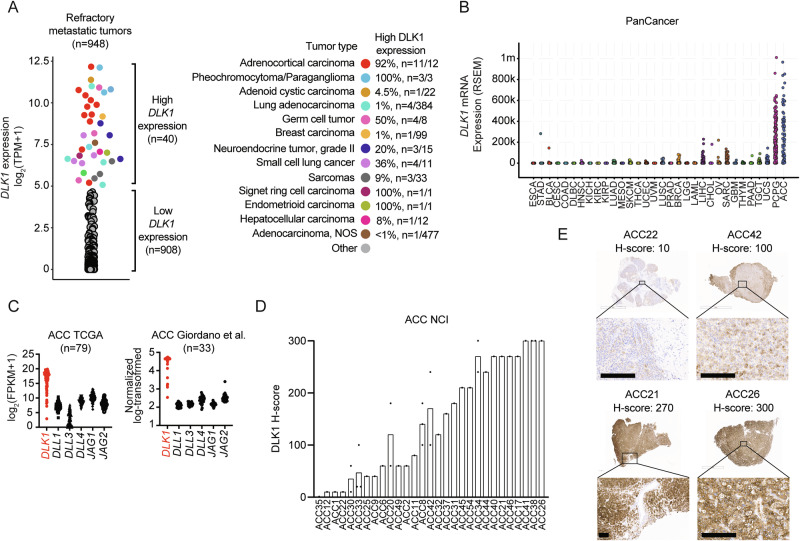
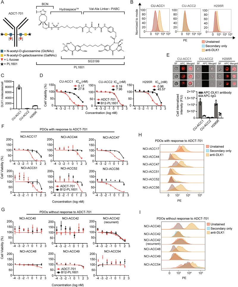
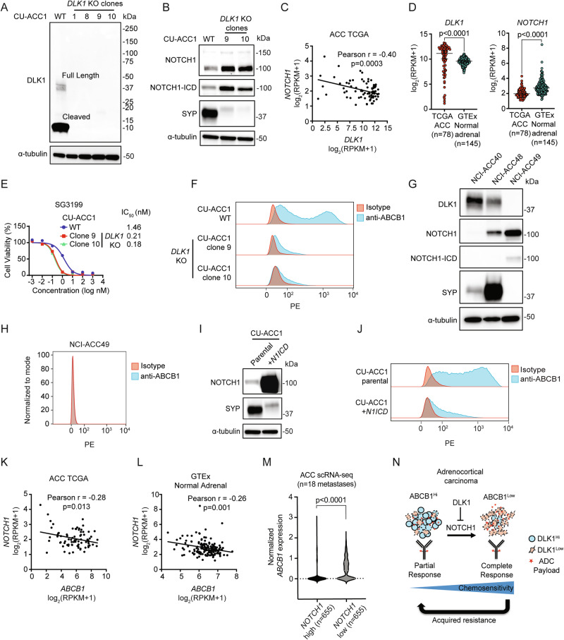
While immunotherapeutic targeting of cell surface proteins is an increasingly effective cancer therapy, identification of new surface proteins, particularly those with biological importance, is critical. Here, we uncover delta-like non-canonical Notch ligand 1 (DLK1) as a cell surface protein with limited normal tissue expression and high expression in multiple refractory adult metastatic cancers including small cell lung cancer (SCLC) and adrenocortical carcinoma (ACC), a rare cancer with few effective therapies. In ACC, ADCT-701, a DLK1 targeting antibody-drug conjugate (ADC), shows in vitro and in vivo activity but is overall limited due to high expression and activity of the drug efflux protein ABCB1 (MDR1, P-glycoprotein). In contrast, ADCT-701 induces complete responses in DLK1+ ACC and SCLC in vivo models with low or no ABCB1 expression. Genetic deletion of DLK1 in ACC dramatically downregulates ABCB1 and increases ADC payload and chemotherapy sensitivity through NOTCH1-mediated transdifferentiation. This work identifies DLK1 as an immunotherapeutic target that regulates tumor cell plasticity and chemoresistance in ACC and supports an active phase I clinical trial targeting DLK1 with an ADC in ACC and neuroendocrine neoplasms (NCT06041516).


Conflict of interest statement

Competing interests: Nitin Roper and Jaydira Del Rivero have received research funding from ADC Therapeutics for this study. The other authors have no competing interests to report.

Figures

Fig. 1
Fig. 1. Identification of DLK1 as the most highly expressed Notch ligand in adrenocortical carcinoma.
DLK1 mRNA expression across adult refractory metastatic cancers (n = 948). Tumor types with high DLK1 expression are highlighted in the colors shown. The percentage of each tumor type with high DLK1 expression is shown on the right. DLK1 mRNA expression in the TCGA PanCancer dataset. C Expression of Notch ligands from two independent ACC bulk RNA-seq datasets. D Quantification of DLK1 IHC staining among ACC NCI tumors (n = 38). Vertical bars with more than one tumor represent the mean H-score. E IHC images from four representative ACC NCI tumors with varying levels of DLK1 expression. Scale bars represent 200 μm. IHC immunohistochemistry. Source data are provided as a Source Data file.
Fig. 2
Fig. 2. ADCT-701, a DLK1 targeting antibody-drug conjugate, has potent in vitro activity in adrenocortical carcinoma.
A Schematic structure of ADCT-701, a DLK1 targeting antibody drug conjugate. B Representative surface expression of DLK1 among ACC cell lines: CU-ACC1, CU-ACC2, and H295R. C DLK1 molecules/cell in ACC cell lines (n = 3 independent experiments). D ADCT-701 cytotoxicity among CU-ACC1, CU-ACC2, and H295R cells. Cells were treated with ADCT-701 and B12-PL1601 (non-targeted control ADC) for 7 days (data representative of n = 3 independent experiments). E Representative imaging flow cytometry images and signal intensity analysis (n = 3 biological replicates) showing cellular internalization of DLK1 antibodies in CU-ACC1, CU-ACC2, and H295R cells. F Cytotoxic activity of ADCT-701 responsive (n = 6) and (G) ADCT-701 non-responsive (n = 6) ACC short-term patient-derived organoids (PDOs). Cells were treated with ADCT-701 and B12-PL1601 for 7 days. Data representative of n = 1 (ACC17, ACC44, ACC56, and ACC49) or n = 2 (ACC47, ACC51, ACC52, ACC40, ACC42, ACC48, and ACC54) independent experiments. Flow cytometry histograms assessing DLK1 among ADCT-701 H responsive and I non-responsive ACC PDOs. Error bars represent mean values ± S.E.M. Drug response curve data are presented as mean values ± S.E.M. Source data are provided as a Source Data file.
Fig. 3
Fig. 3. ADCT-701 induces robust anti-tumor responses in DLK1+ ACC tumors.
A CU-ACC1 and H295R xenograft tumor growth curves after treatment with saline (CU-ACC1: n = 3; H295R: n = 4), B12-PL1601 (CU-ACC1: n = 3; H295R: n = 4), or ADCT-701 (CU-ACC1: n = 3; H295R: n = 3) (1 mg/kg) (initial tumor size reached an average of 100–150 mm3). Additional doses of ADCT-701 indicated by arrows. Error bars represent mean values ± S.E.M. B ACC PDXs 164165, 592788, and POBNCI_ACC004 tumor growth curves after treatment with saline (164165: n = 8; 592788: n = 7; POBNCI_ACC004: n = 5), B12-PL1601 (164165: n = 7; 592788: n = 7; POBNCI_ACC004: n = 5) or ADCT-701 (164165: n = 8; 592788: n = 6; POBNCI_ACC004: n = 5) (1 mg/kg) (initial tumor size reached an average of 100–200 mm3). Each line shown represents an individual mouse within a given experiment. The X symbol indicates the administration of ADCT-701 re-dosing. Arrow indicates unexpected death of 1 POBNCI_ACC004 tumor-bearing mouse prior to endpoint. DLK1 immunohistochemistry with H-scores shown above each individual xenograft or PDX tumor. PR partial response, CR complete response. Scale bars represent 200 μm. Source data are provided as a Source Data file.
Fig. 4
Fig. 4. ABCB1, a drug efflux protein, mediates intrinsic and acquired resistance to ADCT-701.
A Cytotoxicity of SG3199 among ADCT-701 responsive (NCI-ACC44, NCI-ACC51, and NCI-ACC56) and non-responsive (NCI-ACC40, NCI-ACC48, and NCI-ACC54) DLK1+ ACC short-term patient-derived organoids (PDOs). Cells were treated with SG3199 for 3 days (n = 3 independent experiments for ACC44, ACC51, ACC56, and ACC48 PDOs; n = 5 independent experiments for ACC40 and ACC54 PDOs). B Drug transporter mRNA expression in DLK1+ ADCT-701 responder and non-responder PDOs as measured by quantitative RT-PCR. Results were presented as Δcycle threshold (ΔCt). Each color represents an individual PDO. R responder, NR non-responder (n = 3 independent experiments). Unpaired t tests were used to calculate two-tailed p-values. C Flow cytometry histograms assessing ABCB1 and the number of ABCB1 molecules per cell of DLK1+ ADCT-701 responsive (NCI-ACC44, NCI-ACC51, and NCI-ACC56) and non-responsive (NCI-ACC40, NCI-ACC48, and NCI-ACC54) ACC PDOs. D ADCT-701 cytotoxicity in the NCI-ACC40 and NCI-ACC48 PDOs with and without treatment with ABCB1 inhibitors (1 μM valspodar, 10 μM elacridar and 1 μM tariquidar). Cells were treated with ADCT-701 combined with or without ABCB1 inhibitors for 7 days (data representative of n = 3 independent experiments). E Flow cytometry histograms assessing ABCB1 among 164165, 592788 and POBNCI_ACC004 PDXs (data representative of n = 2 independent experiments). F ADCT-701 cytotoxicity in 164165, 592788 and POBNCI_ACC004 PDX-derived organoids. Cells were treated with ADCT-701 for 7 days (data representative of n = 3 independent experiments). G ADCT-701 cytotoxicity in the 164165 and 592788 PDX-derived organoids treated with or without ABCB1 inhibitors (1 μM valspodar, 10 μM elacridar and 1 μM tariquidar). Cells were treated with ADCT-701 combined with or without ABCB1 inhibitors for 7 days (data representative of n = 3 independent experiments). H Volcano plot of differentially expressed genes in control tumors (n = 4) versus post-ADCT-701 acquired resistant tumors (n = 3) in POBNCI_ACC004 PDX. Wald test negative log10 p-values are shown on the y-axis. I Flow cytometry histograms assessing ABCB1 among ADCT-701 resistant and control POBNCI_ACC004 PDX tumors. J ADCT-701 cytotoxicity in the ADCT-701 resistant POBNCI_ACC004 PDX-derived organoid treated with or without ABCB1 inhibitors (1 μM valspodar, 10 μM elacridar and 1 μM tariquidar). Cells were treated with ADCT-701 combined with or without ABCB1 inhibitors for 7 days (data representative of n = 3 independent experiments). Error bars represent mean values ± S.E.M. Source data are provided as a Source Data file.
Fig. 5
Fig. 5. ADCT-701 elicits complete, durable responses in DLK1+ small cell lung cancer tumors without ABCB1 expression.
A Cell surface DLK1 expression by flow cytometry in 3 small cell lung cancer (SCLC) cell lines (H524, H146, and H1436) (data representative of n = 3 independent experiments). B DLK1 molecules/cell relative to DLL3 among SCLC cell lines (n = 3 independent experiments). Error bars represent mean values ± S.E.M. C Flow cytometry histograms assessing ABCB1 in SCLC cell lines (data representative of n = 3 independent experiments). D SG3199 cytotoxicity in SCLC cell lines. Cells were treated with SG3199 for 3 days (data representative of n = 4 independent experiments). E ADCT-701 cytotoxicity among SCLC cell lines. Cells were treated with ADCT-701 or B12-PL1601 for 7 days (data representative of n = 4 independent experiments). F Tumor growth curves of SCLC xenograft models after treatment (1 mg/kg) with saline (H524: n = 5; H146: n = 5; H1436: n = 6), B12-PL1601 (H524: n = 5; H146: n = 4; H1436: n = 7) or ADCT-701 (H524: n = 4; H146: n = 6; H1436: n = 7) when tumor size reached an average of 100–150 mm3. Arrows indicate re-treatment with ADCT-701. Scale bars represent 200 μm. Error bars represent mean values ± S.E.M. Source data are provided as a Source Data file.
Fig. 6
Fig. 6. DLK1 is a major regulator of ABCB1 and chemoresistance in adrenocortical carcinoma.
A Immunoblot analysis of DLK1 and loading control (α-tubulin) proteins in CU-ACC1 cells with and without DLK1 KO. Four single-cell KO clones are shown. B Immunoblot analysis of NOTCH1 signaling, total NOTCH1 and NOTCH1 intracellular domain (ICD), NE marker synaptophysin (SYP), and loading control (α-tubulin) proteins with and without DLK1 KO in CU-ACC1 cells. Two single-cell KO clones are shown. C Correlation between NOTCH1 and DLK1 expression among TCGA ACC tumors. Pearson correlation coefficients with two-tailed p-values are shown. D DLK1 and NOTCH1 expression in TCGA ACC tumors and normal adrenals. Unpaired t tests were used to calculate two-tailed p-values. Error bar represents mean values ± 95% C.I. E SG3199 cytotoxicity in CU-ACC1 parental and DLK1 KO clones. Cells were treated with SG3199 for 3 days (data representative of n = 4 independent experiments). Error bars represent mean values ± S.E.M. F Flow cytometry histograms assessing ABCB1 in CU-ACC1 cells with and without DLK1 KO. G Immunoblot analysis of DLK1, total NOTCH1 and NOTCH1-ICD, SYP, and α-tubulin proteins in DLK1+ NCI-ACC40, DLK1+ NCI-ACC48 and DLK1- ACC49 PDOs. H Flow cytometry histograms assessing ABCB1 in DLK1 negative NCI-ACC49 PDOs. I Immunoblot analysis of total NOTCH1 (to detect the NOTCH1-ICD plasmid expression), SYP, and α-tubulin proteins in CU-ACC1 cells with and without NOTCH1-ICD overexpression. J Flow cytometry histograms assessing ABCB1 in CU-ACC1 cells with and without N1ICD overexpression. K Correlation between NOTCH1 and ABCB1 expression among TCGA ACC tumors and (L) among GTEx normal adrenal tissues. Pearson correlation coefficients with two-tailed p-values are shown. M Single cell RNA-seq data of ABCB1 expression comparing high NOTCH1 to low NOTCH1 expressing cells from 18 ACC metastatic tumors. Unpaired t tests were used to calculate two-tailed p-values. N Model summarizing the findings of the current study. For immunoblots, experiments were performed two times with similar results. Source data are provided as a Source Data file.

Update of

References

    1. Dumontet, C., Reichert, J. M., Senter, P. D., Lambert, J. M. & Beck, A. Antibody-drug conjugates come of age in oncology. Nat. Rev. Drug Discov.22, 641–661 (2023). - PubMed
    1. Kunnimalaiyaan, M. & Chen, H. Tumor suppressor role of Notch-1 signaling in neuroendocrine tumors. Oncologist12, 535–542 (2007). - PubMed
    1. Zhou, B. et al. Notch signaling pathway: architecture, disease, and therapeutics. Signal Transduct. Target. Ther.7, 95 (2022). - PMC - PubMed
    1. Saunders, L. R. et al. A DLL3-targeted antibody-drug conjugate eradicates high-grade pulmonary neuroendocrine tumor-initiating cells in vivo. Sci. Transl. Med.7, 302ra136 (2015). - PMC - PubMed
    1. Puca, L. et al. Delta-like protein 3 expression and therapeutic targeting in neuroendocrine prostate cancer. Sci. Transl. Med.11, 10.1126/scitranslmed.aav0891 (2019). - PMC - PubMed

Publication types

MeSH terms

Associated data

LinkOut - more resources