. 2025 Jul 1;16(1):477.
doi: 10.1038/s41419-025-07795-7.

Activating PIK3CA mutations in adipose-derived stem cells drive mutant-like phenotypes of wild-type cells in macrodactyly

Affiliations

Activating PIK3CA mutations in adipose-derived stem cells drive mutant-like phenotypes of wild-type cells in macrodactyly

Xiao Zhang et al. Cell Death Dis. .

Abstract

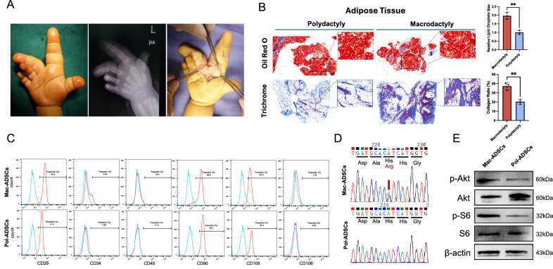
Macrodactyly is a congenital overgrowth disorder characterized by pathological adipose proliferation due to PIK3CA mutations in adipose-derived stem cells (ADSCs). Due to the somatic mosaicism, the affected tissues comprise a mixture of mutant and wild-type cells. However, how PIK3CA mutated ADSCs influence adjacent wild-type cells in macrodactyly remains poorly understood. In this study, we utilized coculture systems to investigate the effects of macrodactylous adipose-derived stem cells (Mac-ADSCs) on normal ADSCs, fibroblasts (FBs), and vascular endothelial cells (VECs). Our study demonstrated that activating PIK3CA mutations in Mac-ADSCs promotes the proliferation, migration, invasion, adipogenesis, and angiogenesis of wild-type ADSCs, FBs and VECs. Furthermore, using RNA sequencing and cytokine arrays, we revealed that these effects are primarily mediated by various secreted paracrine cytokines. These findings demonstrated that activating PIK3CA mutation alters the paracrine characteristics of Mac-ADSCs and reshapes the microenvironment of macrodactyly, driving adjacent wild-type cells to exhibit mutant-like phenotypes. Targeting PIK3CA with BYL-719 could influence the progression of macrodactyly by inhibiting the paracrine signaling of Mac-ADSCs.


Conflict of interest statement

Competing interests: The authors declare no competing interests. Ethics approval and consent to participate: All methods were performed in accordance with the relevant guidelines and regulations. Ethical approval for this study was granted by the Institutional Review Board of Shanghai Ninth People’s Hospital (Approval Number: SH9H-2022-T357-1). Informed consent was obtained from all individual participants included in the study.

Figures

Fig. 1
Fig. 1. Excessive adipose tissue accumulation and PIK3CA gain-of-function of ADSCs in macrodactyly.
A Gross appearance and radiograph showed diffuse tissue hyperplasia in macrodactyly. B Oil Red O and Masson’s trichrome staining revealing diffuse enlargement of adipocytes and localized collagen deposition of adipose tissue in macrodactyly compared with polydactyly. C Flow cytometric analysis of ADSC-specific markers (CD29, CD34, CD45, CD90, CD105, CD106) in Mac-ADSCs and Pol-ADSCs. D Sanger sequencing identifying PIK3CA mutations in Mac-ADSCs but not in Pol-ADSCs. E Western blot showing activation of the PI3K-AKT pathway in Mac-ADSCs. Data are presented as the mean ± SD from three independent experiments. *p < 0.05, **p < 0.01 by unpaired t test.
Fig. 2
Fig. 2. Mac-ADSCs enhance proliferation, migration, invasion and functional changes in wild-type cells.
A Schematic of the coculture system used for functional assays. The figure was created with Figdraw (https://www.figdraw.com/) and is permitted for publication. B Effect of Mac-ADSCs conditioned medium (CM) on proliferation of ADSCs, FBs and VECs assessed by CCK-8 assay. CE Migration and invasion capacities of (C) ADSCs, (D) FBs, and (E) VECs evaluated by Transwell assays. F mRNA levels of collagen markers in FBs when cocultured with different ADSCs. G mRNA levels of adipogenesis markers in ADSCs after 3 days of adipogenic induction. H Oil Red O staining of ADSCs after 12 days of adipogenic induction. I Representative images of angiogenic tube formation by VECs cocultured with different ADSCs. Scale bars: 100 μm. Data are presented as mean ± SD from three independent experiments. ns, not significant, *p < 0.05, **p < 0.01, ***p < 0.001, ****p < 0.0001 by ANOVA analysis.
Fig. 3
Fig. 3. Mac-ADSCs influence wild-type cells through paracrine signaling.
A Venn diagram showing overlap between the differentially expressed genes (DEGs) and the genes encoding secretory proteins in Mac-ADSCs versus Pol-ADSCs. B Heat map of DEGs encoding secretory proteins, with color-coded expression levels. C GO/KEGG pathway analysis of DEGs that encode secretory proteins. D Human cytokine antibody arrays detecting cytokines in the supernatant of Pol-ADSCs (left) and Mac-ADSCs (right). Boxes highlight cytokines with significant differential expression. E Heat map showing the intensities of the dots representing the differentially expressed cytokines (data presented as grayscale values in each group).
Fig. 4
Fig. 4. Knockdown of PIK3CA attenuates the paracrine effects of Mac-ADSCs.
(A) mRNA and (B) protein expression levels of PIK3CA in Mac-ADSCs after shRNA transfection. C Proliferation of ADSCs, FBs, and VECs when cocultured with shRNA-transfected Mac-ADSCs. DF Migration and invasion capacities of (D) ADSCs, (E) FBs, and (F) VECs when cocultured with shRNA-transfected Mac-ADSCs. G, H Adipogenic differentiation potential of wild-type ADSCs evaluated by (G) mRNA expression levels of adipogenic markers and (H) Oil Red O staining after coculturing with shRNA-transfected Mac-ADSCs. I Tube formation capacity of VECs when cocultured with shRNA-transfected Mac-ADSCs. JL Activation status of the PI3K-AKT signaling pathway in (J) ADSCs, (K) FBs, and (L) VECs when cocultured with shRNA-transfected Mac-ADSCs. Scale bars: 100 μm. Data are presented as the mean ± SD from three independent experiments. *p < 0.05, **p < 0.01, ***p < 0.001, ****p < 0.0001 by ANOVA analysis.
Fig. 5
Fig. 5. BYL-719 inhibits the paracrine effects of Mac-ADSCs.
A Dose-response curve used to estimate the IC50 of BYL-719 in Mac-ADSCs. B Effect of BYL-719-treated Mac-ADSCs on proliferation of wild-type ADSCs, FBs, VECs. CE Effect of BYL-719-treated Mac-ADSCs on migration and invasion capacities of (C) ADSCs, (D) FBs, and (E) VECs. F, G Adipogenic potential of wild-type ADSCs assessed by (F) mRNA levels of adipogenic markers and (G) Oil Red O staining after coculture with BYL719-treated Mac-ADSCs. H Tube formation capacity of VECs when cocultured with BYL-719-treated Mac-ADSCs. Scale bars: 100 μm. Data are presented as mean ± SD from three independent experiments. ns, not significant, *p < 0.05, **p < 0.01, ***p < 0.001, ****p < 0.0001 by unpaired t test.
Fig. 6
Fig. 6. Summary of the paracrine effects of Mac-ADSCs.
PIK3CA mutations activate the PI3K-AKT signaling pathway in ADSCs, transforming them into Mac-ADSCs and altering their cytokine secretion profile. The differentially secreted cytokines from Mac-ADSCs influence neighboring wild-type cells by activating their PI3K-AKT signaling pathway and enhancing their proliferation, migration, and invasion abilities. Additionally, these cytokines promote angiogenesis in VECs and adipogenesis in ADSCs. Notably, this paracrine effect can be reversed by the PIK3CA inhibitor BYL-719. This figure was created with Figdraw (https://www.figdraw.com/) and is permitted for publication.

Similar articles

References

    1. Wu J, Tian G, Ji Y, Higgins JP, Lee WPA. Clinical Characteristics of 90 Macrodactyly Cases. J Hand Surg. 2020;45:982.e1–982.e5. - PubMed
    1. Ezaki M. Insights into the pathogenesis of macrodactyly. J Hand Surg Eur Vol. 2019;44:25–31. - PubMed
    1. Lindhurst MJ, Parker VER, Payne F, Sapp JC, Rudge S, Harris J, et al. Mosaic overgrowth with fibroadipose hyperplasia is caused by somatic activating mutations in PIK3CA. Nat Genet. 2012;44:928–33. - PMC - PubMed
    1. Tian W, Huang Y, Sun L, Guo Y, Zhao S, Lin M, et al. Phenotypic and genetic spectrum of isolated macrodactyly: somatic mosaicism of PIK3CA and AKT1 oncogenic variants. Orphanet J Rare Dis. 2020;15:288. - PMC - PubMed
    1. Hanker AB, Kaklamani V, Arteaga CL Challenges for the clinical development of PI3K inhibitors: Strategies to improve their impact in solid tumors. 2020. - PMC - PubMed

MeSH terms

Substances

Supplementary concepts