. 2025 Jul 1;16(1):473.
doi: 10.1038/s41419-025-07643-8.

Mitochondrial transplantation: adaptive bio-enhancement

Affiliations

Mitochondrial transplantation: adaptive bio-enhancement

Xiaomeng Lu. Cell Death Dis. .

Abstract

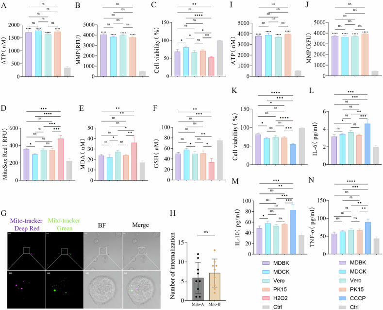
Mitochondria, often referred to the powerhouse of the cell, are essential for cellular energy production, and their dysfunction can profoundly affect various organs. Transplantation of healthy mitochondria can restore the bioenergetics in diseased cells and address multiple conditions, but more potentials of this approach remain unclear. In this study, I demonstrated that the source of transplanted mitochondria is not limited by species, as exhibit no significant responses to mitochondria derived from different germlines. Moreover, I identified that metabolic compatibility between the recipient and exogenous mitochondria as a crucial factor in mitochondrial transplantation, which confers unique metabolic properties to recipient cells, enabling them to combat different diseases. Additionally, my findings indicated competitive interactions among mitochondria with varying functions, with more bioenergetic-active mitochondria yielded superior therapeutic benefits. Notably, no upper limit for the bioenhancement provided by exogenous mitochondria has been identified. Based on these insights, I proposes a novel therapeutic approach-adaptive bioenhancement through mitochondrial transplantation.


Conflict of interest statement

Competing interests: The author declares no competing interests. Ethics approval and consent to participate: The experimental animal facilities have been licensed by the Guangdong Medical Experimental Animal Center (SCXK-2021-0041). All methods were performed in accordance with the relevant guidelines and regulations.

Figures

Fig. 1
Fig. 1. Universality of mitochondrial transplantation.
A Analysis of membrane potential in both cells and animals isolated mitochondria. Ctrl: Mitochondria were subjected to two freeze-thaw cycles at −80 °C and 37 °C to completely disrupt their structure and function, serving as a comparison, n = 3. B ATP production capacity in both cells and animals isolated mitochondria. Ctrl: Mitochondria were subjected to two freeze-thaw cycles at −80 °C and 37 °C, used for comparison, n = 3. C Mitochondria were randomly selected from cells (Vero) and animals (Bull Frog) for electron microscopy to observe their morphology. Scale bar: 500 nm. DG Activity of respiratory chain complexes I and III in both cells and animals isolated mitochondria, n = 3. H Co-localization fluorescence imaging at 24 h of transplanted mitochondria labeled with Mito-Tracker Green (green fluorescence) from Eel, MDCK, CRFK, Sparrow, Vero, and Lizard, alongside AC16, L929, and HepG2 cells labeled with WGA594 (red fluorescence). Blue fluorescence (DAPI) represents the nucleus. Scale bar: 10 μm. IN Immunoreactivity of IL-6, IL-10, and TNF-α in cell culture medium collected 24 h after the transplantation of exogenous mitochondria, n = 3. Ctrl: Untreated group. ns no statistical significance, ****p < 0.0001.
Fig. 2
Fig. 2. Mitochondria with matching metabolic characteristics provide better therapeutic outcomes.
A, I ATP production capacity in isolated mitochondria. Ctrl: Mitochondria subjected to two freeze-thaw cycles at −80 °C and 37 °C, used for comparison, n = 3. B, J Membrane potential in isolated mitochondria. Ctrl: Mitochondria subjected to two freeze-thaw cycles at −80 °C and 37 °C, used for comparison, n = 3. C Effects of mitochondrial transplantation from four different species on the viability of BMDM cells after H2O2 treatment. Ctrl: Untreated group, n = 3. D Mitochondrial ROS fluorescence intensity in BMDM cells 24 h after transplantation of mitochondria from four different species, followed by H2O2 treatment. Ctrl: Untreated group, n = 3. E, F Effects of mitochondrial transplantation from four different species on MDA and GSH levels in BMDM cells after H2O2 treatment. Ctrl: Untreated group, n = 3. G, H Fluorescence imaging and statistical analysis of the internalization of mitochondria A labeled with Mito-Tracker Red and mitochondria B labeled with Mito-Tracker Green in BMDM cells after H2O2 treatment. Original image scale bar: 10 µm; magnified region scale bar: 2 µm, n = 9. K Effects of mitochondrial transplantation from four different species on the viability of AC16 cells after CCCP treatment. Ctrl: Untreated group, n = 3. LN Changes in IL-6, IL-10, and TNF-α levels in AC16 cells after CCCP treatment, following mitochondrial transplantation from four different species. Ctrl: Untreated group, n = 3. ns no statistical significance, *p < 0.05, **p < 0.01, ***p < 0.001, ****p < 0.0001.
Fig. 3
Fig. 3. Generation of more potent pluripotent hybrid mitochondria through mitochondrial fusion.
A Co-localization fluorescence imaging 24 h after the fusion of HL1 cells labeled with WGA647 and H9C2 cells labeled with WGA594. Scale bar: 20 µm. B Flow cytometric sorting of hybrid cells 24 h after the fusion of HL1 cells labeled with WGA647 and H9C2 cells labeled with WGA488. C, D Western blot analysis of the mitochondrial fusion and fission proteins Mfn1, Opa1, and Drp1 in HL1, H9C2, and HL1 + H9C2 cells on the second day after cell fusion, n = 3. E ATP production capacity in isolated mitochondria, Ctrl: Mitochondria subjected to two freeze-thaw cycles at −80 °C and 37 °C, used for comparison, n = 3. F JC-1 membrane potential detection of isolated mitochondria. Ctrl: Mitochondria subjected to two freeze-thaw cycles at −80 °C and 37 °C, used for comparison, n = 3. G Effects of transplantation of three different types of mitochondria on the viability of AC16 cells after H2O2 treatment. Ctrl: Untreated group, n = 3. H Effects of transplantation of three different types of mitochondria on mitochondrial ROS levels in AC16 cells after H2O2 treatment. Ctrl: Untreated group, n = 3. I, J Effects of transplantation of mitochondria from three different species on MDA and GSH levels in AC16 cells after H2O2 treatment. Ctrl: Untreated group, n = 3. ns no statistical significance, *p < 0.05, **p < 0.01, ***p < 0.001, ****p < 0.0001.
Fig. 4
Fig. 4. Competitive internalization of mitochondria: the fittest functions best.
A, H ATP production capacity in isolated mitochondria. Ctrl: Mitochondria subjected to two freeze-thaw cycles at −80 °C and 37 °C, used for comparison, n = 3. B, I Membrane potential in isolated mitochondria. Ctrl: Mitochondria subjected to two freeze-thaw cycles at −80 °C and 37 °C, used for comparison, n = 3. C Effects of mitochondria transplantation from four different species on the viability of HepG2 cells after CCCP treatment. Ctrl: Untreated group, n = 3. D Effects of mitochondria transplantation from four different species on mitochondria ROS in HepG2 cells after CCCP treatment. Ctrl: Untreated group, n = 3. E Detection of DNA damage repair in HepG2 cells after CCCP treatment following mitochondria transplantation from four different species. Ctrl: Untreated group, n = 3. F, G Fluorescence imaging and statistical analysis of the internalization of Lizard mitochondria labeled with Mito-Tracker Deep-Red and Bull Frog mitochondria labeled with Mito-Tracker Green in HepG2 cells after CCCP treatment. Original image scale bar: 10 µm, magnified region scale bar: 2 µm, n = 6. J Effects of mitochondria transplantation from four different species on the viability of BMDM cells after LPS treatment. Ctrl: Untreated group, n = 3. KM Changes in IL-6, IL-10, and TNF-α levels in BMDM cells after LPS treatment following mitochondria transplantation from four different species. Ctrl: Untreated group, n = 3. N Detection of DNA damage repair in BMDM cells after LPS treatment following mitochondria transplantation from four different species, n = 3. O, P Fluorescence imaging and statistical analysis of the internalization of Lizard mitochondria labeled with Mito-Tracker Green, Sparrow mitochondria labeled with Mito-Tracker Deep-Red, and Salmon mitochondria labeled with Mito-Tracker Red in BMDM cells after LPS treatment. Original image scale bar: 10 µm, magnified region scale bar: 5 µm, n = 6. ns no statistical significance, *p < 0.05, **p < 0.01, ***p < 0.001, ****p < 0.0001.
Fig. 5
Fig. 5. Mitochondrial Transplantation Enhances Physical Function in Mice.
A, B Evaluation of isolated mitochondrial functions through ATP production and mitochondrial membrane potential (MMP) assays, with mitochondria subjected to two freeze-thaw cycles at −80 °C and 37 °C to completely disrupt their structure and function as a control, n = 3. C Biodistribution of exogenous mitochondria in the mouse liver, kidney, and heart detected by in vivo imaging 24 h after intravenous tail vein injection of mitochondria. The blank group consists of untreated LPS-Ctrl group mice. D, E Assessment of motor function in acute inflammation model mice and normal model mice 3 days after the transplantation of two different types of mitochondria, with untreated groups as control, n = 6. FH Changes in body weight of acute inflammation model mice and normal model mice before and 1 week after the transplantation of two different types of mitochondria, with untreated groups as control, n = 6. I Grip strength measurements in acute inflammation model mice and normal model mice 3 days after the transplantation of two different types of mitochondria, with untreated groups as control, n = 6. ns no statistical significance, *p < 0.05, **p < 0.01, ***p < 0.001, ****p < 0.0001.
Fig. 6
Fig. 6. Mitochondrial Transplantation Enhances Biological Potency in Mice and BMDM cells.
AC Changes in serum levels of IL-6, TNF-α, and IL-10 in acute inflammation model mice and normal model mice 1 week after transplantation of two different mitochondria, with untreated groups as control, n = 3. DF Evaluation of kidney and liver function via serum Cr, ALT, and AST levels in acute inflammation model mice and normal model mice 1 week after transplantation of two different mitochondria, with untreated groups as control, n = 3. G, H Measurement of serum MDA levels and SOD activity in acute inflammation model mice and normal model mice 1 week after transplantation of two different mitochondria, with untreated groups as control, n = 3. IK Analysis of JNK and P-JNK, p65, and P-p65 protein expression levels in liver and kidney tissues via Western blot 1 week after transplantation of two different mitochondria in acute inflammation model mice and normal model mice, with untreated groups as control, n = 3. L Effects of two different mitochondria on the viability of LPS-treated BMDM cells 24 h after transplantation, with untreated groups as control, n = 3. M Quantitative analysis of mitochondrial ROS levels in LPS-treated BMDM cells 24 h after transplantation of two different mitochondria, with untreated groups as control, n = 3. ns no statistical significance, *p < 0.05, **p < 0.01, ***p < 0.001, ****p < 0.0001.

Similar articles

References

    1. Emani SM, Piekarski BL, Harrild D, Del Nido PJ, McCully JD. Autologous mitochondrial transplantation for dysfunction after ischemia-reperfusion injury. J Thorac Cardiovasc Surg. 2017;154:286–9. - PubMed
    1. Gollihue JL, Rabchevsky AG. Prospects for therapeutic mitochondrial transplantation. Mitochondrion. 2017;35:70–9. - PMC - PubMed
    1. McCully JD, Cowan DB, Emani SM, Del Nido PJ. Mitochondrial transplantation: from animal models to clinical use in humans. Mitochondrion. 2017;34:127–34. - PubMed
    1. McCully JD, Cowan DB, Pacak CA, Toumpoulis IK, Dayalan H, Levitsky S. Injection of isolated mitochondria during early reperfusion for cardioprotection. Am J Physiol Heart Circ Physiol. 2009;296:H94–105. - PMC - PubMed
    1. Gorick C, Debski A. Mitochondrial transplantation for ischemic heart disease. Nat Nanotechnol. 2024;19:1247–1248. - PubMed

LinkOut - more resources