. 2025 Jul 1;16(1):5695.
doi: 10.1038/s41467-025-60632-5.

Orthosteric STING inhibition elucidates molecular correction of SAVI STING

Affiliations

Orthosteric STING inhibition elucidates molecular correction of SAVI STING

Tao Xie et al. Nat Commun. .

Abstract

While the progression of STING activators into the clinic has been successful, the discovery and clinical progression of STING inhibitors remain elusive. Questions persist about the molecular properties needed to distinguish between a STING activator and inhibitor, particularly within SAVI disease, a monogenic autoinflammatory disease that renders STING constitutively active, and how different conformations correlate to function. In this work, we use an orthosteric STING activator and inhibitor from the same chemical series to discover that STING M271 is a critical residue for molecular activation that can be leveraged as a unique molecular signature for pharmacological or genetically driven activation and inhibition. Furthermore, we demonstrate how the therapeutic requirements of a molecular corrector of SAVI STING differs from an orthosteric STING inhibitor, and why this is important for the SAVI disease population.


Conflict of interest statement

Competing interests: The authors declare no competing interests. Requests for materials should be addressed to Tao Xie.

Figures

Fig. 1
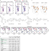
Fig. 1. STING bound diABZI-based agonist and inhibitor are nearly indistinguishable in crystal structures.
a diABZI-i inhibits cGAMP-induced IFNβ in PBMCs (n = 3 | 1 donor) and diABZI-a1 induces IFNβ in PBMCs (n = 3 | 3 donors, representative donor displayed). Data represented as mean with SD in graph. Source data are provided as a Source Data file. b Superposition of crystal structures (side view) of STING bound to diABZI-a1 (gray) and diABZI-i (purple). c diABZI-a1 and diABZI-i superposition over the STING binding pocket. d Cartoon depicting apical wing distance and beta sheet lid between STING monomers. e Superposition of crystal structures (top view) of STING bound to diABZI-a1 (gray) and diABZI-i (purple). f THIQi inhibits cGAMP-induced IFNβ in PBMCs (n = 3 | 1 donor). Data represented as mean with SD in graph. Source data are provided as a Source Data file. g Crystal structure overlay (top view) of STING bound to THIQi (orange) and diABZI-i (purple). h Apical wing distance measurements from structures in this study and reference structures collected from the PDB.
Fig. 2
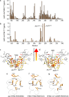
Fig. 2. NMR distinguishes between STING activation and inhibition via M271CH3 chemical shift in 1H-13C HSQC spectrum.
a Overlay of 1H-13C HSQC spectra of STING155-341 with and without diABZI-a1 and diABZI-i. b Overlay of 1H-13C HSQC spectra of THIQi-bound STING155-341 with diABZI-a1 and diABZI-i-complexed STING155-341. c Crystal structures focused on V155 and M271 residues of STING155-341 bound with diABZI-a1 and diABZI-i. d Chemical structures of diverse STING agonists used for this study. e Overlay of 1H-13C HSQC of STING155-341 recorded in the absence (red) and presence (blue) of various antagonists and agonists highlighting M271CH3. f Tabulated M271CH3 1H and 13C chemical shift differences between apo and bound STING.
Fig. 3
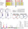
Fig. 3. Structural basis for large chemical shift changes of M271CH3 and A277CH3 upon agonist binding.
Backbone amide (a) and methyl (b) chemical shift differences between STING155-341-THIQi and STING155-341-ABZI. Chemical shift differences are mapped onto (c) STING155-341-THIQi and (d) STING155-341-ABZI structures. M271CH3 and A277CH3 exist underneath the binding pocket and are distant from the CDN binding pocket yet show significant chemical shifts differences, indicating different conformations between these two complexes. Hydrophobic interaction network in (e) apo-STING155-341, (f) THIQi-bound STING155-341, and (g) 2’, 3’-cGAMP bound STING155-341.
Fig. 4
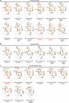
Fig. 4. The evolutionary conserved M271 residue mediates SAVI STING V155M-driven IFN induction.
a V155 and M271-focused phylogenetic sequence alignment. b, c Comparison of 1H-13C HSQC spectra among STING WT, SAVI STING V155M and SAVI STING G158A. 1H-13C HSQC of STING155-341-THIQi in the absence (d) and presence (e) of BMS-025 and of STING V155M155-341-THIQi in the absence (f) and presence (g) of BMS-025. I and A in d-g represent the inactive and active conformations, respectively. h M271-focused mutational analysis using HEK293 IFN-driven luciferase reporter cell line with variable amounts of plasmid transiently transfected (n = 3 biological replicates). Data represented as mean with SEM in graph. Source data are provided as a Source Data file. Statistical significance determined by two-way ANOVA against WT with adjusted p value depicted (*- p ≤ 0.05, **- p ≤ 0.01, ***- p ≤ 0.001, ****- p ≤ 0.0001). M271V (25 ng), p = 0.0357. M271V (50 ng), p = 0.0061. M271L (50 ng), p = 0.0007.
Fig. 5
Fig. 5. M271CH3 chemical shift correction is necessary but not sufficient for STING molecular correction of V155M SAVI.
a,  b Overlay of 1H-13C HSQC of STING155-341 (red) and STING155-341 V155M recorded in the absence (blue) and presence (green) of various antagonists and agonists. c Tabulated M271CH3 1H and 13C chemical shift differences between apo and bound STING V155M. d Comparison of basal IFN-driven luciferase levels between WT THP-1 and STING V155M THP-1 cell lines (n = 4 biological replicates). THP1-DualTM KI STING (Invivogen, cat# thpd-m155) harbor either the WT allele or V155M point mutation and have the endogenous STING alleles knocked out. Data represented as mean with SD in graph. Source data are provided as a Source Data file. e Agonism and antagonism behavior of diABZI-a1, THIQi, and diABZI-i on basal IFN levels in STING V155M THP-1 cell line (n = 3 biological replicates); representative replicate shown. Data represented as mean with SD in graph. Source data are provided as a Source Data file. f Tabulated IFN-driven luciferase EC50 and IC50 values in WT THP-1 (with and without cGAMP stimulation) and STING V155M THP-1 (no stimulation).
Fig. 6
Fig. 6. Conformational states of the hydrophobic dimer interface captured in different STING structures.
a State 1 (S1) adopts a similar conformation to that seen in THIQi-complexed STING155-341 (Fig. 3f) and STING LDB (PDB 4EMU, Fig. 3e). b State 2 (S2) is similar to that observed in 2’, 3’-cGAMP-bound STING155-341 structure (Fig. 3g). c State 3 (S3) remains a conformation between S1 and S2. Compounds highlighted with red color are inhibitors, and others are agonists.

Similar articles

References

    1. Decout, A., Katz, J. D., Venkatraman, S. & Ablasser, A. The cGAS-STING pathway as a therapeutic target in inflammatory diseases. Nat. Rev. Immunol.21, 548–569 (2021). - PMC - PubMed
    1. Li, S. et al. STING-induced regulatory B cells compromise NK function in cancer immunity. Nature610, 373–380 (2022). - PMC - PubMed
    1. Liu, Y. et al. Activated STING in a vascular and pulmonary syndrome. N. Engl. J. Med. 371, 507–518 (2014). - PMC - PubMed
    1. Fosbenner, D. et al. Modulators of stimulator of interferon genes (STING). Patent No. WO2019069270A1 (World Intellectual Property Organization, 2019).
    1. Ramanjulu, J. M. et al. Design of amidobenzimidazole STING receptor agonists with systemic activity. Nature564, 439–443 (2018). - PubMed

LinkOut - more resources