Clinical Trial
. 2025 Jul 2;16(1):6060.
doi: 10.1038/s41467-025-60737-x.

Intraoperative nerve-specific fluorescence visualization in head and neck surgery: a Phase 1 trial

Affiliations
Clinical Trial

Intraoperative nerve-specific fluorescence visualization in head and neck surgery: a Phase 1 trial

Yu-Jin Lee et al. Nat Commun. .

Abstract

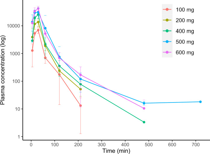
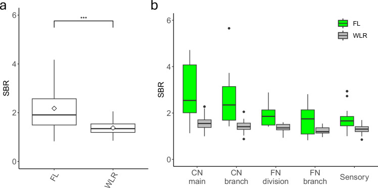
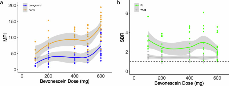
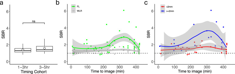
Iatrogenic nerve injury is a surgical complication with significant morbidity. This clinical trial, now complete, investigates the systemic administration of bevonescein, which selectively binds to nerves, in a single-arm, prospective multi-center, dose-escalation Phase 1 trial in adult patients with head and neck neoplasms undergoing parotidectomy or thyroidectomy in the United States. Twenty-seven participants are enrolled in the trial and receive the systemic agent. The primary outcome is safety with no dose-limiting toxicity among the 27 patients, but a single adverse event was identified that was possibly related to the study drug (vomiting). Secondary outcomes include the pharmacokinetics, optimal dose, and timing of bevonescein. The half-life of bevonescein is 29-72 min, and the optimal dose is 500 mg by objective measures, with the fluorescence signal-to-background ratio (SBR; 2.1 ± 0.8) significantly higher compared to white light (1.3 ± 0.2; p = 0.003). The fluorescent SBR of nerves between the early (1-3 h) versus late (3-5 h) timing cohorts is not statistically different. Here, we present data of a nerve imaging agent showing that preoperative intravenous infusion of bevonescein is well tolerated. This trial is registered at ClinicalTrials.gov (NCT04420689) and is sponsored by Alume Biosciences (San Diego, CA).


Conflict of interest statement

Competing interests: Q.T.N. is a co-founder and interim-CEO of Alume. B.J.B. is a co-founder and Chief Medical Officer of Alume. A patent for the technology described in this manuscript has been filed with the United States Patent and Trademark Office (USPTO) by The Regents of the University of California (Q.T.N. and B.J.B.). The other authors declare no competing interests.

Figures

Fig. 1
Fig. 1. Pharmacokinetics of bevonescein (presented in log scale).
This demonstrates the dose-dependent pharmacokinetics of bevonescein, consistent with other systematically administered proteins. Detection at levels below 10 ng/mL was not included in the dataset. Each color represents different dose cohorts and error bars represent standard deviation where n ≥ 3. A sample size of the subjects for each dose group was the following: 100 mg, n = 3 patients; 200 mg, n = 3 patients; 400 mg, n = 3 patients; 500 mg, n = 14 patients; 600 mg, n = 3 patients. The center is defined as the mean. Source data are provided as a Source Data file.
Fig. 2
Fig. 2. Comparison of SBR in WLR and FL images (all cohorts).
a The SBR of nerves in FL images was significantly higher (p < 0.001) compared to the paired WLR images using two-sided Wilcoxon rank sum test with the sample size n = 109 for each group. *** indicates p-value < 0.001. b The SBR of FL images were significantly higher compared to the paired WLR images across the nerve categories on a two-sided Wilcoxon rank sum test (p-value < 0.01 for all categories). Adjustments were not made for multiple comparisons. “CN main” includes the facial nerve main trunk, spinal accessory nerve, the hypoglossal nerve, and the vagus nerve (n = 28 unique nerves for each FL and WLR). “CN branch” includes recurrent laryngeal nerve and any branches off the main cranial nerve (n = 17 unique nerves for each FL and WLR). “FN division” includes the upper and lower divisions of the facial nerve (n = 13 unique nerves for each FL and WLR). “FN branch” includes branches from the divisions of the facial nerve, such as the buccal, marginal mandibular branches (n = 27 unique nerves for each FL and WLR). “Sensory” includes the greater auricular nerve (n = 22 unique nerves for each FL and WLR). Standard boxplots are shown with median as the bold center line, mean as a diamond, the first and third interquartile range (IQR), and the highest and lowest values within 1.5*IQR as hinges, and outliers as additional points. Green represents FL data, and gray represents WLR data. Source data is provided as a Source Data file. SBR signal to background ratio, FL fluorescence, WLR white light reflectance, CN cranial nerve, FN facial nerve.
Fig. 3
Fig. 3. The optimal dose of bevonescein is 500 mg.
A The MPI of nerves and background in FL images were evaluated, and the background was significantly higher at the 600 mg dose compared to the 500 mg dose. The blue line and dots represent the background MPI, and the orange line and dots represent the nerve MPI. The centers of the lines represent the mean value. The gray shaded error regions represent the standard error. B The SBR of FL images at the 600 mg dose decreased compared to the SBR at the 500 mg dose. The green line and dots represent the SBR for FL images and the dark gray line and dots represent the SBR for WLR images. The centers of the lines represent the mean value. The gray shaded error regions represent the standard error. Source data is provided as a Source Data file. MPI – mean pixel intensity; SBR – signal to background ratio; FL – fluorescence; WLR – white light reflectance.
Fig. 4
Fig. 4. Intraoperative nerve images at 500 mg bevonescein dose.
Paired intraoperative nerve images from 4 different patients at 500 mg bevonescein dose. WLR images (AD) and the same field of view seen with fluorescence signal overlayed on WLR (EH). Nerves on the surgical field surface (solid yellow arrows) appear yellowish/green compared to adjacent non-nerve tissue (reddish). There is white surgical gauze visible in the left lower quadrant of A. Nerve not on the surgical field surface (dashed yellow arrows) are more easily discernible in F, G, H compared to B, C, D. WLR – white light reflectance; Paired –white light reflectance with fluorescence overlay. The Visualization Scoring System utilized the WLR and Paired images. The SBR analysis utilized the WLR and FL (grayscale) images.
Fig. 5
Fig. 5. Effect of time from infusion to intraoperative imaging on SBR in patients who received 500 mg of bevonescein.
a There was no significant difference in SBR of the key nerves when comparing the two timing cohorts: bevonescein administered 1–3 h (n = 36 unique nerves) vs 3–5 h (n = 58 unique nerves) prior to incision. “ns” = not significant using two-sided Wilcoxon rank sum test. Standard boxplots are shown with median as the bold center line, mean as a diamond, the first and third interquartile range (IQR), and the highest and lowest values within 1.5*IQR as hinges, and outliers as additional points. b The peak SBR intensity was at 350 min from study drug administration to the time the image was captured. The green line and dots represent the SBR for FL images, and the dark gray line and values represent the SBR for WLR images. The centers of the lines represent the mean value. The gray shaded error regions represent standard error. c The effect of time from infusion of 500 mg bevonescein to intraoperative imaging on SBR on nerve width. The presented data included all patients who received the 500 mg dose (including those from the dose-escalation cohort and the dose-timing cohort). The red line and dots represent the SBR for FL images for nerves less than 2 mm, and the blue line and dots represent the SBR for FL images for nerves greater than or equal to 2 mm. The centers of the lines represent the mean value. The gray shaded error regions represent the standard error. Source data is provided as a Source Data file.

Similar articles

References

    1. Lee, Y. J. et al. Intraoperative fluorescence-guided surgery in head and neck squamous cell carcinoma. Laryngoscope131, 529–534 (2021). - PubMed
    1. Ottolino-Perry, K. et al. Intraoperative fluorescence imaging with aminolevulinic acid detects grossly occult breast cancer: a phase II randomized controlled trial. Breast Cancer Res. BCR23, 72 (2021). - PMC - PubMed
    1. Sibinga et al. Intraoperative molecular fluorescence imaging of pancreatic cancer by targeting vascular endothelial growth factor: a multicenter feasibility dose-escalation study. J. Nucl. Med. Publ. Soc. Nucl. Med.64, 82–89 (2023). - PMC - PubMed
    1. Stibbe, J. A. et al. First-in-patient study of OTL78 for intraoperative fluorescence imaging of prostate-specific membrane antigen-positive prostate cancer: a single-arm, phase 2a, feasibility trial. Lancet Oncol.24, 457–467 (2023). - PubMed
    1. de Valk, K. S. et al. Dose-finding study of a CEA-targeting agent, SGM-101, for intraoperative fluorescence imaging of colorectal cancer. Ann. Surg. Oncol.28, 1832–1844 (2021). - PMC - PubMed

Publication types

Associated data