. 2025 Jul 1;16(1):5673.
doi: 10.1038/s41467-025-60876-1.

Adhesin Als4112 promotes Candida auris skin colonization through interactions with keratinocytes and extracellular matrix proteins

Affiliations

Adhesin Als4112 promotes Candida auris skin colonization through interactions with keratinocytes and extracellular matrix proteins

Guolei Zhao et al. Nat Commun. .

Abstract

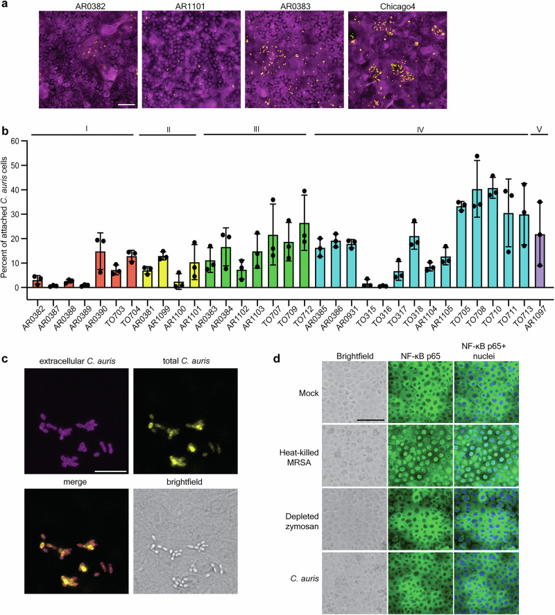
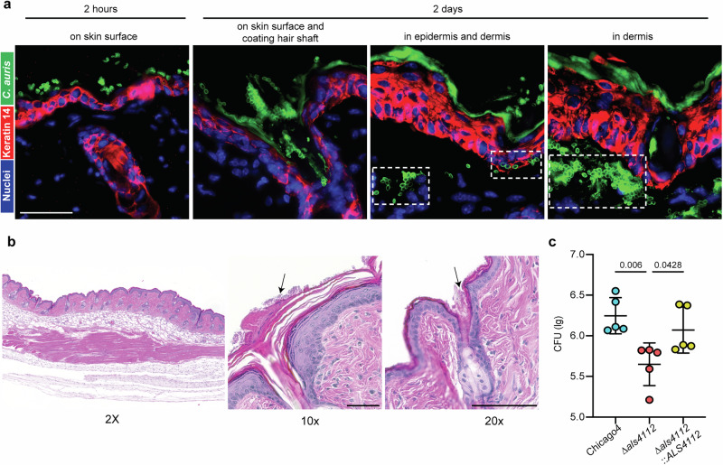
Candida auris is a fungal pathogen notorious for persistent skin colonization and transmission in healthcare settings. Here, we show that a C. auris conserved adhesin, Als4112, is required for skin colonization via keratinocyte attachment and direct interactions with host extracellular matrix proteins, especially basement membrane proteins such as laminin. Deletion of ALS4112 reduces skin colonization in mouse models of epicutaneous and systemic infection. In addition, coating plastic and catheter surfaces with collagen I or III inhibits C. auris attachment and biofilm formation. Our study highlights the critical role of Als4112 in C. auris colonization and virulence, and explores potential strategies to reduce the pathogen's adherence to abiotic surfaces and thus its spread in healthcare settings.


Conflict of interest statement

Competing interests: The authors declare no competing interests.

Figures

Fig. 1
Fig. 1. C. auris adheres to human keratinocyte cells.
a Representative immunofluorescence images showing adherence of AR0382 (Clade I), AR1101 (Clade II), AR0383 (Clade III), and Chicago4 (Clade IV) cells to human keratinocytes. Magenta = keratinocytes, yellow = C. auris. Scale bar, 100 μm. b Adherence of 33 C. auris clinical isolates from five clades to keratinocytes, measured by the proportion of cells remaining attached after washing. Values represent the mean ± s. d., calculated from three biological replicates. c C. auris cells are not internalized. Chicago4 cells adhered to the surface of keratinocytes were detected using an FITC-conjugated anti-Candida antibody. Total C. auris cells were detected with calcofluor white stain following cell membrane permeabilization. Images are presented in pseudo-color. Scale bar, 50 μm. Data are representative of three independent experiments yielding similar results. d C. auris does not induce inflammatory signaling. Keratinocytes were unstimulated or stimulated with heat-killed S. aureus strain USA300, depleted zymosan, or C. auris Chicago4 cells for six hours, and nuclear translocation of the NF-κB subunit p65 was detected by fluorescence microscopy. The nuclei were stained with Hoechst (blue). The middle panel shows the NF-κB staining (green), and the right panel shows the overlay. Images are presented in pseudo-color. Scale bar, 100 μm. Data are representative of three independent experiments yielding similar results. Statistical differences were assessed using one-way ANOVA (b). Source data are provided as a Source Data file.
Fig. 2
Fig. 2. Als4112 is the major adhesin mediating keratinocyte adherence.
a The 1595 insertional mutants in the Chicago4 strain background were screened for adhesion defects by measuring the proportion of cells remaining attached to keratinocytes after three PBS washes. Each dot represents one mutant. Mutants with a Z-score below −1.8 were considered to have a significant adhesion defect. b Validation of keratinocyte adherence for the three insertional mutants identified in (a). c Keratinocyte adherence of representative wild-type strains from four clades, ∆als4112, and complement strains generated in the same background. d Keratinocyte adherence of Clade I wild-type AR0382 and mutants lacking one of 13 adhesin-encoding genes in the AR0382 background. e Adherence of Chicago4 and ∆iff0675 to keratinocytes was measured by the proportion of cells remaining attached after washing. f Domain architecture of Als4112 based on the Clade I primary sequence. g Domain architecture of Iff0675 based on the Clade I primary sequence. Values represent the mean ± s. d., calculated from three biological replicates (be). Statistical differences were assessed using one-way ANOVA with Dunnett’s multiple comparisons test (bd), or two-sided unpaired t test (e). P values are indicated in the figures. Source data are provided as a Source Data file.
Fig. 3
Fig. 3. Modulating ALS4112 expression levels regulates C. auris adherence to keratinocytes.
a ALS4112 transcript abundance is associated with adhesion to keratinocytes in the same 33 C. auris isolates from (1b). Log2 fold change values are expressed relative to AR0383 and normalized to Actin. Each point signifies the mean of three biological replicates. Pearson correlation analysis revealed a strong positive association (r = 0.7441, 95% CI [0.5383, 0.8661]), with F(1, 31) = 38.46, P < 0.0001. b Representative immunofluorescence images showing keratinocyte adherence of Chicago4, ∆als4112, and complement strains generated in the Chicago4 background, as well as AR0382 and AR0382 pTEF1-ALS4112 strains. Magenta = keratinocytes, yellow = C. auris. Images are presented in pseudo-color. Scale bar, 100 μm. c Overexpression of ALS4112 using pTEF1 promoter significantly enhanced keratinocyte adhesion in the poorly adhesive AR0382 background. d pTEF1 promoter significantly increased the transcript level of ALS4112 in AR0382 background, as measured by qRT-PCR. e Chicago4 cells pretreated with anti-NDV-3A serum exhibited reduced adherence to keratinocytes compared to cells pretreated with control serum. Values represent the mean ± s. d., calculated from three biological replicates (ce). Statistical differences were assessed using one-way ANOVA with Dunnett’s multiple comparisons test (c and d) or two-sided unpaired t test (e). P values are indicated in the figures. Source data are provided as a Source Data file.
Fig. 4
Fig. 4. Als4112 mediates C. auris adherence to ECM proteins.
a The number of cells attached to human ECM protein prints, counted with CellProfiler. Values represent the mean number of C. auris cells attached across technical replicates of the same ECM print. b Fluorescence microscopy images showing adhesion of AR0383 Eno1-RFP cells to human ECM protein prints, highlighted by circles. Yellow dots represent C. auris cells, shown in pseudo color. Scale bar, 100 μm. c In situ incubation of C. auris on mouse skin shows co-localization with laminin, but not collagen I or III. Scale bar, 50 μm. d The number of AR0383 Eno1-RFP and AR0383 ∆als4112 cells that remained attached to laminin-coated glass coverslips after washing were counted. The percentage of attached cells for each strain relative to the total number of attached cells was plotted. Values represent the mean ± s. d., calculated from three biological replicates. Statistical differences were assessed using one-way ANOVA with Dunnett’s multiple comparisons test (a) or two-sided unpaired t test (d). P values are indicated in the figures. Source data are provided as a Source Data file.
Fig. 5
Fig. 5. Als4112 facilitates C. auris colonization of host skin.
a Immunofluorescence staining was performed to visualize the localization of C. auris cells (green) in the skin 2 h or 2 days post infection. The keratin 14 signal (red) marked the epidermis and Hoechst was used to stain the nuclei. Scale bar, 25 μm. b H&E staining of murine skin infected with C. auris. Arrows point to fungal cells on the skin surface and coating the hair shaft. Scale bar, 100 μm (c) C. auris bioburden after 2 days of incubation on murine skin in vivo. Data are presented as CFU/g of tissue, normalized to the delivered inocula. n = 5 mice. Statistical difference was determined using one-way ANOVA with Dunnett’s multiple comparisons test. P values are indicated in the figures. Source data are provided as a Source Data file.
Fig. 6
Fig. 6. Als4112 promotes C. auris virulence during systemic infection.
a Immunosuppressed mice were infected intravenously with 5 × 10⁷ C. auris cells. Ten infected mice for each group were monitored for survival for 21 days. b Kidney and c Heart of the C. auris-infected mice were sectioned and stained with H&E. Representative images are from a single experiment in which two mice per group were selected for H&E staining, with similar histopathological findings observed across animals. Stained slides were imaged at 2X and 20X resolution using Olympus bright field microscopy. The lower panel for each organ represents the magnified area of colored boxes from 2X panel. Scale bar, 500 μm (2X) and 50 μm (20X). Statistical differences were determined using the Log-rank (Mantel-Cox) test (a). P values are indicated in the figures. Source data are provided as a Source Data file.
Fig. 7
Fig. 7. Coating surfaces with collagen I or III inhibits C. auris colonization.
a Top: Brightfield images showing AR0383 cells remaining attached to uncoated or collagen-coated 96-well plates after three washes with PBS. Bottom: Images processed by ImageJ for cell counting with CellProfiler. Scale bar, 100 μm. b Adhesion of AR0382, AR1101, AR0383, and Chicago4 cells to uncoated or specific collagen-coated 96-well plates, measured by the proportion of cells remaining attached after washing. Values represent the mean ± s. d., calculated from three biological replicates. c Biofilms were grown on uncoated or specific collagen-coated 96-well plates in RPMI at 37 °C for 24 h. Quantification of biofilm formation was determined with crystal violet staining. d Adhesion of AR0383 to catheter was measured by the proportion of cells remaining attached after washing. Superplots showing the summary of data from biological replicates, incorporating all associated technical replicates for statistical analysis (c and d). e Uncoated or collagen III-coated polyethylene central venous catheters were inserted into the jugular veins of rats and inoculated intraluminally with C. auris AR0383 strain. CFU was used to assess C. auris cells colonized on the luminal catheter surface 24 h after infection. Values represent the mean ± s. d., calculated from two independent experiments, incorporating all associated technical replicates for statistical analysis. n = 3 rats. Statistical differences were determined using one-way ANOVA with Dunnett’s multiple comparisons test (bd) or two-sided unpaired t test (e); P values are indicated in the figures. Source data are provided as a Source Data file.

Similar articles

Cited by

References

    1. Satoh, K. et al. Candida auris sp. nov., a novel ascomycetous yeast isolated from the external ear canal of an inpatient in a Japanese hospital. Microbiol. Immunol.53, 41–44 (2009). - PubMed
    1. Lockhart, S. R. et al. Simultaneous emergence of multidrug-resistant Candida auris on 3 continents confirmed by whole-genome sequencing and epidemiological analyses. Clin. Infect. Dis.64, 134–140 (2017). - PMC - PubMed
    1. Chow, N. A. et al. Tracing the evolutionary history and global expansion of Candida auris using population genomic analyses. MBio11, e03364–19 (2020). - PMC - PubMed
    1. Mulet Bayona, J. V., Tormo Palop, N., Salvador García, C., Guna Serrano, M. D. R. & Gimeno Cardona, C. Candida auris from colonisation to candidemia: A four-year study. Mycoses66, 882–890 (2023). - PubMed
    1. Pacilli, M. et al. Regional emergence of Candida auris in Chicago and lessons learned from intensive follow-up at 1 ventilator-capable skilled nursing facility. Clin. Infect. Dis.71, e718–e725 (2020). - PMC - PubMed

MeSH terms

LinkOut - more resources