. 2025 Jul 1;16(1):5960.
doi: 10.1038/s41467-025-61145-x.

The molecular basis for acetylhistidine synthesis by HisAT/NAT16

Affiliations

The molecular basis for acetylhistidine synthesis by HisAT/NAT16

Matti Myllykoski et al. Nat Commun. .

Abstract

Acetylhistidine has been detected in human blood, but its origin and function are not known. It is formed when the acetyl group of acetyl-CoA is transferred to the α-amino group of histidine. Here we identify the intracellular NAT16 as the human histidine acetyltransferase (HisAT) responsible for histidine acetylation in vitro and in vivo. A NAT16 variant (p.Phe63Ser) present in over 5% of the population was previously found to correlate with reduced plasma levels of acetylhistidine and increased risk of kidney disease. Our biochemical analysis of HisAT/NAT16 Phe63Ser shows reduced affinity for Histidine supporting a model where this variant has less acetylhistidine catalysis leading to lower blood level of acetylhistidine. We find that HisAT adopts a double-GNAT (Gcn5-related N-Acetyltransferase) fold where the N-terminal domain binds acetyl-CoA and with distinct active site conformation allowing the binding of histidine in between the two domains. We detect similar structures from across living organisms and find that the HisAT structure is conserved in several archaeal and bacterial species. In sum, NAT16 is the human histidine acetyltransferase utilizing a rare double-GNAT structure to steer plasma acetylhistidine levels with potential impact for kidney function.


Conflict of interest statement

Competing interests: The authors declare no competing interests.

Figures

Fig. 1
Fig. 1. Purified NAT16 acetylates histidine in vitro.
a Purified human NAT16 was studied with a DTNB assay in the presence of Ac-CoA and substrate candidates. The molecules that produced clear positive signals are shown in addition to selected negative hits. The Ac-CoA concentration was 300 µM, indicating that the histidine and both methylhistidine reactions reached the maximum during the 30-min reaction time. The assay consisted of four parallel technical replicate reactions and two control reactions without enzyme. Each substrate candidate was probed once. The individual data points and the mean for the parallels are shown. Source data are provided as a Source Data file. b Schematic reaction showing the NAT16 catalyzed conversion of histidine and Ac-CoA to acetylhistidine.
Fig. 2
Fig. 2. NAT16 is responsible for acetylhistidine synthesis in human cells.
a A schematic showing the generation of HAP1 NAT16 knockout and overexpressing cell lines. Canva was used to generate the figure. b Cell fractionation of HAP1 WT cells and HAP1 NAT16 KO cells, both with NAT16-V5 overexpression to determine the cellular localization of the protein. Fractions of whole lysate (WL), cytosolic fraction (Cyt), organellar fraction (Org) and the nuclear fraction (Nuc) were analyzed by Western blotting. Different antibodies were included as controls for the different subcellular compartments: anti-GAPDH as a cytosolic control, anti-Calnexin an organellar control and anti-Histone H4 a nuclear control. Anti-V5 was used to detect NAT16-V5. The data are representative of three independent experiments. c Plots showing the relative abundance of selected metabolites detected in HAP1 cells with normal levels of NAT16 (WT), no NAT16 (KO), or overexpressed NAT16 (WT + NAT16; KO + NAT16). Each group had five replicates and the individual points, their mean, and standard deviation are shown. Note that some of the Y-axes were split for clarity. The significance was estimated with a two-tailed t test coupled to Benjamini-Hochberg procedure with false positive rate at 0.05. Significance of the differences between the groups WT and KO, WT and WT + NAT16, and KO and KO + NAT16 are shown. The p values were as follows, Acetylhistidine: WT vs. KO: 0.000357, WT vs. WT + NAT16: 0.00000466, KO vs. KO + NAT16: 0.0000000147, Histidine: WT vs. KO: 0.534, WT vs. WT + NAT16: 0.985, KO vs. KO + NAT16: 0.390, N-acetylarginine: WT vs. KO: 0.157, WT vs. WT + NAT16: 0.00000852, KO vs. KO + NAT16: 0.0000000104, Acetyllysine: WT vs. KO: 0.219, WT vs. WT + NAT16: 0.00000989, KO vs. KO + NAT16: 0.00000669. The relative abundances are not comparable between different metabolites. Source data are provided as a Source Data file.
Fig. 3
Fig. 3. The role of HisAT in controlling serum acetylhistidine levels.
a A model showing the relationship between the F63 (rs34985488-G) and S63 (rs34985488-A) variants and serum acetylhistidine levels. Canva was used to generate the figure. b Enzyme kinetics results show an increased apparent histidine KM for the HisAT S63 vs F63 variants. The assays measured v0 at variable Ac-CoA concentrations (between 2 µM and 250 µM) at fixed histidine concentration (5 mM) and at variable histidine concentrations (between 20 µM and 4.5 mM) with fixed Ac-CoA (50 µM), both with four independent experiments. The individual KM values, their mean, and the standard deviation are shown. The two-tailed p values were determined with Welch’s unequal variances t test. For Ac-CoA KM, p value was 0.0885 (difference between means 6.79, 95% confidence interval −1.60 to 15.2) and for His KM, p value was 0.0228 (difference between means 240, 95% confidence interval 62 to 417). Source data are provided as a Source Data file.
Fig. 4
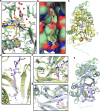
Fig. 4. HisAT crystal structure.
a HisAT adopts a double-GNAT fold composed of a central β-sheet and surrounding helices. The secondary structure elements are presented as a cartoon model where the α-helices and the loops are shown in green and the β-strands as light orange. The stick models of the substrate histidine (His) in magenta and the co-substrate analog S-ethyl-CoA (S-Et-CoA) in yellow are also shown. The N- and C-termini, the secondary structure elements, and the ligands are labeled. b A surface representation of the HisAT structure with the first GNAT domain (until res. 184) shown in pink and the second GNAT domain shown in blue. The stick representations of histidine and S-ethyl-CoA are shown in magenta and yellow, respectively. c Histidine shown in pink bound to the substrate-binding site, (d) arginine (Arg) shown in blue bound to the substrate-binding site, and e the Ac-CoA analog S-ethyl-CoA bound to the Ac-CoA binding site. The substrate and the co-substrate interacting residues are labeled and highlighted in green, and the surrounding waters are depicted as red spheres.
Fig. 5
Fig. 5. Acyl chain binding and structural comparison between human HisAT and other double-GNAT enzymes.
a Binding mode of myristoylhistidine (Myr-His) and the residues that form a hydrophobic binding pocket identified in the HisAT crystal structure. b Surface model of the acyl chain binding pocket with oxygen and nitrogen atoms colored red and blue, respectively. c Overlay of the overall crystal structures of HisAT in green and human NMT1 (PDB-ID 5O9U) in yellow. The alignment R.M.S.D of the Cα was 3.1 Å. d Overlay of the acyl chains of acylated peptide (Myr-pep) in NMT1 structure and acylated histidine (Myr-His) in HisAT structure. e Closeup of the overlay of NMT1 and HisAT active site structures showing the overlap of the catalytic C-terminal Gln496 of NMT1 and the histidine binding Trp246 of HisAT. f Overlay of HisAT and archaeal Ta0821 (PDB-ID 3C26). The alignment R.M.S.D. of the Cα was 2.6 Å. g Overlay of the β-hairpin structures at the active sites of HisAT and Ta0821.
Fig. 6
Fig. 6. Analysis of the structural relationships of HisAT-type and NMT-type double-GNAT proteins.
a Dendrogram of the structural relationships of selected protein structures determined by Dali server. The proteins are identified by a number included in Supplementary Table 9, Uniprot ID, possible PDB ID, and letter E for eukaryote, A for archaea, and B for bacteria. b Overlay (R.M.S.D. 2.4 Å) of the structures of human HisAT in green and a similar protein from Thorarchaeota archaeon (Uniprot ID A0A524CVK5) in blue. c Close-up of the overlay of the conserved active site residues of human HisAT and the archaeal protein. d Overlay (R.M.S.D. 3.8 Å) of the structures of human HisAT in green and a protein from Chloroflexi bacterium (Uniprot ID A0A2W6C360) in brown. e Close-up of the overlay of the conserved active site residues of human HisAT and the bacterial protein. f Structure-based sequence alignment of selected regions of the proteins in the HisAT-type group.

Similar articles

References

    1. Brosnan, M. E. & Brosnan, J. T. Histidine metabolism and function. J. Nutr.150, 2570S–2575S (2020). - PMC - PubMed
    1. Moro, J., Tomé, D., Schmidely, P., Demersay, T.-C. & Azzout-Marniche, D. Histidine: A systematic review on metabolism and physiological effects in human and different animal species. Nutrients12, 1414 (2020). - PMC - PubMed
    1. Wade, A. M. & Tucker, H. N. Antioxidant characteristics of L-histidine. J. Nutr. Biochem.9, 308–315 (1998).
    1. Liao, S.-M., Du, Q.-S., Meng, J.-Z., Pang, Z.-W. & Huang, R.-B. The multiple roles of histidine in protein interactions. Chem. Cent. J.7, 44 (2013). - PMC - PubMed
    1. Thalacker-Mercer, A. E. & Gheller, M. E. Benefits and adverse effects of histidine supplementation. J. Nutr.150, 2588S–2592S (2020). - PubMed

LinkOut - more resources