. 2025 Jul 1;16(1):5510.
doi: 10.1038/s41467-025-61048-x.

Wnt-directed CXCL12-expressing apical papilla progenitor cells drive tooth root formation

Affiliations

Wnt-directed CXCL12-expressing apical papilla progenitor cells drive tooth root formation

Mizuki Nagata et al. Nat Commun. .

Abstract

The tooth root is a critical component of the tooth anchored to surrounding alveolar bones. Tooth root formation is driven by cells in the apical papilla (AP) that generate new dentin-forming odontoblasts at the root-forming front. Mesenchymal stem cells have been isolated from AP for regenerative use; however, how AP cells physiologically coordinate tooth root formation remains undefined. We find that CXCL12+ cells emerge in AP under hypoxic environments at the onset of tooth root formation. Using Cxcl12-creER-based cell-lineage analysis, we further find that CXCL12+ AP cells contribute not only to odontoblasts but also to cementum-forming cementoblasts of the elongating root, while showing plasticity to alveolar bone osteoblasts under regenerative conditions. Canonical Wnt inactivation inhibits odontoblast fates of CXCL12+ AP cells and induces substantial root truncation, with their aberrant fibroblast fates suppressed by TGF-β receptor inhibitor galunisertib. Therefore, CXCL12+ AP cells maintain odonto-cementogenic fates in a Wnt-dependent manner, identifying these cells as pivotal dental mesenchymal progenitor cells driving tooth root formation with substantial plasticity.


Conflict of interest statement

Competing interests: The authors declare no competing interests.

Figures

Fig. 1
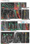
Fig. 1. CXCL12+ cells emerge in the hypoxic apical papilla at the onset of tooth root formation.
ac HIF1α expression. Mandibular first molar (M1) sections at P0 (a) and P6 (b). a’, b’ High magnification. Yellow: HIF1α, gray: DIC/DAPI. c Quantification of HIF1α expression (mean intensity) from P0 to P15, n = 3 mice each time point. dh Cxcl12-GFP expression. M1 sections of Cxcl12GFP/+ mandibles at P0 (d), P3 (e), P6 (f, g). Green: Cxcl12-GFP, cyan: E11, gray: DIC/DAPI. (h): Quantification of Cxcl12-GFP+ DP cells from P0 to P25, n = 3 mice each time point. in Short chase analysis of Cxcl12-creER+ cells. i M1 sections of Cxcl12GFP/+; Cxcl12-creER; R26RtdTomato mandibles at P5 (pulsed at P3). i’ High magnification of distal M1. j Flow cytometry analysis of P5 CD45neg tooth bud cells isolated from Cxcl12GFP/+; Cxcl12-creER; R26RtdTomato mice (pulsed at P3). k M1 sections of Gli1GFP/+; Cxcl12-creER; R26RtdTomato mandibles at P5 (pulsed at P3). ln Immunostaining for CK5 (l), PDGFRα (m), SOX9 (n) in distal M1 of Cxcl12-creER; R26RtdTomato at P5 (pulsed at P3). Green: Cxcl12-GFP (i), Gli1-GFP (k), red: Cxcl12CE-tdT, cyan: CK5/PDGFRα/SOX9, gray: DIC/DAPI. o Cell proliferation. EdU was administered twice (6 and 3 h) to Cxcl12-creER; R26RtdTomato mice (pulsed at P3) before analysis at P8. Arrowheads: EdU+Cxcl12CE-tdT+ AP cells. Scale bar: 500 μm (a, b, d, e, f, i, k, o), 50 μm (d’, e’, f’, i’, k’, l, m, n, o’), 20 μm (a’, b’, g). M1: mandibular first molar, DP: dental papilla, DF: dental follicle, HERS: Hertwig’s epithelial root sheath. All data are mean ± SD. Representative images of at least three independent biological samples are shown in the figures. Source data are provided as a Source Data file.
Fig. 2
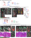
Fig. 2. Cxcl12-creER+ AP cells function as odonto-cemento-progenitor cells.
Cell fate analysis of Cxcl12-creER+ AP cells during tooth root formation. Mandibular first molar (M1) sections. a, During active tooth root formation, after 7 days of chase at P10. Col1a1(2.3 kb)-GFP; Cxcl12-creER; R26RtdTomato (a) and Cxcl12-creER; R26RtdTomato (b) molars (pulsed at P3). (a’, b’): High magnification of distal M1. Arrowheads in (a): Col1a1-GFP+Cxcl12CE-tdT+ odontoblasts. Green: Col1a1-GFP, red: Cxcl12CE-tdT, cyan: E11, gray: DIC/DAPI. cf At the completion of tooth root formation, after 22 days of chase at P25. c, d Col1a1(2.3 kb)-GFP; Cxcl12-creER; R26RtdTomato (c) and Osteocalcin (3.8 kb) (Oc)-GFP, Cxcl12-creER; R26RtdTomato (d) molars (pulsed at P3). Arrowheads in (c’): Col1a1-GFP+Cxcl12CE-tdT+ odontoblasts, arrows in (d): Oc-GFP+Cxcl12CE-tdT+ cementoblast. Green: Col1a1-GFP (c) or Oc-GFP (d), red: Cxcl12CE-tdT, gray: DIC/DAPI. Cxcl12-creER; R26RRGBow/RGBow molars (pulsed at P3). Arrowheads: same-color clones of odontoblasts. Green: GFP, red: mOrange2, blue: mKate2. (f): EdU label-retention assay of Cxcl12-creER+ cells. Cxcl12-creER; R26RtdTomato mice (pulsed at P3) were serially pulsed with EdU every 8 h for 3 days from P3 to P6. Arrows: EdU-retaining Cxcl12CE-tdT+ cells at the neurovascular bundle. Green: S100, red: Cxcl12CE-tdT, cyan: EdU, gray: DIC/DAPI. g Long-term cell fate after 6 months of chase. Oc-GFP; Cxcl12-creER; R26RtdTomato molars. Asterisk: absence of tdT+ alveolar bone osteocytes. Arrows: Oc-GFP+Cxcl12CE-tdT+ cementoblasts. Green: Oc-GFP, red: Cxcl12CE-tdT, gray: DIC/DAPI. Scale bar: 500 μm (a, c, f, g), 200 μm (g’, g”), 50 μm (a’, b, c’, d, e, f’). DP: dental papilla/pulp, DF: dental follicle, HERS: Hertwig’s epithelial root sheath, Od: odontoblasts, D: dentin, C: cementum, PDL: periodontal ligament, AB: alveolar bone. Representative images of at least three independent biological samples are shown in the figures.
Fig. 3
Fig. 3. Injury/insult-induced plasticity of CXCL12+ AP derivatives cell fates.
ae Impact of periodontal drill-hole injury on Cxcl12-creER+ AP derivatives. Surgery at 8–10 weeks-old Oc-GFP; Cxcl12-creER; R26RtdTomato mice (pulsed at P3), analysis at 14 days after the procedure. a Experimental scheme of mandibular periodontal drill-hole surgery model. A 1.0 mm diameter drill hole is created on the buccal surface of the mandible to remove the alveolar bone and the cementum. b, c Mandibular first molar (M1) frontal section, left mandible (Control) (b), right mandible (Surgery) (c). Asterisk in (b’): absence of tdTomato+ alveolar bone osteocytes. Arrowheads in (c’): Oc-GFP+Cxcl12CE-tdT+ cementoblasts newly formed within the defect. Arrows in (c’): Oc-GFP+Cxcl12CE-tdT+ osteoblasts newly formed within the defect. d, e Quantification of Oc-GFP+Cxcl12CE-tdT+ cementoblasts (d) and alveolar bone osteoblasts (e). n = 4 mice per each group. fj Impact of ligature-induced periodontitis on Cxcl12-creER+ AP derivatives. Intervention at 12 weeks old Oc-GFP; Cxcl12-creER; R26RtdTomato mice (pulsed at P3), analysis at 14 days after installment. f Experimental scheme of maxillary ligature-induced periodontitis model. A 6-0 silk suture was inserted between the first and second molar on the left side. Both ends of the suture string were knotted to prevent ligature loss. gi Maxillary first and second molar (M1&M2) sagittal section, right maxilla (Control) (g, h), left maxilla (Ligature) (i, j). Asterisk in (b’): absence of tdTomato+ alveolar bone osteocytes. Arrows in (i"): tdTomato+ alveolar bone osteoblasts in fibrotic lesion. Green: Oc-GFP, red: Cxcl12CE-tdT, gray: DIC/DAPI. Scale bars: 500 μm (g, h, i, j), 200 μm (b, c), 50 μm (b’, c’, g’, g”, i’, i”). B: buccal side, L: lingual side, D: dentin, C: cementum, PDL: periodontal ligament, AB: alveolar bone. *P < 0.05, Two-tailed, one-way ANOVA followed by the Mann-Whitney U test. All data are mean ± SD. Exact P-value is indicated in the figures. Representative images of at least three independent biological samples are shown in the figures. Source data are provided as a Source Data file.
Fig. 4
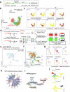
Fig. 4. Single-cell RNA-seq characterization of CXCL12+ AP cells and their differentiation potential.
ac Heterogeneity of Cxcl12-GFP+ cells. Single-cell RNA-seq analysis of Cxcl12-GFP+ cells isolated from Cxcl12GFP/+ molars at P6. a Workflow of cell dissociation, FACS isolation of Cxcl12-GFP+ cells, and scRNA-seq. b UMAP plot of major classes of Cxcl12-GFP+ cells (Cluster 0–7, 7,528 cells). Pooled from n = 3 mice. c Feature plots of representative DF (Runx2, Acta2), DP (Sox9, Sp7), apical papilla/follicle (Wif1, Smoc2) and Schwann cell (Plp1) markers, and canonical Wnt (Ctnnb1) marker. High expression: violet, Low expression: yellow. dj Putative cell origin and cell-cell interaction of Cxcl12-creER+ AP cell derivatives. d Workflow of integrative scRNA-seq analysis, merging datasets of Cxcl12CE-tdT+ cells at P6 and P25 (pulsed at P3). DP: dental pulp, PDL: periodontal ligament. e, f UMAP plot of the merged dataset (Cluster 0–14, 5869 cells). e Colored by each dataset (Cxcl12CE-tdT+ at P6: 358 cells, Col1a1GFP+ and Cxcl12CE-tdT+ cells at P25: 5,511 cells), (f): major classes of Cxcl12CE-tdT+ cells, osteoblast (C1), cementoblast (C6), PDL (C0), apical papilla/pulp (C4), dental pulp (C3, C7), stromal (C2), hematopoietic (C5, C10, C12), epithelial (C9) cells. g Feature plots of the representative pulp (Sox9), apical papilla/pulp (Wif1), PDL (Scx), cementoblast (Pthlh), osteoblast (Bglap) markers and tdTomato. h CellChat intercellular communication analysis. A chord diagram demonstrates the intercellular interaction network via the Wnt signaling pathways. The lines denote intercellular interaction strengths, linking sender cells (blunt end) with receiver cells (arrowhead). The color bars in the outer circle indicate cell states sending outgoing signals, while those in the inner circles represent cell types receiving incoming signals. i VeloVAE analysis after removing tdTomato- cell populations (C1,5,9-14). Black arrows: dynamic velocity vectors. The orientation of black arrows denotes the developmental direction from progenitors to their derivatives. j scVelo-computed initial state and end point probability. Initial cell state probability (upper panel) and terminal cell state probability (lower panel). Arrowhead: putative points of root cells in AP cells.
Fig. 5
Fig. 5. Canonical Wnt signaling maintains odontogenic fates of CXCL12+ AP cells.
Impact of canonical Wnt inactivation in Cxcl12-creER+ AP cells on cell fates and tooth root formation. Cxcl12-creER; Ctnnb1fl/+; R26RtdTomato (AP-Ctnnb1 cHet). Cxcl12-creER; Ctnnb1fl/fl; R26RtdTomato (AP-Ctnnb1 cKO) mice were pulsed at P3. a Diagram of conditional Ctnnb1 deletion in Cxcl12-creER+ AP cells. bh Cell proliferation and periodontal ligament cell differentiation. Mandibular first molars (M1) sections. b, c, f EdU assays at 7 days of chase at P10. EdU was administered twice (6 and 3 h) before analysis. Quantification of EdU+Cxcl12CE-tdT+ cells is shown in (f). d, e, g, h POSTN staining at 22 days of chase at P25. Quantification of Cxcl12CE-tdT+ odontoblasts (g) and POSTN+Cxcl12CE-tdT+ periodontal fibroblasts (h). n = 4 mice per group (f, g), n = 4 mice (AP-Ctnnb cHet), n = 4 mice (AP-Ctnnb cKO). Green: EdU (ac), POSTN (d, e), red: Cxcl12CE-tdT, gray: DIC/DAPI. Scale Bars: 500 μm (be), 50 μm (b’e’). ip 3D-µCT analysis at 6 months. i Composite 3D surface model overlay, superimposition registered on mandibles (upper) and mandibular molars (lower). jp Quantitative 3D-µCT analysis of M1&M2 root length (j, k), M1 root thickness (l), M1 dentin thickness (m), M1 crown length (n), M1 crown width (o), and M1 eruption height (p). M1: mandibular first molar, M2: mandibular second molar. n = 5 mice per each group. *P < 0.05, **P < 0.01, Two-tailed, one-way ANOVA followed by the Mann-Whitney U test. All data are mean ± SD. Exact P-value is indicated in the figures. Source data are provided as a Source Data file.
Fig. 6
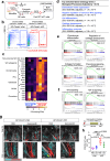
Fig. 6. Canonical Wnt signaling prevents premature differentiation of CXCL12+ AP cells.
Identification of mechanisms underlying canonical Wnt inactivation-induced CXCL12+ AP cell fate shifts by RNA-req. a, b Workflow of cell dissociation, FACS isolation of Ctnnb1Het (Control) and ∆Ctnnb1 Cxcl12-tdT+ cells and bulk RNA-seq. b FACS sorting of Cxcl12-creER+ AP cells at P10 (pulsed at P3). Blue box: Cxcl12CE-Ctnnb1Het-P3 (Ctnnb1Het) cells isolated from AP-Ctnnb1 cHet molars, red box: Cxcl12CE-∆Ctnnb1-P3 (∆Ctnnb1) cells isolated from AP-Ctnnb1 cKO molars. cf RNA-seq analysis results. c Heatmaps of representative 20 differentially expressed genes (DEGs) associated with non-canonical Wnt signaling, Wnt antagonist, chondrocyte, periodontal ligament (PDL), osteoblast, and dental pulp/odontoblast (DP/Od) markers, as well as chemokine. Left 4 lanes: Ctnnb1Het, right 4 lanes: ∆Ctnnb1. PDL: periodontal ligament, DP: dental pulp, Od: odontoblast. Yellow: higher expression, purple: lower expression, n = 4 mice per each group. d Top enriched GO terms – biological processes (GO-BP) overrepresented in DEGs (Adjusted p < 0.01, Fisher’s exact test). e, f GSEA analysis. Representative 4 pathways (e) and 2 up-regulated signaling pathways (f) in Cxcl12CE-∆Ctnnb1-P3 cells. NES: normalized enrichment score (Adjusted p < 0.001, weighted Kolmogorov-Smirnov test). g, h Administration of TGF-β receptor inhibitor galunisertib (Gal) to AP-Ctnnb1 cHet and cKO mice. Gal or PBS was administrated into AP-Ctnnb1 cHet or cKO from P8 to P20. g Mandibular 1st molar of AP-Ctnnb1 cHet and cKO at P25 (pulsed at P3). Scale bars: 500 μm (upper panels), 100 μm (lower panels). h Quantification of POSTN+tdTomato+ cells/tdTomato+ dental pulp cells. n = 3 mice (AP-Ctnnb cHet + PBS), n = 4 mice (AP-Ctnnb cHet + Gal), n = 6 mice (AP-Ctnnb cKO + PBS), n = 4 mice (AP-Ctnnb cKO + Gal), *P < 0.05, **P < 0.01, ***P < 0.001, Two-tailed, one-way ANOVA followed by the Mann-Whitney U test. All data are mean ± SD. Exact P-value is indicated in the figures. Source data are provided as a Source Data file.
Fig. 7
Fig. 7. Wnt-directed CXCL12-expressing AP progenitor cells drive tooth root formation.
Cxcl12+ AP mesenchymal progenitor cells differentiate into odontoblasts, dental pulp cells, PDL cells, and cementoblasts via canonical Wnt signaling during tooth root development. The TGF-β signaling pathway is a potential downstream target of canonical Wnt signaling in CXCL12+ AP cells. During periodontal injury response, descendants of Cxcl12+ AP cells were recruited to the bone defect and differentiated into osteoblast-lineage cells.

References

    1. Nanci, A. Ten Cate’s Oral Histology. (Elsevier Health Sciences, 2017).
    1. Cho, M. I. & Garant, P. R. Development and general structure of the periodontium. Periodontol 200024, 9–27 (2000). - PubMed
    1. Ten Cate, A. R. The development of the periodontium-a largely ectomesenchymally derived unit. Periodontol 200013, 9–19 (1997). - PubMed
    1. Luan, X., Ito, Y. & Diekwisch, T. G. Evolution and development of Hertwig’s epithelial root sheath. Dev. Dyn.235, 1167–1180 (2006). - PMC - PubMed
    1. Nanci, A. & Bosshardt, D. D. Structure of periodontal tissues in health and disease. Periodontol 200040, 11–28 (2006). - PubMed

MeSH terms

LinkOut - more resources