. 2025 Jul 1;15(1):20877.
doi: 10.1038/s41598-025-04923-3.

Synergistic strategy of riboflavin and lipoids to bioengineer resin dentin hybrid layer

Affiliations

Synergistic strategy of riboflavin and lipoids to bioengineer resin dentin hybrid layer

Wong Er-Vin et al. Sci Rep. .

Abstract

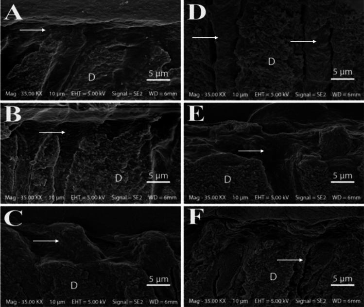
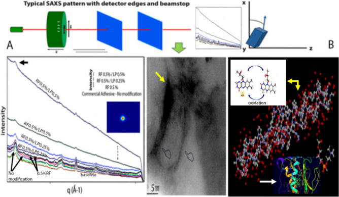
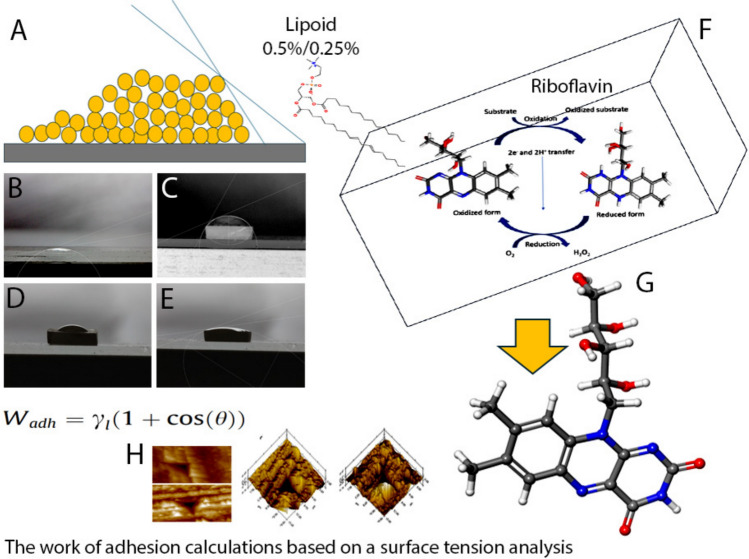
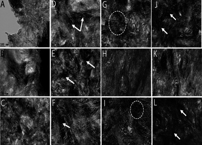
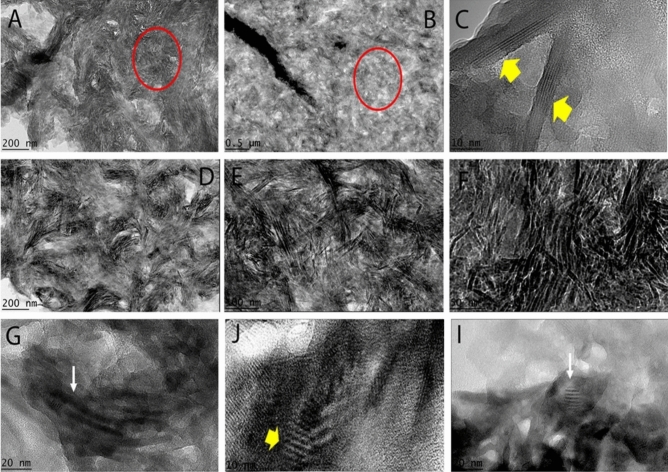
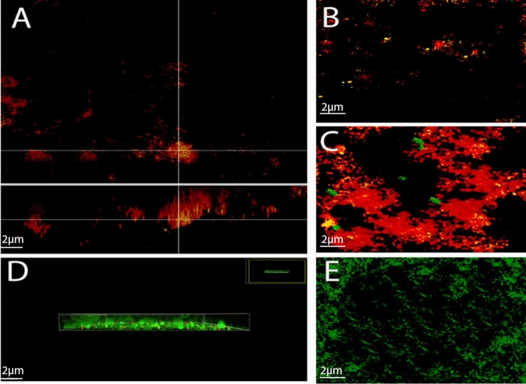
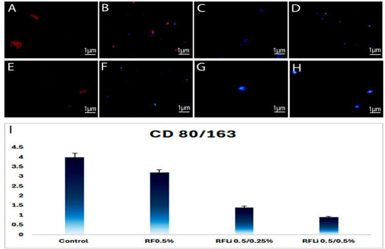
The study aimed to synthesize and evaluate the effects of an experimental adhesive system containing different concentrations of riboflavin in combination with Lipoid/phosphatidylcholine solutions on the dentin-bonding interface. Lipoid solutions were prepared in riboflavin (RF) experimental self-etching adhesive, (Ct0, RF0.5%, RFLi0.5%/0.25%, RFLi0.5%/0.5%). Resin-dentin slabs were prepared for hybrid layer evaluation and microtensile strength was tested. Interfacial leakage was also determined using silver tracer nanoleakage. The mechanical properties of collagen fibrils were evaluated. The adhesive was assessed for contact angle and degree of conversion. A biofilm based on Streptococcus mutans, Actinomyces naeslundii, and Streptococcus sanguis was used to evaluate fluorescence in-situ hybridization for antimicrobial analysis. Collagen was examined using a transmission electron microscope with in-situ hybridization. Human macrophages were stained for immunofluorescence. Resin tags were detectable in all adhesive specimens. RFLi preserved adhesive bond strength after long-term aging. Sizes and dispersion of fibrils in RF0.5%, RFLi0.5%/0.25%, RFLi0.5%/0.5% were substantially larger (p < 0.05). Contact angle values exhibited significant differences (p < 0.05). Both Ei and Hi were impacted by different adhesives. RFLi 0.5%/0.5% group exhibited an increase in the degree of conversion. RFLi0.5%/0.5% displayed well-preserved collagen fibrils. Confocal images showed the presence of dead bacteria amongst RFLi0.5%/0.25%/RFLi0.5%/0.5% groups. CD80 + markers on macrophages were detected in the RFLi0.5%/0.25%/RFLi0.5%/0.5% groups. RFLi0.5%/0.5% modified adhesives show enhanced bonding to dentin and can be expected to prolong the long-term integrity of the resin-dentin interface.


Conflict of interest statement

Declarations. Competing interests: The authors declare no competing interests. Ethics approval: The project obtained permission from the Institutional Review Board of International Medical University Kuala Lumpur. Obtained written informed consent from all persons who contributed extracted teeth for the laboratory in-vitro experiments approved by the Institutional Research Ethical Committee (537/2021). Consent for Publication: Consent for publishing has been obtained from all authors. All authors read and approved the manuscript.

Figures

Fig. 1
Fig. 1
Schematic representation of experiments performed.
Fig. 2
Fig. 2
Representative SEM images of resin tags and hybrid layer detectable in all the adhesive specimen groups. (A) The control adhesive on dentin was penetrated with the adhesive showing a resin-dentin hybrid layer (white arrow). Similarly, the application of (B) RF0.5%, (C) RFLi0.5%/0.25% and (E) RFLi0.5%/0.5% to dentin resulted in the presence of a visibly intact resin-infiltrated hybrid layer (white arrow). (D) The specimens generated with the RFLi0.5%/0.5% adhesive showed lengthy resin tags (white arrow) that penetrated deeply. In some of the specimens using (F) control adhesives, only brief resin tags (white arrow) were created while bonding towards dentin (D: intertubular sound dentin).
Fig. 3
Fig. 3
Representative confocal laser microscopy images taken at the resin dentin interface for all experimental groups. The resin-dentin interface formed by the (A-B) control adhesive system revealed the occurrence of commonly seen resin tags. The resin permeated the dentin, forming thick hybrid layers in both (3D-E) RFLi0.5%/0.25% and (3 F) RFLi0.5%/0.5% groups modified adhesives. None of the (C) RF0.5% or (D-E) RFLi0.5%/0.25% (gaps seen between resin tags due to mineral precipitation inside the interface have been identified with white arrows) and (F) RFLi 0.5%/0.5% groups depicted resin-dentin interface gaps. The creation of a hybrid layer containing resin tags was detected in all specimens (HL identified with white arrows), with the length of the tags being more prominent in (J) RFLi0.5%/0.5% and (3I) RFLi0.5%/0.25% groups adhesives as compared to (Fig. 3G; arrow represents the direction of resin tags) control, and RF0.5% groups (H; arrow pointing towards hybrid layer); (K-L) lateral angles of RFLi0.5%/0.5% groups indicating a comprehensive penetration of adhesive. (red rhodamine dye within adhesive and resin tags; dentin is represented as green fluorescence; RT = resin tags; HL = hybrid layer).
Fig. 4
Fig. 4
In the backscattered SEM images of (A) control group, which is the adhesive without riboflavin and lipoid, a significant amount of silver was deposited along the resin-dentin interface and within the dentinal tubules. The (B) RF0.5% adhesive groups displayed a slightly lower amount of silver deposition as observed in the resin-dentin interface. As for (C) RFLi0.5%/0.25% and (D) RFLi0.5%/0.5% groups were concerned, no silver deposition is observed in the resin-dentin interfaces; (E) commercial control adhesive.
Fig. 5
Fig. 5
(A) Display of scatter patterns and intensity vs. q-range plots for the dentin specimens modified with different adhesive treatments using SAXS. The colored graphical lines depict SAXS/WAXD patterns for collagen matrices (sizes and dispersion of fibrils) seen in controls, RF0.5%, RFLi0.5%/0.25%, RFLi0.5%/0.5%. The RF experimental groups displayed substantially larger changes within the patterns. The inset within the SAXS graphical streams show the deconvolved (blue profile) collected at a point representing deionised pattern of acquisition. The patterns represented (n = 2 per group) groups from control from the lowest intensity to higher intensities within no modification, RF0.5%, > RFLi0.5%/0.25%, > RFLi0.5%/0.5% (labeled). The shifts in X-ray diffraction peaks (black arrow) indicate changes in collagen fibril dimensions due to chemical changes and the crosslinking effect (p < 0.05) (D space of collagen fibril within RFLi0.5%/0.5%; yellow arrow). (B) The figure displays the most suitable positions assumed by the riboflavin molecule and lipoid as it is docked into collagen and enzymes (yellow arrow). The inset image pointed by yellow is the free radical oxidation by the presence of RF molecules. Schematic representation of the collagen network and the positioning of the simulation for an individual chain showing a simplified depiction of the collagen fibril network and the intermolecular cross-links connecting adjacent collagen molecules. The hydroxyproline residues of the molecule are denoted by fixed grey spheres. The blue and red denote the MD-modelled curves, corresponding to the colour scheme of amino acids and intramolecular hydrogen, respectively. The riboflavin binding mechanism has been confirmed to successfully bind to the catalytic sites of MMP-2 and −9 enzymes (white arrow). The blue, yellow and green ribbons resented when the enzyme’s active site was blocked by the crosslinking agent.
Fig. 6
Fig. 6
(A) Macromolecular adhesion angle; contact angle images estimated using wetting angle analysis exhibit statistical differences for (B) control (C) RF0.5% (D) RFLi0.5%/0/25% (E) RFLi0.5%/0/5%; (F) numerical model for the RFLi formulation (G) RF model and (H) Nanoindentations for AFM of resin dentin hybrid layer.
Fig. 7
Fig. 7
Representative TEM images exhibited different levels of structural alterations after 3 months of storage. (A-C) non-cross-linked and (D-F) RF0.5% cross-linked specimens. The specimens treated with a modified (D-F) RF0.5% adhesive exhibited well-preserved collagen fibril structure as pointed by white arrows. Specimens treated with a (G-I) RFLi0.5%/0.25% exhibited a thicker collagen network modified with the permeation enhancer exhibiting a well-preserved and compact collagen network as indicated by a dotted circle. Specimens that were treated with the permeation enhancer (J-L) RFLi0.5%/0.5% displayed a similar well-preserved thick structure of collagen fibrils with distinct collagen crossbanding (white arrows).
Fig. 8
Fig. 8
High magnification transmission electron microscopy (TEM) imaging, showing that the collagen fibrils structure in dentin specimens crosslinked with RFLi0.5%/0.5% remained intact and exhibited a consistent periodicity pattern even after 3 months of storage. (A-C) Control; (D-F) RFLi0.5%/0.25%; (G-I) RFLi 0.5%/0.5%. In a higher magnification of the TEM images, it can be seen even clearer where the crosslinking is being formed, shown by the red circles and yellow arrows.
Fig. 9
Fig. 9
FISH accurately identified almost all bacteria that were stained as streptococci. The combined pictures in the"orthogonal section display mode"clearly showed the presence of dead bacteria amongst the (A) RFLi0.5%/0.25% and (B-C) RFLi0.5%/0.5% groups. There was a considerable rise in the number of bacteria on (D-E) control specimens. (green live bacteria; red dead bacteria).
Fig. 10
Fig. 10
Confocal laser scanning microscopy images of invading cells that express CD163 (red) and CD80 (blue). (A) Control group and the (B) RF0.5% groups, the macrophages showed a strong CD163 + phenotype, indicating the presence of M2 markers. In contrast, the CD80 + markers were also detected in the (C-D) RFLi0.5%/0.25% and (E–H) RFLi0.5%/0.5% groups. (I) M1/M2 ratio was considerably lower in the RFLi0.5%/0.25% and RFLi0.5%/0.5% groups compared to the control, and RF0.5% groups (p < 0.05). Values greater than 1.0 indicate a pro-inflammatory M1-type reaction, whereas values lower than 1.0 indicate an anti-inflammatory M2-type response.

References

    1. Perdigão, J. Current perspectives on dental adhesion: (1) Dentin adhesion - not there yet. Jpn. Dent. Sci. Rev.56, 190–207 (2020). - PMC - PubMed
    1. Mohammad, A. S., Julia, V., Anna, V. & Steven, M. M. Functional role of inorganic trace elements in dentin apatite tissue—part III: Se, F, Ag, and B. J. Trace Elem. Med. Biol.10.1016/j.jtemb.2022.126990 (2021). - PubMed
    1. Pashley, D. H. et al. State of the art etch-and-rinse adhesives. Dent. Mater.27, 1–16 (2011). - PMC - PubMed
    1. Tjäderhane, L. Dentin bonding: Can we make it last?. Op. Dent.40, 4–18 (2015). - PubMed
    1. Betancourt, D. E., Baldion, P. A. & Castellanos, J. E. Resin-dentin bonding interface: Mechanisms of degradation and strategies for stabilization of the hybrid layer. Intern. J. Biomater.10.1155/2019/5268342 (2019). - PMC - PubMed

LinkOut - more resources