. 2025 Jul 2;15(1):22840.
doi: 10.1038/s41598-025-05907-z.

Unlocking the potential of phytochemicals in inhibiting SARS-CoV-2 MPro protein - an in silico and cell-based approach

Affiliations

Unlocking the potential of phytochemicals in inhibiting SARS-CoV-2 MPro protein - an in silico and cell-based approach

Khushboo Singh et al. Sci Rep. .

Abstract

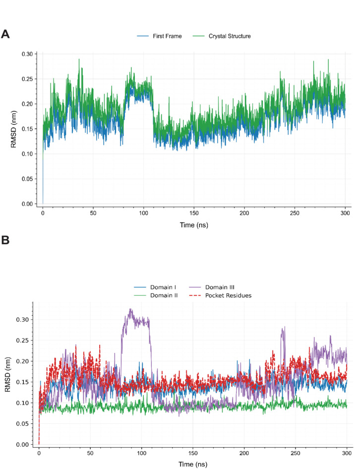
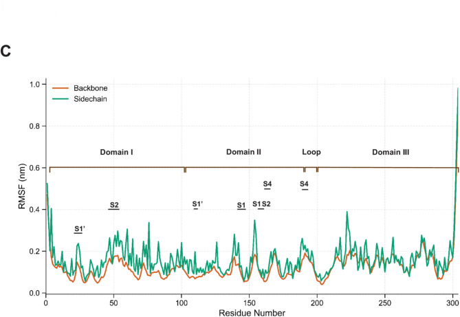
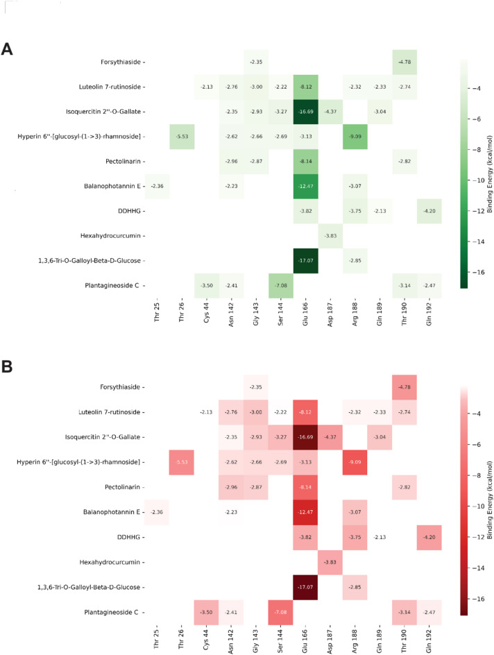
The main protease (MPro) of SARS-CoV-2 plays a crucial role in viral replication and is a prime target for therapeutic interventions. Phytochemicals, known for their antiviral properties, have been previously identified as potential MPro inhibitors in several in silico studies. However, the efficacy of these remains in question owing to the inherent flexibility of the MPro binding site, posing challenges in selecting suitable protein structures for virtual screening. In this study, we conducted an extensive analysis of the MPro binding pocket, utilizing molecular dynamics (MD) simulations, principal component analysis (PCA) and free energy landscape (FEL) to explore its conformational diversity. Based on pocket volume and shape-based clustering, five representative protein conformations were selected for virtual screening. Virtual screening of a library of ~ 48,000 phytochemicals suggested 39 phytochemicals as potential MPro inhibitors. Based on subsequent MM-GBSA binding energy calculations and ADMET property predictions, five compounds were advanced to cell-based viral replication inhibition assays, with three compounds (demethoxycurcumin, shikonin, and withaferin A) exhibiting significant (EC50 < 10 μm) inhibition of SARS-CoV-2 replication. Our study provides an understanding of the binding interactions between these phytochemicals and MPro, contributing significantly to the identification of promising MPro inhibitors. Furthermore, beyond its impact on therapeutic development against SARS-CoV-2, this research highlights a crucial role of proper nutrition in the fight against viral infections.


Conflict of interest statement

Declarations. Competing interests: The authors declare no competing interests.

Figures

Fig. 1
Fig. 1
Structure of SARS-CoV-2 MPro. [Left] Cartoon representation of one protomer showing three domains: Domain I (blue, residues 8-101), Domain II (yellow, residues 102–184), and Domain III (green, residues 201–303). The catalytic residues His41 and Cys145 are shown as sticks. [Right] Surface representation of the substrate-binding pocket with key subsites (S1, S1’, S2, and S4) that serve as binding locations for inhibitors.
Fig. 2
Fig. 2
Structural stability and flexibility analysis of SARS-CoV-2 MPro during 300 ns MD simulation. (A) Global RMSD of Cα atoms compared to initial frame and crystal structure, showing distinct conformational phases. (B) Domain-specific RMSD revealing differential flexibility: Domain II remains stable, Domain I shows moderate flexibility, Domain III exhibits pronounced conformational changes, and pocket residues display temporal correlation with Domain III movements. (C) RMSF analysis comparing backbone and sidechain atom flexibility.
Fig. 2
Fig. 2
Structural stability and flexibility analysis of SARS-CoV-2 MPro during 300 ns MD simulation. (A) Global RMSD of Cα atoms compared to initial frame and crystal structure, showing distinct conformational phases. (B) Domain-specific RMSD revealing differential flexibility: Domain II remains stable, Domain I shows moderate flexibility, Domain III exhibits pronounced conformational changes, and pocket residues display temporal correlation with Domain III movements. (C) RMSF analysis comparing backbone and sidechain atom flexibility.
Fig. 3
Fig. 3
Conformational analysis of SARS-CoV-2 MPro. (A) PCA projection onto the first two principal components (PC1 and PC2) showing major protein motions during the 300 ns simulation, with points colored by simulation time progression (blue→purple). (B) [Left] FEL revealing three major conformational states (Basins 1–3), where deeper blue regions represent energetically favorable conformations. [Right] Structural overlay of representative conformations from each basin (Basin 1: steel blue, Basin 2: plum, Basin 3: golden), highlighting regions with significant conformational differences.
Fig. 4
Fig. 4
Binding pocket conformations of SARS-CoV-2 MPro identified through volume-based clustering. (A-E) Surface representations of representative structures from five distinct clusters (Clusters 1–5), showing the variable geometry of the binding site. The catalytic residues His41 (pink) and Cys145 (green) are highlighted, with binding subsites (S1, S1’, S2, S2’, S4) labeled in red.
Fig. 5
Fig. 5
Binding modes of top-scoring non-glycoside phytochemicals with SARS-CoV-2 MPro. Left panels (A, C,E, G) show 3D surface representations of the binding pocket with compounds in stick format; right panels (B, D,F, H) display 2D interaction diagrams. Binding subsites (S1, S1’, S2, S2’, S4) are labeled in red. Compounds shown: (A-B) SAC binding to Cluster 3 conformation, (C-D) SAL binding to Cluster 3 conformation, (E-F) AHDPH binding to Cluster 3 conformation, and (G-H) Shikonin binding to Cluster 5 conformation.
Fig. 6
Fig. 6
Binding interactions of SARS-CoV-2 MPro and phytochemicals with high docking scores in at least one protein conformation. Left panels (A,C,E,G) show 3D surface representations of the binding pocket with compounds in stick format; right panels (B, D,F, H) display 2D interaction diagrams. Binding subsites (S1, S1’, S2, S4) are labeled in red. Compounds shown: (A-B) Cynarin binding to Cluster 1conformation, (C-D) Demethoxycurcumin binding to Cluster 2 conformation, (E-F) Hexahydrocurcumin binding to Cluster 3 conformation, and (G-H) Withaferin A binding to Cluster 5 conformation.
Fig. 7
Fig. 7
Interaction energy heatmaps between MPro residues and top ten phytochemicals. (A) Van der Waals interaction energies (green scale) and (B) Coulombic interaction energies (red scale). Values shown are in kcal/mol, with more negative values (darker colors) indicating stronger interactions. Several residues, including His41, Gly143, and Glu166, form strong interactions with multiple phytochemicals.
Fig. 8
Fig. 8
Antiviral activity and cytotoxicity of phytochemicals against SARS-CoV-2. (A) Dose-response curve for Remdesivir (control), showing viral inhibition (blue) and cell viability (red), EC50 = 3 µM. (B) Evaluation of Shikonin (EC50 = 10 µM), Demethoxycurcumin (EC50 = 8.8 µM), and Withaferin A (EC50 = 2.8 µM). FA denotes the fraction of cells affected; GRI represents the predicted compound response at an infinite concentration.

Similar articles

References

    1. Wu, F. et al. Author correction: A new coronavirus associated with human respiratory disease in China. Nature580, E7 (2020). - PMC - PubMed
    1. Gorbalenya, A. E. et al. The species severe acute respiratory syndrome-related coronavirus: classifying 2019-ncov and naming it SARS-COV-2. Nat. Microbiol.5, 536–544 (2020). - PMC - PubMed
    1. Galan, L. E. et al. Phase 2 randomized study on chloroquine, hydroxychloroquine or Ivermectin in hospitalized patients with severe manifestations of SARS-COV-2 infection. Pathog Glob Health. 115, 235–242 (2021). - PMC - PubMed
    1. Vallejos, J. et al. Ivermectin to prevent hospitalizations in patients with covid-19 (IVERCOR-covid19) a randomized, double-blind, placebo-controlled trial. BMC Infect. Dis.21, 635 (2021). - PMC - PubMed
    1. Dhawan, B. N. Anti-viral activity of Indian plants. Proc. Natl. Acad. Sci. India Sect. B Biol. Sci.82, 209–224 (2012). - PMC - PubMed

MeSH terms

LinkOut - more resources