. 2025 Jul 1;15(1):20467.
doi: 10.1038/s41598-025-05435-w.

CXCL8 is essential for cervical cancer cell acquired radioresistance and acts as a promising therapeutic target in cervical cancer

Affiliations

CXCL8 is essential for cervical cancer cell acquired radioresistance and acts as a promising therapeutic target in cervical cancer

Qinghong Hu et al. Sci Rep. .

Abstract

Acquired radioresistance critically challenges cervical cancer radiotherapy management. Clinically relevant radioresistant cell models remain scarce, and CXCL8's role in cervical cancer-despite its tumorigenic/therapy-resistant associations in other cancers-is poorly characterized. Two radioresistant cervical cancer cell strains were established. mRNA-seq and bioinformatics analysis of radiosensitivity regulators identified CXCL8 as a key mediator. In vitro, assays of cell viability, clone formation, apoptosis and cell cycle were conducted following transient transfection of cervical cancer radiotherapy-resistant cell strains with knockdown of CXCL8, as well as subsequent addition of exogenous CXCL8 to cervical cancer parental cell strains. Radioresistant cervical cancer cell lines (Hela-RR/Siha-RR) were established through clinical protocol-mimicking irradiation, validated via proliferation/clonogenic/cell cycle assays. mRNA-seq identified 50 co-upregulated and 54 co-downregulated genes in resistant strains, with CXCL8 among top differentially expressed genes (IL11, CXCL8, MMP1, HSPA8, CA9, PPFIA4, EDN2, GUCY1A2, EFNA3, TNFAIP6). qRT-PCR confirmed CXCL8, TNFAIP6, SRNA8 and PPFIA4 dysregulation. Cox regression analysis of 96 candidate radiosensitivity regulators prioritized CXCL8 among eight key genes in cervical cancer. GEPIA2 and immunohistochemistry revealed CXCL8 overexpression in tumors. Functional studies demonstrated CXCL8 knockdown sensitized resistant cells to radiation, while exogenous CXCL8 induced resistance in parental lines.

Keywords: CXCL8; Cervical cancer; Radioresistance; Radiosensitization; Tumor microenvironment.


Conflict of interest statement

Declarations. Competing interests: The authors declare no competing interests. Institutional review board approval: This study was approved by the Ethics Committee of The First Affiliated Hospital of Zhengzhou University. Informed consent: N/A. Registry and registration number: N/A. Animal studies: N/A.

Figures

Fig. 1
Fig. 1
Acquired radiotherapy-resistant cell lines have enhanced clone formation after radiation exposure. A Representative images of colony formation assays. B Quantitative comparison of clonogenic capacity between Hela and Hela-RR cell lines. C Quantitative comparison of clonogenic capacity between Siha and Siha-RR cell lines. All data are presented as mean ± standard deviation. Statistical significance was determined by two-tailed Student’s t-test. (Significance markers: *P < 0.05, **P < 0.01, ***P < 0.001; ns = not significant (P > 0.05).)
Fig. 2
Fig. 2
Acquired radiotherapy-resistant cell lines have enhanced cell survival after radiation exposure. A Time-dependent cell proliferation curves of Hela and Hela-RR cells without irradiation. B Time-dependent cell proliferation curves of Siha and Siha-RR cells without irradiation. C Time-dependent cell proliferation curves of Hela and Hela-RR cells after irradiation with a single 6 Gy dose of 6 MeV X-rays. D Time-dependent cell proliferation curves of Siha and Siha-RR cells after irradiation with a single 4 Gy dose of 6 MeV X-ray. All data are presented as mean ± standard deviation. Statistical significance was determined by two-tailed Student’s t-test.(Significance markers: *P < 0.05, **P < 0.01, ***P < 0.001; ns = not significant (P > 0.05).)
Fig. 3
Fig. 3
Acquired radiotherapy-resistant cell lines are more resistant to apoptosis after radiation exposure. A Representative images of apoptosis assay of HeLa-RR and HeLa cells 24 h post-irradiation with a single 6 Gy dose of 6 MeV X-rays. B Representative images of apoptosis assay of SiHa-RR and SiHa cells 24 h post-irradiation with a single 4 Gy dose of 6 MeV X-rays. C Quantitative comparison of resistant capacity to apoptosis after radiation exposure between HeLa-RR and HeLa cells. D Quantitative comparison of resistant capacity to apoptosis after radiation exposure between SiHa-RR and SiHa cells. All data are presented as mean ± standard deviation. Statistical significance was determined by two-tailed Student’s t-test.(Significance markers: *P < 0.05, **P < 0.01, ***P < 0.001; ns = not significant (P > 0.05).)
Fig. 4
Fig. 4
Cell cycle assay results of acquired radiotherapy-resistant cell lines corresponding parental cell lines after radiation exposure. A Representative images of cell cycle assay results of HeLa and HeLa-RR cells, unirradiated or 12 h post-irradiation with a single 6 Gy dose of 6 MeV X-rays. B Representative images of cell cycle assay results of SiHa and SiHa-RR cells, unirradiated or 12 h post-irradiation with a single 4 Gy dose of 6 MeV X-rays. C Quantitative comparison of G0/G1, S and G2/M phase between HeLa-RR and HeLa cells in different treatment groups (unirradiated, 1 h after irradiation, and 12 h after irradiation). D HeLa and HeLa-RR cells in each phase is shown in the histogram in different treatment groups. E Quantitative comparison of G0/G1, S and G2/M phase between SiHa-RR and SiHa cells in different treatment groups (unirradiated, 1 h after irradiation, and 12 h after irradiation). F HeLa and HeLa-RR cells in each phase is shown in the histogram in different treatment groups.All data are presented as mean ± standard deviation. Statistical significance was determined by two-tailed Student’s t-test.(Significance markers: *P < 0.05, **P < 0.01, ***P < 0.001; ns = not significant (P > 0.05).)
Fig. 5
Fig. 5
Acquired radiotherapy-resistant cell lines are more invasive after radiation exposure. A Representative images of cell invasion capacity assay of HeLa cells after radiation with a single 6 Gy dose of 6 MeV X-rays. B Representative images of cell invasion capacity assay of HeLa-RR cells after radiation with a single 6 Gy dose of 6 MeV X-rays. C Representative images of cell invasion capacity assay of SiHa cells after radiation with a single 4 Gy dose of 6 MeV X-rays. D Representative images of cell invasion capacity assay of SiHa-RR cells after radiation with a single 4 Gy dose of 6 MeV X-rays. E Quantitative comparison of capacity to invasive after radiation exposure between HeLa-RR and HeLa cells. F Quantitative comparison of capacity to invasive after radiation exposure between SiHa-RR and SiHa cells. All data are presented as mean ± standard deviation, Statistical significance was determined by two-tailed Student’s t-test. (Significance markers: *P < 0.05, **P < 0.01, ***P < 0.001; ns = not significant (P > 0.05).)
Fig. 6
Fig. 6
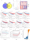
Differential mRNA expression profiles of radiotherapy-resistant cell lines compared to their parental counterparts by mRNA-seq. A Scatter plots of differential expression mRNA (Hela-RR vs. Hela; Siha-RR vs. Siha). B Volcano plots of differential expression mRNA (Hela-RR vs. Hela; Siha-RR vs. Siha). C Hotspot plots of differential expression mRNA (Hela-RR vs. Hela; Siha-RR vs. Siha). D Venn diagram displaying the differential up-regulated mRNAs of two cervical cancer acquired radiotherapy resistant cell lines compared to their respective corresponding parental cell lines. E Venn diagram displaying the differential down-regulated mRNAs of two cervical cancer acquired radiotherapy resistant cell lines compared to their respective corresponding parental cell lines. F Quantitative comparison of qRT-PCR result of mRNA differential levels of HSPA8, TNFAIP6, CXCL8, PPFIA4 in Hela-RR and Hela, Siha-RR and Siha cells. Statistical significance was determined by two-tailed Student’s t-test. (Significance markers: *P < 0.05, **P < 0.01, ***P < 0.001; ****P < 0.0001; ns = not significant (P > 0.05).)
Fig. 7
Fig. 7
Differential expression of CRRF-associated prognostic genes in cervical cancer. A Venn maps for taking differential expression gene intersections with CRRFs by using the TCGA-CESC dataset. B Volcano plots of differential expression gene by using the TCGA-CESC dataset with a threshold filter of FDR < 0.05 and |log2FC| > 2. C Heat maps of differential expression gene by using the TCGA-CESC dataset with a threshold filter of FDR < 0.05 and |log2FC| > 2. D KM curves for the one-factor cox significant factor top8: CXCL8, IFI30, HK2, SPP1, IGF1, PAX9, SLC22A3, and ABCB1. EG Lasso-cox analysis to construct the model: E Trajectory of change of each independent variable, where the log value of the independent variable lambda is shown on the horizontal axis and the coefficients of the independent variables are shown on the vertical axis. F Confidence intervals under each lambda. G Regression coefficients of Signature.R (4.2.1) were used for statistics and visualization. Kaplan–Meier analysis, log-rank test and Cox’s proportional hazards regression model were applied to analyze the corresponding data. The combination score was determined using SynergyFinder. (Significance markers: *P < 0.05, **P < 0.01, ***P < 0.001; ****P < 0.0001; ns = not significant (P > 0.05).)
Fig. 8
Fig. 8
CXCL8 expression is higher in cervical cancer than in adjacent normal tissues by GEPIA2 analysis and immunohistochemical verification. A CXCL8 expression in cervical cancer by GEPIA2 analysis. B Survival analysis of CXCL8 expression in cervical cancer by GEPIA2 analysis. C Immunohistochemical verification of CXCL8 expression in cervical cancer tissues. D Immunohistochemical verification of CXCL8 expression in adjacent normal tissues. E The expression of CXCL8 in tumor tissues from cervical cancer patients and corresponding adjacent normal tissue was examined by immunohistochemical analysis (n = 12). All 12 paired IHC images for CXCL8 expression are presented in Supplementary Fig. 5. Statistical significance of CXCL8 expression in cervical cancer tissues and corresponding adjacent normal tissue was determined by paired t-test. (Significance markers: *P < 0.05, **P < 0.01, ***P < 0.001; ****P < 0.0001; ns = not significant (P > 0.05).)
Fig. 9
Fig. 9
CXCL8 knockdown restores radiotherapy sensitivity in acquired radioresistant cervical cancer cell lines. A Quantitative comparison of clonogenic capacity of Hela-RR cell lines with or without CXCL8 knockdown after irradiation. B Quantitative comparison of clonogenic capacity of Siha-RR cell lines with or without CXCL8 knockdown after irradiation. C Time-dependent cell proliferation curves of Hela-RR and Siha-RR cells with or without CXCL8 knockdown after irradiation. D Quantitative comparison of resistant capacity to apoptosis after radiation exposure with or without CXCL8 knockdown in Hela-RR lines. E Quantitative comparison of resistant capacity to apoptosis after radiation exposure with or without CXCL8 knockdown in Siha-RR lines. F Quantitative comparison of G0/G1, S and G2/M phase with or without CXCL8 knockdown in Hela-RR lines in different treatment groups (unirradiated and 12 h after irradiation). G Quantitative comparison of G0/G1, S and G2/M phase with or without CXCL8 knockdown in Siha-RR cells in different treatment groups (unirradiated and 12 h after irradiation).All data are presented as mean ± standard deviation. Statistical significance was determined by two-tailed Student’s t-test.(Significance markers: *P < 0.05, **P < 0.01, ***P < 0.001; ns = not significant (P > 0.05).)
Fig. 10
Fig. 10
Exogenous CXCL8 induces radiotherapy resistance in parental cell lines. A Quantitative comparison of clonogenic capacity of Hela cell lines with or without exogenous CXCL8 after irradiation. B Quantitative comparison of clonogenic capacity of Siha cell lines with or without exogenous CXCL8 after irradiation. C Time-dependent cell proliferation curves of Hela and Siha cells with or without exogenous CXCL8 after irradiation. D Quantitative comparison of resistant capacity to apoptosis after radiation exposure with or without exogenous CXCL8 in Hela lines. E Quantitative comparison of resistant capacity to apoptosis after radiation exposure with or without exogenous CXCL8 in Siha lines. F Quantitative comparison of G0/G1, S and G2/M phase with or without exogenous CXCL8 in Hela lines in different treatment groups (unirradiated and 12 h after irradiation). G Quantitative comparison of G0/G1, S and G2/M phase with or without exogenous CXCL8 in Siha cells in different treatment groups (unirradiated and 12 h after irradiation).All data are presented as mean ± standard deviation. Statistical significance was determined by two-tailed Student’s t-test.(Significance markers: *P < 0.05, **P < 0.01, ***P < 0.001; ns = not significant (P > 0.05).)

Similar articles

Cited by

References

    1. Yadav, G., Srinivasan, G. & Jain, A. Cervical cancer: novel treatment strategies offer renewed optimism. Pathol. Res. Pract.254, 155136 (2024). - PubMed
    1. Observatory, G. C. Cancer over time.
    1. Siegel, R., Miller, K. & Wagle, N. Cancer statistics, 73, 17–48 (2023). (2023). - PubMed
    1. Jemal, A., Bray, F. & Center, M. Global Cancer Stat.61, 69–90 (2011). - PubMed
    1. Green, J. A. et al. Survival and recurrence after concomitant chemotherapy and radiotherapy for cancer of the uterine cervix: a systematic review and meta-analysis. Lancet358, 781–786 (2001). - PubMed

MeSH terms