

Application of virtual monochromatic images in bone mineral density measurements: a phantom study
- PMID: 40596116
- PMCID: PMC12218281
- DOI: 10.1038/s41598-025-06014-9
Application of virtual monochromatic images in bone mineral density measurements: a phantom study
Abstract
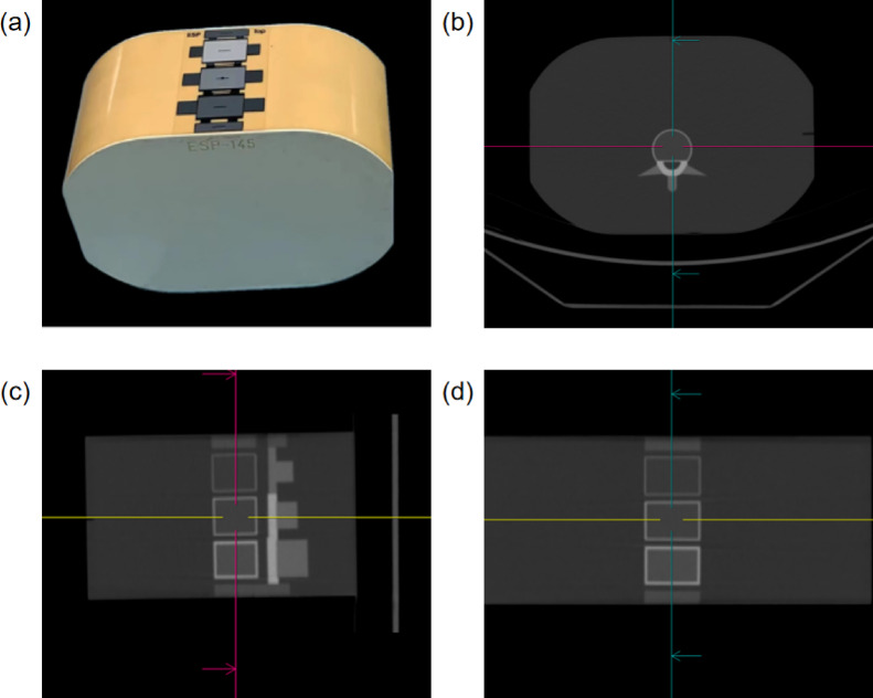
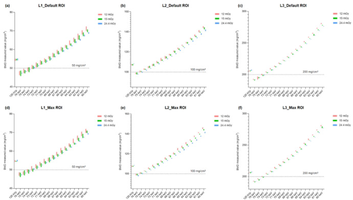
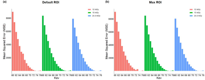
Quantitative CT (QCT) measures bone mineral density (BMD), but beam hardening effect can cause errors and instability in BMD measurements. The purpose of this phantom study is to explore whether the application of virtual monochromatic images (VMIs) in QCT can reduce the measurement error of BMD, and determine the VMI levels that can minimize the error with different radiation doses and regions of interest (ROI) sizes. Utilizing a spine phantom with established hydroxyapatite concentrations, we performed scans with a CT scanner employing rapid tube voltage switching in both single-energy and dual-energy modes. A range of radiation doses (12 mGy, 15 mGy, and 24.4 mGy) was administered, and dual-energy data were reconstructed into VMIs spanning 60-76 keV in 1-keV increments. BMD was measured using regions of interest (ROIs) of varying sizes, and measurements were systematically recorded and compared across all protocols. Our findings indicate that both radiation doses and ROI sizes significantly impacted BMD measurements (P < 0.001). Importantly, BMD values exhibited a consistent decline with increasing VMI levels (rs: - 0.990 to - 0.998, P < 0.001), while measurements derived from 120-kVp images tended to be slightly elevated compared to actual hydroxyapatite concentrations (P < 0.001). Among the evaluated VMIs, the 74-keV level provided the most reliable BMD measurements while ensuring acceptable accuracy. In conclusion, the integration of VMIs in QCT significantly minimizes measurement errors in BMD assessments relative to traditional 120-kVp imaging methods, highlighting their potential to enhance diagnostic accuracy in clinical settings.
Keywords: Bone mineral density; Phantom; Quantitative computed tomography; Virtual monochromatic image.
© 2025. The Author(s).
Conflict of interest statement
Declarations. Competing interests: The authors declare no competing interests.
Figures
References
-
- Mao, S. S. et al. Thoracic quantitative computed tomography (QCT) can sensitively monitor bone mineral metabolism: Comparison of thoracic QCT vs lumbar QCT and dual-energy X-ray absorptiometry in detection of age-relative change in bone mineral density. Acad. Radiol.24, 1582–1587 (2017). - DOI - PubMed
MeSH terms
Substances
Grants and funding
LinkOut - more resources
Full Text Sources
Medical
