. 2025 Jul 1;15(1):22017.
doi: 10.1038/s41598-025-05264-x.

Biochemical and structural characterization of a reactive intermediate deaminase A homolog from Streptococcus sanguinis

Affiliations

Biochemical and structural characterization of a reactive intermediate deaminase A homolog from Streptococcus sanguinis

Alexa B Benedict et al. Sci Rep. .

Abstract

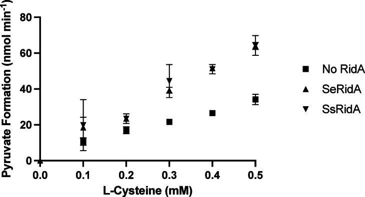
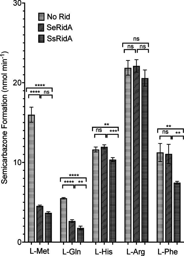
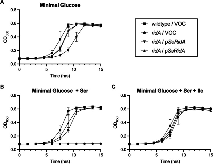
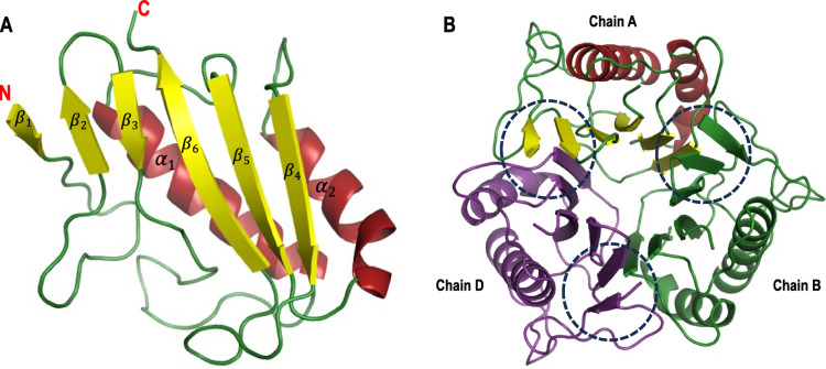
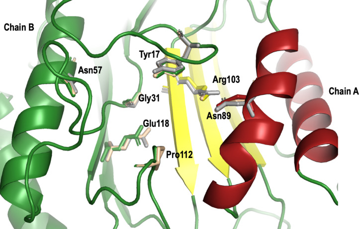
2-Aminoacrylate (2AA) is a short-lived enamine generated as a catalytic intermediate in the dehydration of serine by serine/threonine dehydratase enzymes. 2AA is a metabolic stressor capable of inactivating important pyridoxal phosphate dependent enzymes in a cell. Detoxification of 2AA in a cell is catalyzed by members of the Reactive intermediate deaminase (Rid) family of proteins, which is conserved across all domains of life. We recently identified a RidA homolog, SSA_0809, hereafter SsRidA, in Streptococcus sanguinis with 50% protein sequence identity to a RidA from Salmonella enterica. 2AA deaminase activity assay revealed that SsRidA is capable of enzymatic deamination of 2AA to pyruvate. Furthermore, L-amino acid oxidase assays showed SsRidA has significant activity against several with imino-amino acids similar to the S. enterica RidA. In addition, functional complementation analysis found that SsRidA restored growth of S. enterica ridA mutant in minimal media constituted to increase 2AA stress in the cell. Finally, the crystal structure of SsRidA revealed a homotrimeric protein with active sites at the interface of two interacting monomers. Structure analysis also showed the presence of active site arginine residue along with an active site water molecule implicated in catalysis.

Keywords: Streptococcus sanguinis; 2-Amino acrylate; Enamine deaminase; Metabolic stress; RidA.


Conflict of interest statement

Declarations. Competing interests: The authors declare no competing interests.

Figures

Fig. 1
Fig. 1
SsRidA has 2AA deaminase activity in vitro. Increased pyruvate formation in reactions containing SsRidA indicates that SsRidA is capable of catalyzing 2AA deamination. Each coupled assay contained NADH (0.25 mM), PLP (30 µM), 5LDH (5 U per 300 μl reaction), and CdsH (0.19 μM) in 100 mM Tris–HCl (pH 8.0). RidA proteins (SeRidA or SsRidA) were present at 0.27 μM when indicated. Reactions were performed in triplicate in a 96-well quartz plate and were initiated with addition of L-cysteine at varying concentrations. The plate was shaken for 5 s before first read. The rate of pyruvate formation was calculated using Beer’s law, the extinction coefficient of NADH (6220 M−1 cm−1), and the absorbance at 340 nm from 0 to 30 s. Graph was constructed in Prism 9 (GraphPad). Error bars represent standard deviation.
Fig. 2
Fig. 2
SsRidA displays imino acid deaminase activity in vitro. Each coupled assay contained 50 mM potassium pyrophosphate buffer (pH 8.7), neutralized semicarbazide (100 mM), bovine liver catalase (1 μg/μL), and C. adamanteus L-amino acid oxidase (LOX) (0.5 μg/μL), 10 µM RidA protein when indicated, and 10 μM L-amino acids (L-methionine, L-glutamine, L-histidine, L-arginine, or L-phenylalanine). Reactions were performed in duplicate in a 96-well quartz plate and were initiated with the addition of 10 μM indicated L-amino acid. The plate was shaken for 5 s before first read and absorbance of semicarbazone was measured for 10 min at 248 nm. Graph was constructed in Prism 9 (GraphPad). Error bars represent standard deviation. Significance of this assay was determined using Student’s t test. Asterisk indicates statistical significance (p < 0.05) while ns indicates not significant.
Fig. 3
Fig. 3
SsRidA rescues serine sensitivity of Salmonella enterica ridA mutant. S. enterica ridA mutants harboring one of three plasmids were grown at 37 °C in a 96-well plate with shaking in (A) minimal glucose media, (B) minimal glucose media with serine (5 mM), or (C) serine and isoleucine. The S. enterica wild type carrying empty vector only control (squares) (VOC), or S. enterica ridA mutant harboring empty vector (circles), S. enterica ridA (pSeRidA) (inverted triangles) or S. sanguinis ridA (pSsRidA) (triangles). Graph was constructed in Prism 10 (GraphPad). Error bars represent standard deviation.
Fig. 4
Fig. 4
Overall structure of SsRidA. (A) Cartoon representation of SsRidA monomer structure. The monomer core is composed of two α-helices (red) packed against a mixed beta sheet consisting of six β-strands (yellow) and the loops connecting the α-helices and β-strands are shown in green. (B) Cartoon representation of overall structure of SsRidA homotrimer with active sites shown within dashed circles. Individual interacting monomers of SsRidA homotrimer is colored independently. Chain A was used as representative of SsRidA monomer structure. SsRidA cartoon representation was created using PyMol molecular graphics program, version 2.5.5; Schrödinger LLC (https://www.pymol.org/) with ray tracing set at mode 2.
Fig. 5
Fig. 5
Comparison of the active site of SsRidA with its homolog from E. coli. Superposition of SsRidA active site with active sites from 1QU9 (grey) and 2UYK (wheat) showing the geometry of 7 invariant residues. Active site residues contributed from two adjacent chains, Asn89 and Arg103 from chain A and Tyr17, Gly31, Asn57, Pro112 and Glu118 from chain B are clearly visible in this structure. SsRidA is represented as cartoon structure and follows the same color coding seen in Figs. 1A,B. Active site invariant residues (numbering based on SsRidA) are represented as sticks and the active site residues of SsRidA follows the same color coding of respective chains. Root mean square deviation (RMSD) values between SsRidA (Chain A) and its E. coli homologs 1QU9 (Chain A) and 2UYK (Chain A) are 0.96 and 0.92 respectively for 124 aligned residues. SsRidA cartoon and active site residue stick representations were created using PyMol molecular graphics program, version 2.5.5; Schrödinger LLC (https://www.pymol.org/) with ray tracing set at mode 2. RMSD values were calculated using RCSB pairwise structural alignment tool.

Similar articles

References

    1. Lambrecht, J. A., Flynn, J. M. & Downs, D. M. Conserved YjgF protein family deaminates reactive enamine/imine intermediates of pyridoxal 5’-phosphate (PLP)-dependent enzyme reactions. J. Biol. Chem.287(5), 3454–3461 (2012). - PMC - PubMed
    1. Borchert, A. J., Ernst, D. C. & Downs, D. M. Reactive imines and enamines in vivo: Lessons from RidA paradigm. Trends. Biochem. Sci.44(10), 849–860. 10.1016/j.tibs.2019.04.011 (2019). - PMC - PubMed
    1. Irons, J. L., Gerdes, S., Hodge-Hanson, K. & Downs, D. M. RidA proteins protect against metabolic damage by reactive intermediates. Microbiol. Mol. Biol. Rev.84(3), e00024-e120 (2020). - PMC - PubMed
    1. Fulton, R. L. & Downs, D. M. Modulators of a robust and efficient metabolism: Perspectives and insights from the Rid superfamily of proteins. Adv. Microb. Physiol.83, 117–179 (2023). - PMC - PubMed
    1. Morishita, R. et al. Ribonuclease activity of rat liver perchloric acid-soluble protein, a potent inhibitor of protein synthesis. J. Biol. Chem.274(29), 20688–20692 (1999). - PubMed

MeSH terms

Substances

LinkOut - more resources