. 2025 Jul 1;15(1):21269.
doi: 10.1038/s41598-025-05905-1.

Early evaluation of anti-angiogenic effects with gadolinium(III) labeled APN/CD13 specific binding peptides magnetic resonance imaging

Affiliations

Early evaluation of anti-angiogenic effects with gadolinium(III) labeled APN/CD13 specific binding peptides magnetic resonance imaging

Sheng Han et al. Sci Rep. .

Abstract

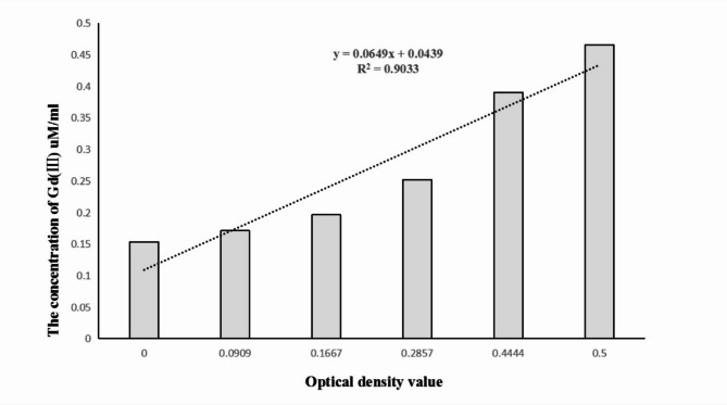
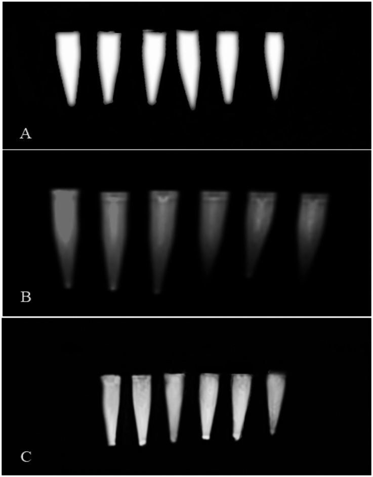
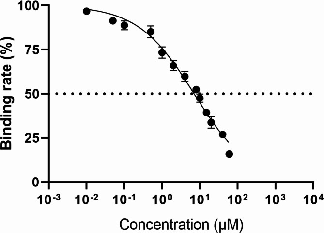
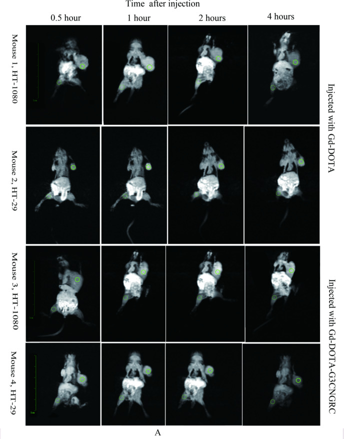
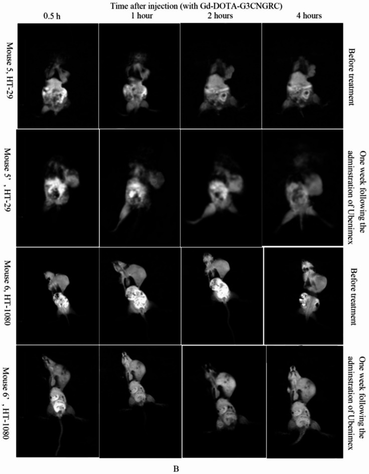
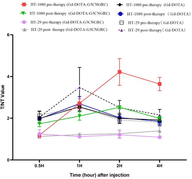
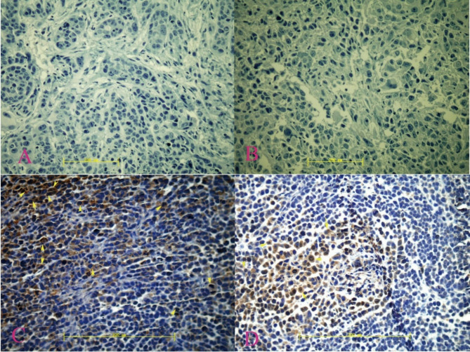
Anti-angiogenesis has been recognized as a crucial strategy in anti-tumor therapy, and the early assessment of its efficacy is equally significant. In this study, we developed a magnetic resonance (MR) probe specifically targeting angiogenesis to facilitate targeted imaging for the early evaluation of anti-angiogenic effects. We synthesized DOTA-G3CNGRC, conjugated it with gadolinium(III), and subsequently evaluated the labeled probe in vitro. The tumor-bearing mouse models of HT-29 (negative for CD13 expression) and HT-1080 (positive for CD13 expression) were successfully established. Magnetic resonance imaging was conducted via intraperitoneal injection of labeled probes and Gd-DOTA, both before and after treatment with ubenimex at a dose of 0.5 mg/kg/day for seven consecutive days. The average signal intensity ratio of the transplanted tumor (target tissue, T) to the left hind leg (non-target tissue, NT) was determined using the region of interest technique (ROI), while changes in tumor size were meticulously recorded. Additionally, APN/CD13 expression levels in transplanted tumors were assessed both prior to and following treatment. The labeling rate of probes was 88.99%. The IC50 of the probes was 7.03 µM. The T/NT ratio of HT-1080 was significantly higher than that of HT-29 (P < 0.001, n = 5). Following treatment, the T/NT ratio of the HT-1080 transplanted tumors was significantly reduced (P < 0.001, n = 5), accompanied by a notable decrease in CD13 expression and negligible changes in the sum of the long and short diameters (P = 0.39, n = 5). The research findings revealed that Gd-DOTA-G3CNGRC can serve as a highly specific gadolinium-based magnetic resonance imaging probe for monitoring the efficacy of anti-angiogenic therapy.

Keywords: APN/CD13; Angiogenesis; Anti-angiogenesis; Asn-Gly-Arg (NGR) peptide; Gadolinium(III).


Conflict of interest statement

Declarations. Competing interests: The authors declare no competing interests.

Figures

Fig. 1
Fig. 1
The standard curve illustrated the relationship between the concentration of Gd(III) and the optical density (OD) value of the xylenol orange mixture. The absorbance (x) at a wavelength of 570 nm, after combining xylenol orange with varying concentrations of gadolinium(III), shows a strong correlation with the concentration of gadolinium(III) (y).
Fig. 2
Fig. 2
Magnetic resonance imaging of Gd-DOTA-G3CNGRC. Gd-DOTA (A) and Gd-DOTA-G3CNGRC (C) both exhibited high-intensity signals, while the ammonium acetate solution (B) displayed low-intensity signals.
Fig. 3
Fig. 3
Cell binding experiment of DOTA-G3CNGRC. In HT-1080 cells, the binding of 99mTc-DOTA-G3CNGRC to the CD13 receptor was inhibited by DOTA-G3CNGRC in a dose-dependent manner.The concentration of the DOTA-G3CNGRC peptide ranged from 0.01 to 60.00 µM. The IC50 value of Gd-DOTA-G3CNGRC was determined to be 7.03 µM (95% CI: 6.47 µM to 7.62 µM, n = 5).
Fig. 4
Fig. 4
Magnetic resonance imaging of tumor-bearing mouse model. Following intraperitoneal injection of Gd-DOTA or Gd-DOTA-G3CNGRC (A), high signal intensity was observed in HT-1080 xenograft tumors (Mouse 1 and Mouse 3), with peak times occurring at 1 h and 2 h post-injection, respectively. In contrast, HT-29 xenograft tumors exhibited high signal intensity only after Gd-DOTA injection (Mouse 2), with peak time consistent with that of HT-1080 xenografts. However, no significant high signal was observed after Gd-DOTA-G3CNGRC injection (Mouse 4). Before and after treatment (B), the signal intensity of the post-treatment HT-29 xenografts (mouse 5’) exhibited no significant change compared to pre-treatment (mouse 5), despite a marked increase in tumor size. In contrast, the signal intensity of the post-treatment HT-1080 xenografts (mouse 6’) significantly decreased compared to pre-treatment (mouse 6), while the tumor size showed no notable variation.The solid and dashed green circles represent the ROI sampling points.
Fig. 4
Fig. 4
Magnetic resonance imaging of tumor-bearing mouse model. Following intraperitoneal injection of Gd-DOTA or Gd-DOTA-G3CNGRC (A), high signal intensity was observed in HT-1080 xenograft tumors (Mouse 1 and Mouse 3), with peak times occurring at 1 h and 2 h post-injection, respectively. In contrast, HT-29 xenograft tumors exhibited high signal intensity only after Gd-DOTA injection (Mouse 2), with peak time consistent with that of HT-1080 xenografts. However, no significant high signal was observed after Gd-DOTA-G3CNGRC injection (Mouse 4). Before and after treatment (B), the signal intensity of the post-treatment HT-29 xenografts (mouse 5’) exhibited no significant change compared to pre-treatment (mouse 5), despite a marked increase in tumor size. In contrast, the signal intensity of the post-treatment HT-1080 xenografts (mouse 6’) significantly decreased compared to pre-treatment (mouse 6), while the tumor size showed no notable variation.The solid and dashed green circles represent the ROI sampling points.
Fig. 5
Fig. 5
T/NT-time curves of HT-1080 and HT-29 xenografts after injection with different contrast agents. In the HT-1080 and HT-29 xenograft tumor models, the peak T/NTratio occurred at 1 h post Gd-DOTA injection, with no significant difference in T/NT ratios observed between the two xenografts before or after treatment. In contrast, Gd-DOTA-G3CNGRC exhibited a T/NT peak at 2 h post-injection in the HT-1080 model, accompanied by significant differences before and after treatment, whereas no distinct peak was observed in the HT-29 model either before or after treatment.
Fig. 6
Fig. 6
Immunohistochemical staining of ANP/CD13 expression in transplanted tumors. HT-29 grafts (A, B) exhibited no significant expression of ANP/CD13, whereas HT-1080 grafts demonstrated high expression of ANP/CD13 prior to treatment with ubenimex (indicated by arrows). Expression of ANP/CD13 was markedly reduced at 7 days post-treatment with ubenimex (indicated by arrows) (20×).

Similar articles

References

    1. Kuai, J., Han, C. & Wei, W. Potential regulatory roles of GRK2 in endothelial cell activity and pathological angiogenesis. Front. Immunol.12, 698424 (2021). - PMC - PubMed
    1. Crippa, L. et al. A new chromogranin A-dependent angiogenic switch activated by thrombin. Blood121, 392–402 (2013). - PMC - PubMed
    1. Liu Chao, Castillo Alesha, B. Targeting Osteogenesis-Angiogenesis coupling for bone repair. J. Am. Acad. Orthop. Surg.26 (7), e153–e155 (2018). - PubMed
    1. Griffioen, A.W. & Dudley, A.C. The rising impact of angiogenesis research. Angiogenesis25, 435–437 (2022). - PMC - PubMed
    1. Lidonnici Jacopo, S., Massimo, M. & Oberkersch Roxana, E. Cancer-Induced metabolic rewiring of tumor endothelial cells. Cancers14 (11), 2735 (2022). - PMC - PubMed