. 2025 Jul 1;15(1):21009.
doi: 10.1038/s41598-025-06906-w.

Biogenic design of silicious architectures on Moso bamboo culm

Affiliations

Biogenic design of silicious architectures on Moso bamboo culm

Makoto Kazama et al. Sci Rep. .

Abstract

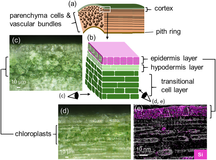
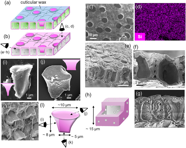
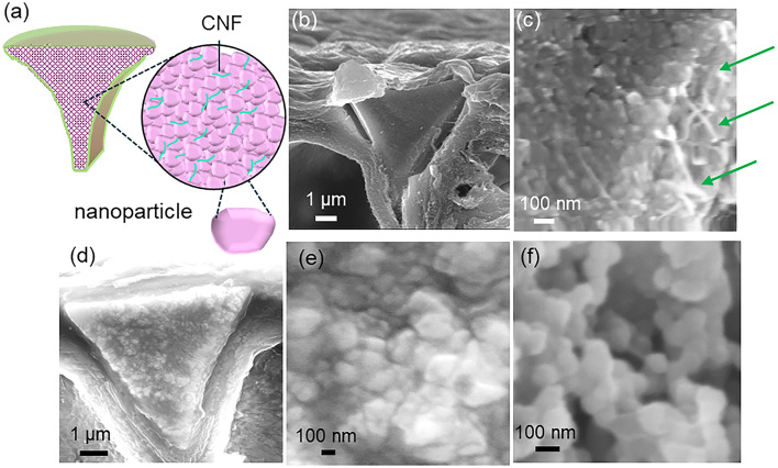
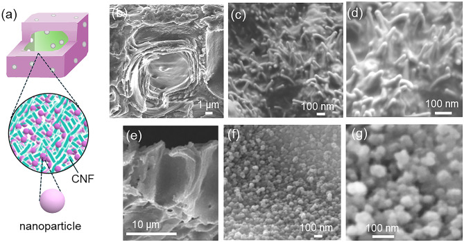
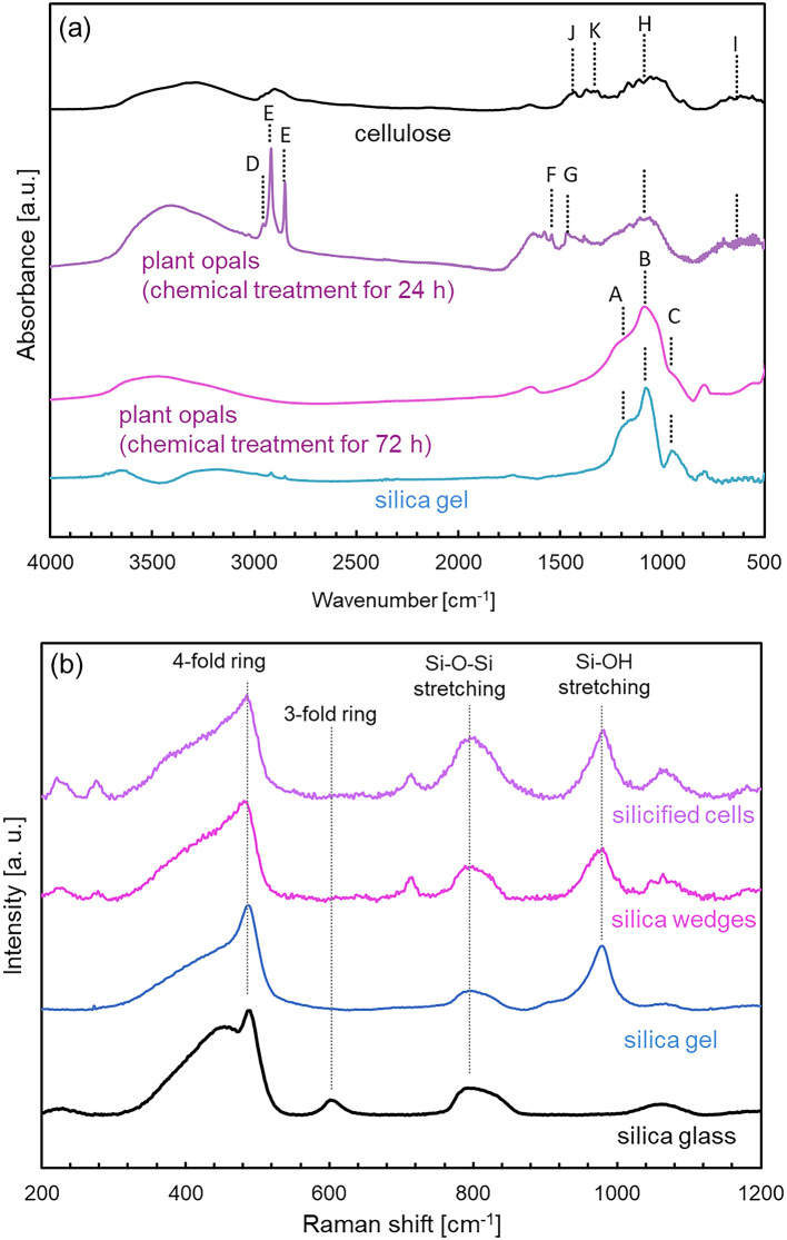
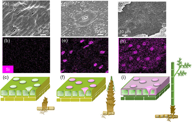
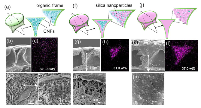
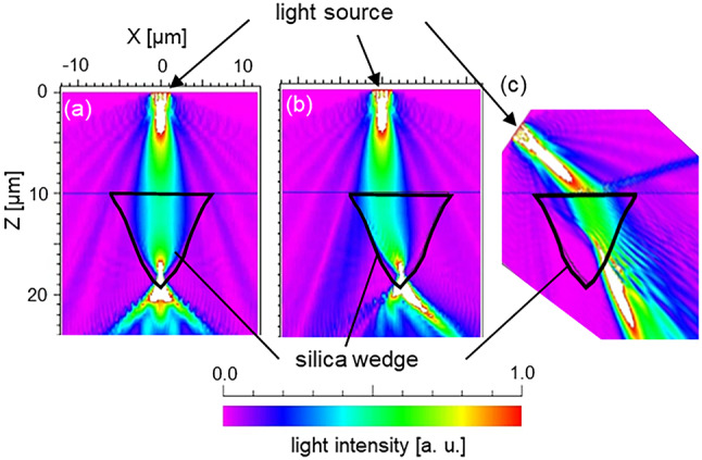
Biosilicas that are produced in vascular plants (plant opal), such as Poaceae, have a variety of shapes and functions and are regarded as an excellent model for the architectural design of artificial amorphous materials. In this work, we studied the micro- and nanostructures and mechanical and optical functions of plant opals on the bamboo culm, which is available as an important natural material. The surface of the culm wall is totally covered with silicified epidermal cells containing silica wedges. The biogenic silicious architectures, such as silicified cell walls and wedges, are composed of nanoscale particles ~ 20-80 nm in diameter with cellulose nanofibrils. Silica wedges, which have a relatively low organic content and relatively high hardness and Young's modulus, are initially formed on cellulose nanofibrils in an organic frame as a scaffold within a few weeks after the emergence of a bamboo shoot. Several months after the formation of the wedges, the epidermal cell walls, which protect the culm surface, are lightly silicified with cellulose nanofibrils. According to a numerical simulation, the silica wedges would have an optical function delivering sunlight to chloroplasts located under the epidermal cells.


Conflict of interest statement

Competing interests: The authors declare no competing interests.

Figures

Fig. 1
Fig. 1
Illustrations (a, b) and optical (c, d) and scanning electron micrograph (SEM) images with the elemental (Si) mapping (e) of the cross sections of a culm wall of moso bamboo. The cross sections perpendicular (c) and parallel (d, e) to the growth direction of the culm. Elliptical grains observed in the parenchyma are transitional layer cells (d, e).
Fig. 2
Fig. 2
Schematic illustrations (a, b, h, l) and SEM images (c, eg, ik) with elemental (Si) mapping (d) of the epidermal layers before (a) and after calcination (bl). The surface (ad), the epidermal cells (eh), and the wedges (il). Pink areas indicate the presence of silica. Eye symbols indicate the direction of observation.
Fig. 3
Fig. 3
Schematic illustration (a) and SEM images (bf) of the silica skeletons for the wedge before (ac) and after partial removal of organic matter with chemical treatments with hypochlorite (df). Green arrows indicate CNFs. The treatment periods were 1 (e) and 10 min (f).
Fig. 4
Fig. 4
Schematic illustrations (a) and SEM images (bg) of the silica skeleton for the silicified cells before (ad) and after (eg) partial removal of organic matter with a chemical treatment with hypochlorite.
Fig. 5
Fig. 5
FT-IR absorption (a) and Raman scattering spectra (b) of plant opals and commercially available silica gel (Kanto Chemical). A typical FT-IR spectrum of cellulose (Fujifilm Wako Pure Chemical) is shown in (a). We used plant opals of the epidermal layer after removal of organic matter by a chemical treatment with hypochlorite and a mixture of sulfuric and nitric acids for (a) and (b), respectively. A: the Si–O–Si stretching TO mode (1200 cm−1), B: the Si-O-Si stretching LO mode (1060 cm−1), C: the Si–OH stretching mode (950 cm−1), D: the N–H stretching mode (2950 cm−1), E: the C–H stretching mode (2848 and 2917 cm−1), F: the N–H stretching mode (1540 cm−1), G: the C–N stretching mode (1460 cm−1), H: the C–O stretching mode (1030–1050 cm−1), I: the C–OH stretching mode (660 cm−1), J: the C–H bending mode (1430 cm−1), K: the C–H bending mode (1315 cm−1). Broad absorption bands around 3200–3500 and 1600 cm−1 are assigned to the H–O–H bending and the O–H stretching modes, respectively. A typical Raman spectrum of silica glass and assignments of the signals are shown in (b).
Fig. 6
Fig. 6
SEM (a, d, g) and elemental (Si) mapping (b, e, h) images and schematic illustrations (c, f, i) of the surface of the epidermal layer of bamboo shoots and culm walls that grew for 1 (ac), 2 (df), and 16 (gi) weeks after emergence.
Fig. 7
Fig. 7
Illustrations (a, f, j), SEM images (b, d, e, g, i, k, m), and elemental (Si) mapping (c, h, l) of the formation process of a wedge-shaped organic frame before (ae) and after (fm) the formation of the silica wedges. Internal structure observed from the cross section (b, g, i, k, m) of a wedge-shaped organic frame after critical drying. We observed the culm of the 1 st (jm), 3rd (fi), and 5th (ae) internodes counting from the base of a bamboo shoot after two weeks from emergence on the ground.
Fig. 8
Fig. 8
Light intensity maps through silica wedges obtained by the BPM. The beams are emitted from the light source located at the central axis of the wedge for the vertical direction (a) and off-axis for the vertical direction (b) and the oblique direction (c). The conditions for the numerical simulation are shown in Fig. S10 in the SI. The light wavelength was set at 660 nm, which is the maximum absorption wavelength for chloroplast. The refractive indices (n) of biosilicas and the matrix of a culm cortex were approximated by the value of amorphous silicas (n = 1.46) and by the average value of water and plant cells (n = 1.425), respectively. The incident beam diameter at the top surface of the wedge (at Z = 10 μm) was ~ 6 μm. The input light was assumed to be a circular beam with uniform intensity within the beam cross section. The vertical beam focuses on the bottom ridge of the wedge, and then the light diffuses under the wedge (a, b). The oblique beam is also introduced into the region under the wedge (c).

Similar articles

References

    1. Wang, T., Antonietti, M. & Cölfen, H. Calcite mesocrystals: ‘morphing’ crystals by a polyelectrolyte. Chem. Eur. J.12, 5722–5730 (2006). - PubMed
    1. Miyajima, R., Oaki, Y., Kogure, T. & Imai, H. Variation in mesoscopic textures of biogenic and biomimetic calcite crystals. Cryst. Growth Des.15, 3755–3761 (2015).
    1. Owari, Y. et al. Ultrastructure of setae of a planktonic diatom, Chaetoceros coarctatus. Sci. Rep.12, 7568 (2022). - PMC - PubMed
    1. Seto, J., Ma, Y., Davis, S. & Cölfen, H. Structure-property relationships of a biological mesocrystal in the adult sea urchin spine. Proc. Natl. Acad. Sci. USA. 109, 3699–3704 (2012). - PMC - PubMed
    1. Ehrlich, H. Chitin and collagen as universal and alternative templates in biomineralization. Int. Geol. Rev.52, 661–699 (2010).

LinkOut - more resources