

Spatiotemporal terahertz modulation enhances NMDAR-mediated miniature EPSCs
- PMID: 40596445
- PMCID: PMC12216413
- DOI: 10.1038/s41598-025-08207-8
Spatiotemporal terahertz modulation enhances NMDAR-mediated miniature EPSCs
Abstract
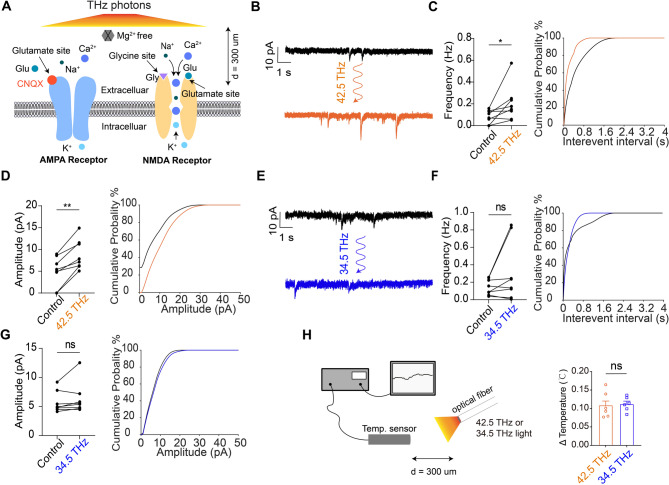
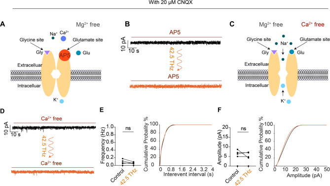
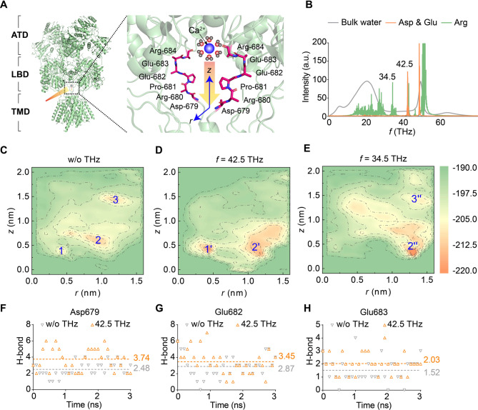
N-Methyl-D-aspartate receptors (NMDAR) are essential for synaptic plasticity and cognitive function, making their modulation a promising strategy for treating disorders like schizophrenia and cognitive impairment. However, methods to selectively modulate NMDAR activity in the lesion's nucleus of the central nervous system remain limited. In this study, using whole-cell patch-clamp recordings, we demonstrated that frequency-specific (42.5 THz) terahertz irradiation significantly enhanced both the frequency and amplitude of NMDAR-mediated miniature excitatory postsynaptic currents (mEPSCs), a response closely linked to Ca²⁺ currents. The mechanism is elucidated via molecular dynamics (MD) simulations, revealing that 42.5 THz irradiation effectively alters the free energy landscape of Ca²⁺ permeating through the NMDAR channel. Specifically, THz photons resonated with key carboxyl groups at the Ca²⁺ binding site, leading to an increase in Ca²⁺ permeability and consequently enhanced mEPSCs. These findings suggest a novel physical therapy approach for treating cognitive deficits and neurological disorders associated with impaired NMDAR function.
Keywords: Molecular dynamics; NMDA receptors; Physical therapy; Terahertz photon; mEPSCs.
© 2025. The Author(s).
Conflict of interest statement
Declarations. Competing interests: The authors declare no competing interests.
Figures
Similar articles
-
Individual NMDA receptor GluN2 subunit signaling domains differentially regulate the postnatal maturation of hippocampal excitatory synaptic transmission and plasticity but not dendritic morphology.Synapse. 2024 Jul;78(4):e22292. doi: 10.1002/syn.22292. Synapse. 2024. PMID: 38813758 Free PMC article.
-
Heterosynaptic Regulation of α2A-Adrenoceptors on Glutamate/GABA Release in the Prefrontal Cortex of Rats.Biomedicines. 2025 May 28;13(6):1322. doi: 10.3390/biomedicines13061322. Biomedicines. 2025. PMID: 40564041 Free PMC article.
-
Topical administration of GLP-1 eyedrops improves retinal ganglion cell function by facilitating presynaptic GABA release in early experimental diabetes.Neural Regen Res. 2026 Feb 1;21(2):800-810. doi: 10.4103/NRR.NRR-D-24-00001. Epub 2024 Jun 26. Neural Regen Res. 2026. PMID: 38934389 Free PMC article.
-
Interventions for preventing weight gain after smoking cessation.Cochrane Database Syst Rev. 2012 Jan 18;1:CD006219. doi: 10.1002/14651858.CD006219.pub3. Cochrane Database Syst Rev. 2012. Update in: Cochrane Database Syst Rev. 2021 Oct 6;10:CD006219. doi: 10.1002/14651858.CD006219.pub4. PMID: 22258966 Updated.
-
Selective noradrenaline reuptake inhibitors for schizophrenia.Cochrane Database Syst Rev. 2018 Jan 25;1(1):CD010219. doi: 10.1002/14651858.CD010219.pub2. Cochrane Database Syst Rev. 2018. PMID: 29368813 Free PMC article.
References
-
- Dupuis, J. P., Nicole, O. & Groc, L. NMDA receptor functions in health and disease: old actor, new dimensions. Neuron111, 2312–2328. 10.1016/j.neuron.2023.05.002 (2023). - PubMed
-
- Suzuki, K., Nosyreva, E., Hunt, K. W., Kavalali, E. T. & Monteggia, L. M. Effects of a ketamine metabolite on synaptic NMDAR function. Nature546, E1–E3. 10.1038/nature22084 (2017). - PubMed
MeSH terms
Substances
Grants and funding
LinkOut - more resources
Full Text Sources
Miscellaneous
