. 2025 Jul 2;15(1):23086.
doi: 10.1038/s41598-025-08491-4.

CD55 may be a new target for colorectal cancer treatment

Affiliations

CD55 may be a new target for colorectal cancer treatment

Qiong Luo et al. Sci Rep. .

Abstract

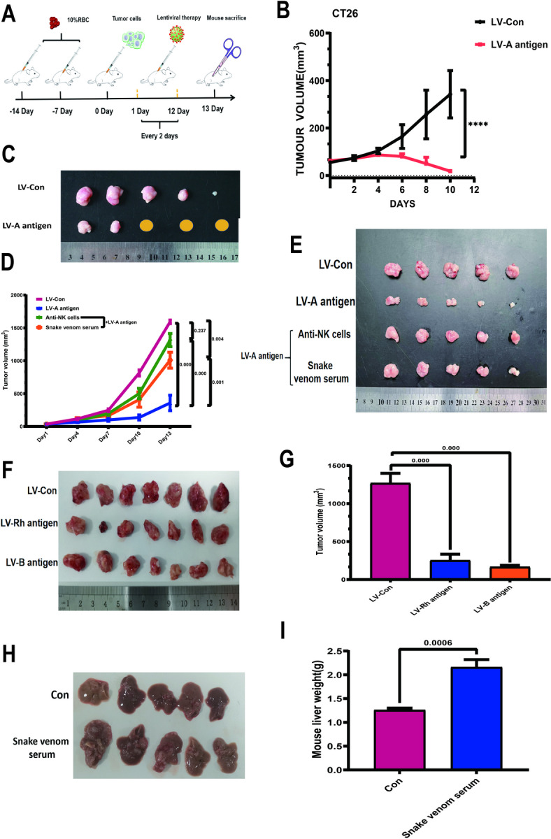
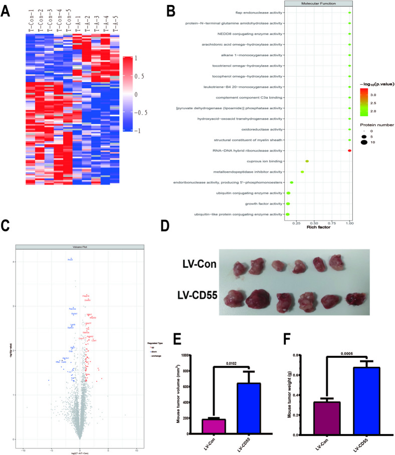
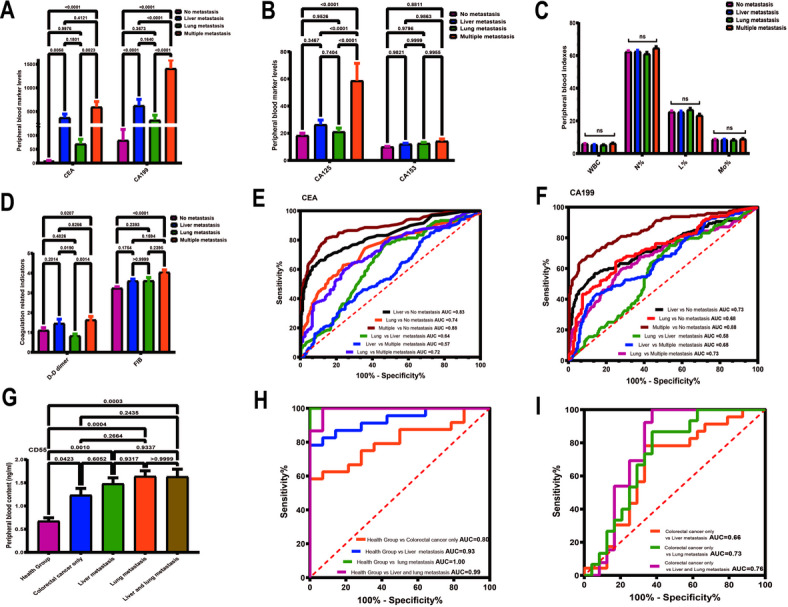
In the early stage, our research group injected lentivirus carrying blood type A antigen into tumors as a drug, and the tumor volume of mice was significantly reduced. We speculate that the complement system plays an important role in anti-tumor therapy, but the specific components are unknown. A mouse model containing blood type antibodies was established. After the mouse axilla formed tumors, lentivirus carrying different blood type antigens was used for treatment. On this basis, NK cell activity was inhibited and the complement system of the body was eliminated. Tumor formation was observed. Flow cytometry was used to observe the changes in tumor tissue and peripheral blood immune cells. Immunohistochemistry was used to observe NK cells in tumors. Proteomics was used to screen differential complement components. Transcriptomics was used to observe the gene expression differences of CT26 cells overexpressing CD55, and RT-PCR was used for verification. The changes of CEA, CA199, CA125, CA153, D-D dimer, total white blood cell count, neutrophil ratio, lymphocyte ratio, monocyte ratio, and fibrinogen in the simple colorectal cancer group, simple liver metastasis group, simple lung metastasis group, and multiple organ metastasis group were retrospectively analyzed, and ROC curves were used for evaluation; ELISA method was used to detect the changes in CD55 content in the healthy group, simple colorectal cancer group, simple liver metastasis group, simple lung metastasis group, and multiple organ metastasis group, and ROC curves were used to evaluate its clinical diagnostic ability. CCK8 and EdU methods were used to detect cell proliferation. Scratch assay and transwell assay were used to observe cell migration. Western blotting was used to observe the changes of MMP9, Vimentin and α-SMA. After the lentivirus carrying blood type A antigen was injected into the axillary tumor of mice, the tumor volume was significantly reduced compared with the control group. The proportion of NK cells in tumor tissue increased significantly. Using antibodies to block NK cell surface activation receptors and eliminate the complement system, both treatments reduced the therapeutic effect. Both blood type B and Rh blood type antigens can significantly reduce tumor volume. Eliminating complement in the body increases the incidence of liver metastasis. The proliferation degree of overexpressed CD55 in tumors is significantly increased. CD55 overexpression can significantly increase the expression of CCL2, CCL3, CCL4, CCL7, CXCL12, TLR7, CSFR1, TCAM, TNF, BTK and MMP9. CEA and CA199 are of certain value in judging liver metastasis, and CD55 has better diagnostic efficacy. Complement significantly inhibits cell proliferation. CD55 significantly promotes CT26 proliferation and migration ability. CD55 is a potential tumor promoter and diagnostic marker for colorectal cancer.

Keywords: ABO blood type; CD55; Colorectal cancer; Complement system; Tumor marker.


Conflict of interest statement

Competing interests: The authors declare no competing interests. Ethical approval and consent to participate: This study was reviewed and approved by the Ethics Committee of Chifeng Hospital(Ethics number: DW2022002 and CK2020004). For the collection of clinical samples, this study strictly adhered to the Declaration of Helsinki, and all individuals participating in the study obtained informed consent and agreed to the publication of the article.

Figures

Fig. 1
Fig. 1
The complement system plays an important role in anti-tumor. (A) Modeling flow chart. 14 days before axillary tumor injection, 10% red blood cell suspension of different blood types was intraperitoneally injected every week. On the third day after axillary tumor injection, lentivirus carrying blood type antigen was used for treatment, and treatment was performed once every 2 days. Mice were killed after 13 days. The flow chart was drawn using WPS Office (PC version 2021, China). (B) Tumor growth curve. Tumor volume was calculated as V = length x width2 × 0.50. Student’s t test was used for testing, and the results are expressed as mean ± SEM, ****P < 0.0001. (C) Mouse tumor images (n = 5 per group). (D, E) Mice producing blood type A antibodies were constructed and then treated with: (1) control virus treatment; (2) lentivirus carrying blood type A antigen treatment; (3) lentivirus carrying blood type A antigen treatment and blocking NK cell activity; (4) lentivirus carrying blood type A antigen treatment and clearing complement. NK cell blocking was performed using NCR1 Antibody. Snake venom serum was used for complement removal, and the dosage was 12.5 µg/mouse /every 2 Days. The tumor diameter was measured and the volume was calculated on days 1, 4, 7, 10, and 13. The tumor was photographed after sacrifice. n = 5 in each group. P < 0.05 was considered significant. (F, G) The effect of B and Rh blood type antigens on tumor treatment was observed according to the principle of A blood type antigen (n = 7 in each group). The tumor was photographed and the volume was calculated after the mice were sacrificed. P < 0.05 was considered significant. (H, I) CT26 tumor cells 5 × 105/mouse were injected into the spleen of mice to simulate the liver metastasis model of colorectal cancer. Snake venom serum was used to remove complement in mice from the second day. The dosage was 12.5 µg/mouse /every 2 Days. The mice were sacrificed on the 14th day, and the liver tissue was photographed and weighed (n = 5 in each group). P < 0.05 was considered significant.
Fig. 2
Fig. 2
CD55 promotes tumor growth in mice. 5 × 105 CT26 tumor cells were injected into the axilla of mice expressing blood type A antibodies, and 3 days later, they were treated with lentivirus carrying blood type A antigen. Mice were killed on the 13th day, and tumor tissues were removed and tested using proteomics (n = 5 per group). Lentivirus without any gene was used as the control group. (A) Heat map between the two groups shows the expression changes of different proteins. (B) GO analysis. (C) Volcano map. (D–F) Complement component CD55 was initially screened. CD55-expressing lentivirus was constructed. Intratumoral injection of mouse axillary tumors was performed (same method as before). Mice were killed on the 13th day, tumor tissues were photographed, and the volume and tumor weight were measured (n = 6 per group). P < 0.05 was considered significant.
Fig. 3
Fig. 3
CD55 modifies tumor cell heterogeneity. CT26 cells were routinely cultured and infected with a CD55-expressing lentivirus at an MOI of 1:5 for transcriptomic analysis. Empty virus was used as the control group. (A) Heat map. (B) GO analysis of downregulated genes. (C) GO analysis of upregulated genes. (D) KEGG analysis of downregulated genes. (E) KEGG analysis of upregulated genes. (F–H) Quantitative Real-time PCR was used to verify the transcriptomic results. GAPDH was used as an internal reference, and each group was repeated 3 times. Student’s t test was used for testing, and the results are expressed as mean ± SEM. P < 0.05 was considered significant.
Fig. 4
Fig. 4
CD55 has diagnostic value for colorectal cancer metastasis. The relationship between clinical test results and tumor metastasis in patients with colorectal cancer was retrospectively analyzed. Data of 1008 patients in Chifeng Municipal Hospital were collected. According to the pathological type, the patients were divided into: simple colorectal cancer group (409 cases), simple liver metastasis group (249 cases), simple lung metastasis group (151 cases), and multiple organ metastasis group (199 cases). (A) Expression of CEA and CA199 in different groups. (B) Expression of CA125 and CA153 in different groups. (C) Changes in white blood cell count, neutrophil ratio, lymphocyte ratio and monocyte ratio. (E) Determination of D-dimer and fibrinogen content. (E, F) The ROC curve method was used to observe the diagnostic efficacy of CEA and CA199 as diagnostic markers in different groups. (G) Peripheral blood samples of 89 subjects were collected and divided into health group (n = 14), colorectal cancer alone group (n = 24), liver metastasis only group (n = 23), lung metastasis only group (n = 13), and multiple organ metastasis group (n = 15). The Elias method was used to detect the content of CD55 in peripheral blood. (H, I) The ROC curve method was used to observe the diagnostic efficacy of CD55 as a diagnostic marker among different groups. One-way ANOVA was used when there were more than two groups. The differences were considered significant when the P value was < 0.05. International units of tumor markers: CEA (ng/ml); CA15-3; (U/mL); CA125 (U/mL); CA199 (kU/L). Abbreviation: WBC (White Blood Cell, unit: 109/L); FIB (fibrinogen, unit: g/L); N% (neutrophil ratio); L% (lymphocyte ratio); M% (monocyte ratio).
Fig. 5
Fig. 5
CD55 increases the proliferation and migration ability of colorectal cancer. Conventional culture of CT26 cells. Add 1 × 104/well cells to a 96-well plate. Add guinea pig serum to a concentration of 5%. Use a medium containing 10% FBS as a control. Use the CCK8 method to measure the absorbance at 450 nm for 2 h, 4 h, 24 h, and 48 h. (B) Use CD55-carrying lentivirus to infect CT26 cells. Add 1 × 104/well cells to a 96-well plate. Use the CCK8 method to measure the absorbance at 450 nm for 2 h, 4 h, 24 h, and 48 h. Use an empty virus as a control. (C) Repeat the above experiment using the Edu method. (D) Use CD55-carrying lentivirus to infect CT26 cells. Evenly place 200 cells in a small dish. Stain with Giemsa 2 weeks later and observe the results. (E) Use CD55-carrying lentivirus to infect CT26 cells. Add the cells to a small dish to ensure 5 × 105/dish. On the second day, a horizontal line was drawn in each well with a pipette tip, and the cells were observed and photographed under a microscope 24 h later. (F) 1 × 104/well was added to the upper chamber of the Transwell chamber, and crystal violet was used to stain and photographed 24 h later. (G, H) CT26 cells were infected with CD55 lentivirus, and the expression of EMT-related proteins was detected by Western Blot. GAPDH was used as an internal reference to analyze the relative gray value. The above experiments involved the sampling of quantitative data with mean ± SEM, and Student’s t-test was used for statistical analysis. At least three replicates were required for each group. P < 0.05 was considered statistically significant.

Similar articles

References

    1. Xi, Y. & Xu, P. Global colorectal cancer burden in 2020 and projections to 2040. Transl Oncol.14 (10), 101174. 10.1016/j.tranon.2021.101174 (2021). - PMC - PubMed
    1. Poturnajova, M. et al. Molecular features and gene expression signature of metastatic colorectal cancer (Review). Oncol. Rep.45 (4), 10. 10.3892/or.2021.7961 (2021). - PMC - PubMed
    1. Roncato, R. et al. The challenge of molecular selection in liver-limited metastatic colorectal cancer for surgical resection: a systematic review and meta-analysis in the context of current and future approaches. Oncol. Res.32 (9), 1407–1422. 10.32604/or.2024.049181 (2024). - PMC - PubMed
    1. Gharib, E. & Robichaud, G. A. From crypts to cancer: a holistic perspective on colorectal carcinogenesis and therapeutic strategies. Int. J. Mol. Sci.25 (17), 9463. 10.3390/ijms25179463 (2024). - PMC - PubMed
    1. He, X. et al. Can immunotherapy reinforce chemotherapy efficacy? A new perspective on colorectal cancer treatment. Front. Immunol.14, 1237764. 10.3389/fimmu.2023.1237764 (2023). - PMC - PubMed