. 2025 Jul 1;8(1):965.
doi: 10.1038/s42003-025-08370-8.

A role of pigment epithelium-derived factor in zinc-mediated mechanism of neurodegeneration in glaucoma

Affiliations

A role of pigment epithelium-derived factor in zinc-mediated mechanism of neurodegeneration in glaucoma

Dmitry V Chistyakov et al. Commun Biol. .

Abstract

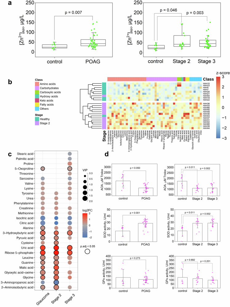
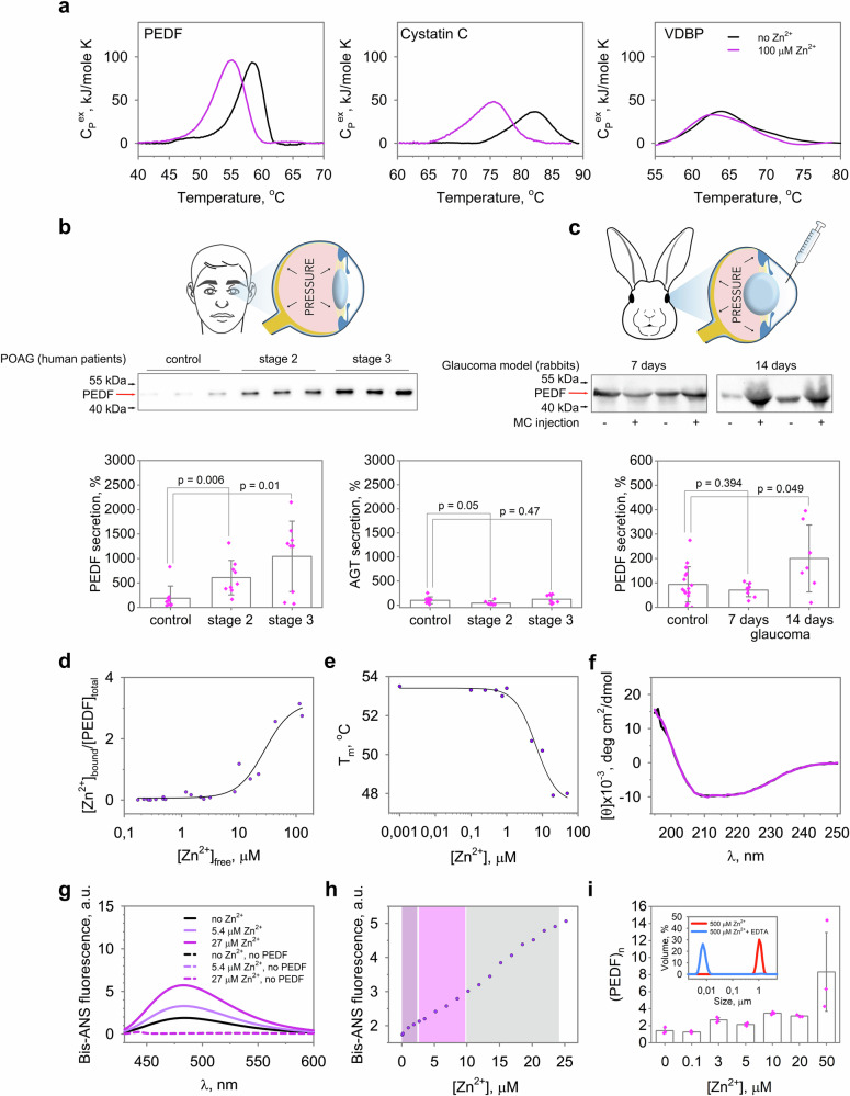
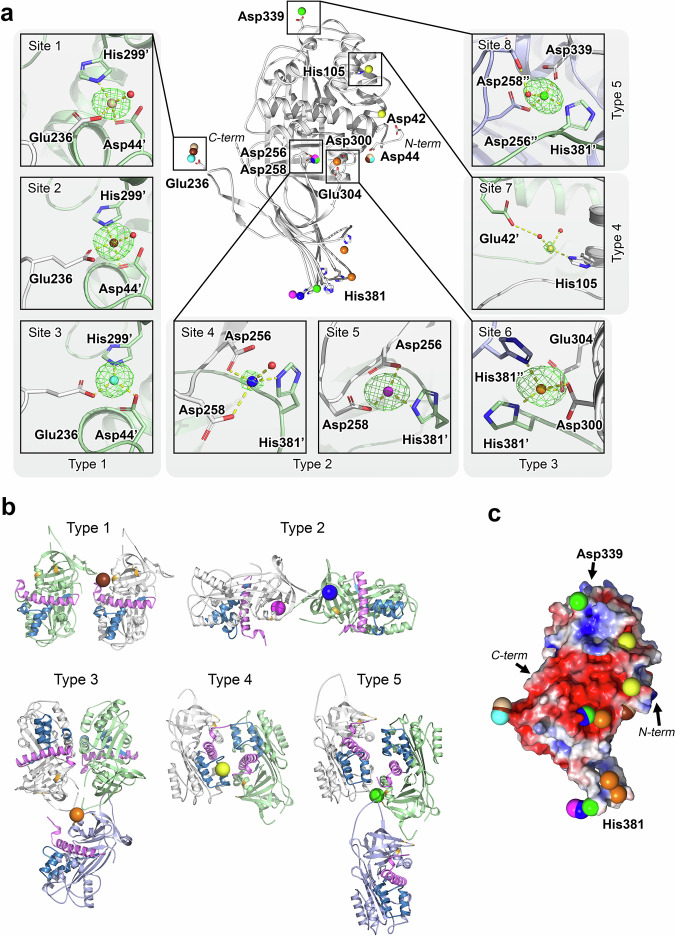
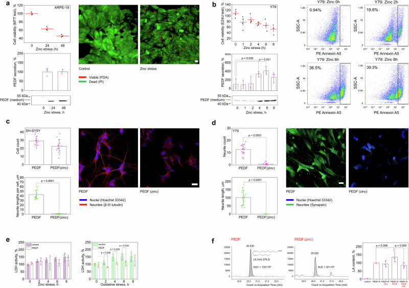
Glaucoma is a neurodegenerative condition involving optic nerve damage and retinal ganglion cells death. Animal studies suggested that the pathway linking these events can be mediated by mobile zinc secreted into the intraretinal space and exerting cytotoxic effects. Whether this mechanism is relevant for human glaucoma and what are the targets of extracellular zinc is unknown. We report that increased zinc content in the aqueous humor and retina is indeed a characteristic of glaucomatous neuropathy, and excess extracellular zinc may be recognized by the key retinal neurotrophic factor PEDF. Biophysical and X-ray crystallographic studies show that PEDF coordinates zinc ions in five types of intermolecular high-affinity sites, leading to a decrease in negative surface charge and reversible oligomerization of the protein, thereby masking the target recognition sites responsible for its neurotrophic and antiangiogenic activities and collagen binding. Notably, PEDF secretion is enhanced in both glaucoma and retinal cell models in response to zinc stress; however, zinc binding negatively affects axogenic, differentiative and prosurvival functions of PEDF by suppressing its ability to activate receptor PEDF-R/PNPLA2. We suggest that glaucomatous neurodegeneration is associated with direct inhibition of PEDF signaling by extracellular zinc, making their complex a promising target for neuroprotective therapy.


Conflict of interest statement

Competing interests: The authors declare no competing interests.

Figures

Fig. 1
Fig. 1. Glaucoma is characterized by altered zinc homeostasis and oxidative stress in patients.
a Total zinc content in AH of participants (Supplementary Data 1) was analyzed using AAS. Zinc concentration significantly increased from as early as stage 2 of POAG (p < 0.05). b, c Identification of low molecular weight zinc chelators in AH by full-scale metabolomic (GC-MS) analysis. b Heat map illustrating changes in the concentration of AH metabolites in stage 2 POAG. Mean-centered and unit variance scaled concentrations (z-scores) for 46 metabolites with p < 0.05 and/or VIP score > 1 are shown. c Zinc-binding metabolites in AH showing alterations in glaucoma. The dot plot illustrates the binary logarithm of fold change (FC), the significance of the variable in prediction (VIP), and the false discovery rate (FDR)-adjusted p-values (p.adj) for the major zinc chelators relative to the control group. The points with p < 0.05 and/or VIP score > 1 are highlighted. The general trend among the most affinity zinc chelators is an increase in amino acids and a decreased in citrate and isocitrate. d Markers of oxidative stress in AH were evaluated by colorimetric methods. Glaucoma is associated with oxidative stress manifested in AH as a significant decrease in AOA with a compensatory increase in Zn2+-dependent SOD (p < 0.05), but without a significant change in GPx activity. The data in a and d are presented as mean and error bars indicate SEM.
Fig. 2
Fig. 2. Glaucoma neuropathy induced by ocular hypertension in an animal model is associated with increased zinc content in AH and retina and oxidative stress.
a Methylcellulose (2%) was injected into the anterior chamber of rabbits, resulting in sustained IOP elevation over 7–14 days (n = 8; p < 0.05 for all time points). b Representative electroretinogram and the mean amplitudes of its a- and b-waves showing a decrease in outer retinal activity on day 14 after methylcellulose injection (n = 6). c Representative micrographs of optic nerve cross-sections, hematoxylin and eosin, magnification 50×. Green and red arrows indicate normal optic disc of the control animal and optic disc excavation on day 14 after methylcellulose injection, respectively. Scale bar represents 200 µm. d Representative micrographs of retinal cross-sections, hematoxylin and eosin, magnification 200×. The designations are as follows: PL, photoreceptor layer, ONL, outer nuclear layer, OPL, outer plexiform layer, INL, inner nuclear layer, IPL, inner plexiform layer, and GCL, ganglion cell layer. Green arrows show viable RGC nuclei, red arrows show pyknotic (apoptotic) RGC nuclei on days 7 and 14 after methylcellulose injection. The scale bar represents 50 µm. e Representative confocal immunofluorescence images of retinal cross-sections obtained before and on day 14 after methylcellulose injection. The specific ganglion cell marker Brn3a was stained with polyclonal antibodies and detected using indirect fluorescence (red); call nuclei were visualized by Hoechst 3342 (blue). The bottom panel shows the results of morphometric evaluation of the total number of viable RGCs based on histologic (n = 9) and immunocytochemical (n = 5) analyses. Scale bar represents 10 µm. f Total zinc content in AH (n = 8) and retina (n = 11) of rabbits with ocular hypertension model was determined by AAS. Zinc concentration increased significantly on the 7th day after methylcellulose injection (p < 0.05). g Determination of oxidative stress markers in AH by colorimetric methods revealed a decrease in AOA and an increase in SOD activity on day 7 after methylcellulose administration (n = 10; p < 0.05). All data are presented as mean and error bars indicate SEM.
Fig. 3
Fig. 3. Ocular neurotrophic factor PEDF is zinc-dependent protein associated with glaucoma.
a Zinc-binding properties of AH proteins PEDF, cystatin C, and VDBP, predicted to contain zinc-coordinating sites by ZincBindPredict, were probed by DSC. PEDF and Cystatin C exhibited a shift in melting temperature in the presence of Zn2+. b The content of signaling factors PEDF and AGT was analyzed in AH of POAG patients and control individuals (taken as 100%) by Western blotting. c PEDF content was determined in AH of animals with or without (taken as 100%) the ocular hypertension model on day 7 (n = 8) and day 14 (n = 8) after induction. A progressive increase in the amount of secreted PEDF was observed during development in both human and experimental glaucoma (p < 0.05). d, e Zinc binding to PEDF was investigated by equilibrium dialysis coupled with AAS and by recording the zinc-dependence of Tm using DSF, showing stoichiometry of 3 and micromolar affinity (solid curves represent the fit of the experimental data using the Hill equation). PEDF structure was analyzed using CD spectroscopy (f) and the fluorescent dye bis-ANS (g) in the presence of 5.4 µM or 27 µM Zn2+. Zinc binding did not affect the secondary structure but increased surface hydrophobicity of PEDF. h Effect of zinc on PEDF structure was monitored using bis-ANS fluorescence assay. Different zinc binding modes were observed at a Zn:PEDF ratio of 1:1 (magenta), at zinc excess 2-4 (pink), and at zinc excess >4 (light gray). i Oligomeric state of PEDF in the presence of zinc was evaluated by DLS. Zinc binding induced the transition of PEDF monomer into dimers (3-5 µM Zn2+), trimers (10–20 µM Zn2+) of larger oligomers (>20 µM Zn2+). In the presence of metal chelator EDTA, the oligomerization induced by high excess of zinc (500 µM) was reversed (inset), indicating its functional significance. The data in b and i are presented as mean and error bars indicate SEM.
Fig. 4
Fig. 4. Crystal structure of PEDF revealed zinc binding sites.
a The crystal structure of PEDF revealed 8 zinc coordination sites located at the intermolecular interface of PEDF dimers and trimers. In the figure, the sites are numbered clockwise and divided into 5 types according to their coordination. Zinc ions corresponding to sites 1–8 are shown in wheat, chocolate, blue, blue, purple, orange, yellow and green colors, respectively. Site 1 was observed in P21212 crystals, while sites 2-8 were found in P212121 crystals. Anomalous difference maps are indicated as green mesh at the 7σ-level. Ions of different colors appeared several times in the figure, showing different interaction interfaces on different monomers within one oligomer. Different colors of protein molecules correspond to different molecules in one oligomer. Zinc coordination bonds are represented by yellow dashed lines. Refer to Supplementary Fig. 9 for additional structural details of the Zn2+-binding sites. b The filling of five types of Zn2+-binding sites leads to the formation of five variants of PEDF dimers and trimers, which affects the accessibility of target recognition sites important for PEDF functions. These include PEDF-R binding and neurotrophic activity (44-mer fragment 78-121, shown in blue), anti-angiogenic activity (34-mer fragment 44-77, shown in purple), and collagen binding (Asp256, Asp258, and Asp300 shown in orange). Refer to Table 3 for details of function inhibition resulting from PEDF di-/tri-merization. c Zinc coordination alters the surface properties of PEDF, such as weakening the negative electrostatic charge (red) of the characteristic region of the protein.
Fig. 5
Fig. 5. Zinc stimulates secretion but suppresses neurotrophic activity of PEDF.
Effect of zinc stress on PEDF secretion was studied in ARPE−19 (a) and Y79 (b) cells. a APRE−19 shows a slow response to zinc stress (half-maximum after 24 h), as confirmed by MTT test, as well as fluorescein diacetate/propidium iodide (FDA/PI) viability assay and confocal microscopy (Scale bar represents 25 μm, n = 3). b Zinc treatment decreased viability (half-maximal after 8 h) and induced apoptosis of Y79 cells, as evidenced by EZ4U test and flow cytometric annexin A5 assay (n = 6). In both cases, zinc stress was accompanied by PEDF release (p < 0.05). The amount of PEDF in the absence (Y79) or at 24 h (ARPE−19) of stress was taken as 100%. c-e Effect of zinc on the neurotrophic activity of PEDF was studied on SH-SY5Y cells (c) and Y79 cells (d-e). c Preincubation of PEDF with zinc reduced its axogenic activity in respect to differentiated SH-SY5Y cells without affecting cell count (Scale bar represents 25 μm, n = 14). d Zinc inhibited the ability of PEDF to stimulate neuronal differentiation of Y79 cells (Scale bar represents 10 μm, n = 21). Neurites and nuclei were stained with anti-β-III tubulin (c) or synapsin (d) antibodies and Hoechst 33342, respectively, and visualized by confocal microscopy. e PEDF attenuated Y79 cell death under oxidative stress but not zinc stress, as shown by LDH assay (n = 3). Leaked LDH activity in the absence of stress was taken as 100%. f Phospholipase activity of PEDF-R in ARPE−19 cell membranes was monitored in the presence of apo or zinc-bound PEDF by quantifying fatty acid release from model phospholipid substrate using HPLC-MS/MS. Extracted ion chromatogram illustrating differences in linoleic acid ([M-H]- LA, m/z 279.2) formation in the presence of apo and zinc-bound PEDF. The histogram on the right illustrates the attenuation of PEDF-mediated enhancement of PEDF-R activity in the presence of zinc (p < 0.05). PEDF-R activity without PEDF/zinc was taken as 100%. All data are presented as mean and error bars indicate SEM.
Fig. 6
Fig. 6. Blockade of PEDF by zinc may contribute to neurodegeneration in glaucoma.
a In the healthy retina, PEDF is a component of normal trophic support of RGCs that is secreted by Müller cells and stimulates phospholipase A2 activity of PEDF-R, resulting in increased production of DHA and upregulation of other neurotrophic factors such as BDNF. b In glaucoma, oxidative stress causes the release of mobile zinc from metallothioneins (MTs) into the extracellular space, which triggers apoptotic signaling in RGCs. Under these conditions, PEDF is intensely secreted by retinal cells but becomes inactive because zinc binding reduces its negative surface charge and promotes oligomerization, which impairs the neuroprotective and regenerative activity of the protein. Along with suppression of other Zn2+-sensitive neurotrophic factors (BDNF, NGF, etc.) that support RGCs growth and survival, this mechanism contributes to neurodegeneration in glaucoma.

References

    1. Toth, K. Zinc in neurotransmission. Annu Rev. Nutr.31, 139–153 (2011). - PubMed
    1. Sensi, S. L., Paoletti, P., Bush, A. I. & Sekler, I. Zinc in the physiology and pathology of the CNS. Nat. Rev. Neurosci.10, 780–791 (2009). - PubMed
    1. Kochanczyk, T., Drozd, A. & Krezel, A. Relationship between the architecture of zinc coordination and zinc binding affinity in proteins-insights into zinc regulation. Metallomics7, 244–257 (2015). - PubMed
    1. Ugarte, M. & Osborne, N. N. Recent advances in the understanding of the role of zinc in ocular tissues. Metallomics6, 189–200 (2014). - PubMed
    1. Zhang, C., Dischler, A., Glover, K. & Qin, Y. Neuronal signalling of zinc: from detection and modulation to function. Open Biol.12, 220188 (2022). - PMC - PubMed